![]()
![]()
![]()
Use LEFT and RIGHT arrow keys to navigate between flashcards;
Use UP and DOWN arrow keys to flip the card;
H to show hint;
A reads text to speech;
41 Cards in this Set
- Front
- Back
|
Anatomy of the "vestibule" |
Rent of bone (inner ear) containing delicate bone chambers, which are filled with endolymph |
|
|
The "Vestibular Apparatus" |
Organs for balance and posture consisting of the article, saccule and semi-circular canals |
|
|
Cochlea |
The auditory apparatus for hearing |
|
|
Sensory structures in article and saccule |
Macula (1 in each) containing hair cells + otolith |
|
|
Sensory structures in ampullae of semi-circular canals |
Crista ampullaris ( 1 in each canal) containing hair cells+capula |
|
|
Sensory structures in cochlea |
Organ of got to (1 in each ear) containing hair cells+basilar and tectorial membranes |
|
|
Sensory interaction of vestibular structures |
Vestibular nerve+cochlear nerve are bundled together into the "auditory nerve". Each auditory nerve later joins the facial nerve to form Cranial Nerve VIII. Each vestibular nerve contains 10,000 rivers. (Each cochlear nerve contains 30,000 rivers). |
|
|
Circuit for control of posture and balance by vestibular apparatus reflexes inv9lves the following CNS components: |
A) vestibular apparatus (utricle-saccule-semi-circular canals) B) vestibular nuclei in brainstem C) flocculo-nodular love of cerebellum D) cranial oculomotor nerves and extraocular muscles of eyes E) motor nerves to postural muscles of skeleton |
|
|
The hair in vestibular apparatus creates... |
Generator potentials when it deodorizer, the hair cell stimulates action potentials in attached vestibular nerve endings |
|
|
Vestibular apparatus hair cell machanism |
When kinocilium bends away from stereocilia, it creates a "notch" region of positive-ion permeability-> positive ions enter hair cell -> inside depolarizes (more +) -> attached vestibular nerve ending depolarizes-> generated volley of action potentials in vestibular nerve |
|
|
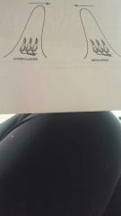
How hair cells signal movement and position in the macular of the utricle and saccule |
A) all cilia of hair cells trapped in gel upon which "floats" the otolith (a stone later of CaCO3 crystals) :) the inertia** of the otolith creates a vending of cilia, depolarizing some -> detects static head position and acceleration** |
|
|
How hair cells detect movement in the crisra ampullaris of the semi-circular canals |
A) the cilia of hair cells in each crista are trapped in the gel of the cupula, which bends in response to flow of endolymph in canals B) the inertia** of endolymph causes bending of the crusts, which causes bending of cilia in the cristae-> detects acceleration** |
|
|
Generator potentials |
Resting potentials in the hair cell creates when the cell let's K+ ion into the cell. There are not action potentials!! They go up or dow, with the vending of cilia. When depilarized, the hair cells relase a transmitter at its synapses with the sensory nerves, beginning a set of action potentials there |
|
|
Inertia |
Pendant of a moving object to keep moving, or an immobile object to stay put |
|
|
Velocity |
(distance per unit time) describes steady movements in a given direction |
|
|
Acceleration |
Is used to describe a change of motion I.e. slowing down or speeding up. Its units are + or - (rate of change of velocity/time). For example, when something drops off a cliff, it speeds up until it hits the ground, due to gravity. The acceleration of the object due to gravity is predictable: it increases its speed 32 ft/second each second it falls |
|

Name these parts of the vestibular apparatus |
A) semi-circular canals detect acceleration and control eye movement reflexes B1) superior canal B2) posterior canal C) ampullae D)horizontal canal E) crusts ampullaris inside F) utricle G) saccule H) cochlear nerve I) facial nerve J) cochlear K) vestibular nerve ganglion |
|
|
Hair cell transducer |
Concerts mechanical force and motion into a change in its membrane potential. Tiny protein cables (tip links) from the tips to,the descending rows of stereocilia of the hair cell pull open a positive-ion channel in the cell membrane of the adjacent stereocilium when the hairs are pulled in one direction-taller away from the shorter rows These positive charges depolarize the cell (more positive inside) and cause the release of transmitter at the synapses to the nerve When hairs are pulled in the opposite direction(taller toward shorter). The tip-links relax, the +ion channels close, and the inside becomes more negative (hyperpolarizes). This causes the hair cell's membrane potential either increases or decreases the number of action potentials that are generated in the attached sensory nerves. Because it can generate an action potential in the attached nerves, the membrane potential of the hair cell is called a generator potential. Hair cells inside the cochlea have the same tip links etc.. But have no kinocilium |
|
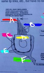
Name parts of hair cell |
A) kinocilium B) stereocilia C) receptor nerve ending D)hair cell body E) synapses F) nerve fiber of vestibular nerve |
|
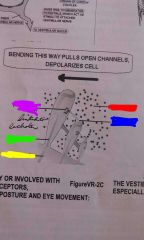
Bending of hair cell |
Purple=positive-ion channel Green=stereocilium Yellow=hair cell body Red= positive ions Blue= tip link to channel |
|
|
A) Otolith B) Kinocilia C) floor of macula D) Vestibular nerve fibers |
|
|
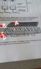
Saccule's macula |
On the side wall of the chamber. The saccule can detect up and down motion |
|
|
Utricle's macula |
On the floor of the chamber. The utricle can detect side-to-side motion |
|
|
A) boney wall of ampulla of semicircular canal B) Cupula C) gel D) hair cells E) endolymph F) vestibular nerve fibers |
|
|
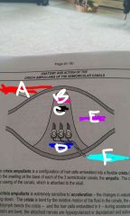
Crista ampullaris |
A configuration of hair cells embedded into a flexible Crista("shelf"), which protrudes into the swelling at the base of each of the three semicircular canals, the ampula. The Crista is attached to the Bony casing of the canals which is attached to the skull. |
|
What does this represent? |
The Crista ampullaris is extremely sensitive to acceleration, the changes in velocity during speeding up or slowing down. The Crista is Bent by the relative motion of the fluid in the canals , the endolymph. The inertia of the endolymph Benz the Crista - and the hair cells embedded in it - during acceleration and deceleration. As the hairs are bent, reattach nerves are hyperpolarized or depolarized and action potentials decrease or increase |
|
|
Auditory canal to the tympanic membrane |
Sound, a series of pressure waves in air (usually), travels through the outer ear down the auditory canal to the tympanic membrane, which marks the beginning of the middle ear. |
|
|
Sound traveling from Tampa neck membrane the oval window of ear |
The tempanic membrane vibrates the ossicles (malleus=hammer, incus=anvil, and stapes=stirrup) which of them bedded in the oval window of the snail-shaped bony structure called the cochlea, which forms the inner ear. |
|
|
The vibrations of the scabies and the membrane of the oval window do what? |
The vibrations of the stirrup, stapes, against the membrane of the oval window set up vibrations in the fluid of the cochlea , the parallels and endolymph. These fluids transmit the vibrations to the flexible basilar membrane, made of tough collagen fibers woven in different directions. The basilar membrane is stiffer at 1 end than the other. |
|
|
What is the result of the basilar membrane being stiffer at 1 and than the other? |
The result is that different frequencies of vibrations cause different parts of the basilar membrane to vibrate. The most flexible and, nearest to the oval window stapes, vibrates most with high frequencies. The opposite, stiffest end, vibrates with low frequencies only. |
|
|
Hair cells sitting on top of the basilar membrane |
Sitting on top of the basilar membrane are two bands of hair cells, the inner hair cells and outer hair cells, the tips of which are trapped in the gel of the tectorial membrane. |
|
|
Inner hair cells |
General information about frequency in the attached auditory nerves. As the basilar membrane flexes, it causes hair cells over the vibrating region to bend thier their hairs and release transmitter to cause depolarization in the attached auditory nerve fibers |
|
|
Outer hair cells |
Can make the basilar membrane more, bouncy, and amplify the sound. They tune the cochlea, to allow us to pay more attention to certain sounds and block out others. It's how we hear in a noisy room. |
|
|
Cochlear hair cells versus macula and cristae ampullaris |
The cochlear hair cells, inner or outer, are different from this hair cells in the macula and crista ampullaris. They have stereocilia no kinocilium, arranged in the same short to tall rows. They have the same mechanisms for making generator potentials - tip links which open positive - ion channels in the hair cell and cause depolarization inside and release of transmitter on the attached auditory nerve |
|
|
The major pathway that action potentials travel from the inner ear is as follows: |
Nurse from basilar membrane hair cells in cochlea - - bundled into the vestibular / auditory nerve - - synapse with the cochlear nuclei in medulla - - which relay to the medial geniculate body in the thalamus - - Associated neurons of the auditory cortex in the temporal lobe, where sound is created. { Associated structures, Superior Olive , brain stem, inferior colliculus, pons/midbrain. As you know, hearing has many functions and there are many areas of the cortex associated with hearing besides frequency and amplitude detection comma directionality of sound, language, music all activate different areas of the cortex.} |
|
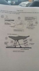
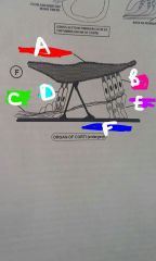
Organ of Corti |
Figures he shows the cochlea and round as a straight tube combo with the basilar membrane running inside. Figured as a cross-section of the cochlear tube, showing the three compartments inside the bone and position of the organ of Corti, the name for the complete structure of basilar and tectorial membrane, hair cells and supporting cells. Figure it out and ask show the organ of Corti magnified. The organ of Corti, and large, consisting of hair cells resting on basilar membrane and hairs embedded in tectorial membrane. And attached auditory nerve endings. |
|
|
A) Tectorial membraneB) Outer hair cellsC) Auditory nerve fibersD) inner hair cellsE) Supporting cellsF) Basilar membrane |
|
|
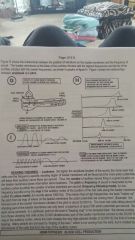
Figure G shows the relationship between the position of vibrations on the basilar membrane in the frequency of the sound. The basilar membrane at the base of the cochlea vibrates with highest frequencies and the tip of the cochlea vibrates with the lowest frequencies comma as shown in graph a figure 8. Figure I shows the relationships between amplitude and pitch |
|
|
Hearing theories - - loudness : |
The higher the amplitude, louder, of a sound, the more inner hair cells over the frequency-specific vibrating region of basilar membrane will be flexed and the more action potentials will leave their auditory nerve fibers per second. Thus, the number of auditory nerves firing in a given location on the basilar membrane gives information about loudness. |
|
|
Hearing theories - - pitch or frequency |
Pitch or frequency of sound interrupted by the auditory cortex by either the number of action potentials per second comma (frequency following model, for low frequency sounds) or by the map in the auditory cortex of the position of the hair cells along the basilar membrane (called the place model, for sounds about 1000 Hz). This means that a high frequency of the brain "makes up" the pitch from its map of where on the basilar membrane the action potentials are coming from! |
|
|
Hearing Theory, hearing map example |
The thinnest and of the basilar membrane vibrates if the pitch is about 20000 Hertz. The inner hair cells sitting above that vibrating part of the basilar membrane can only send a maximum of about 500 Action potentials per second. But each nerve connects to a particular point in the brains "hearing map" in the temporal lobe: the nerves coming from those vibrating hair cells at the 20,000 vibrations / sex part of the basilar membrane connect to the 20,000 Hertz map in the auditory cortex, where the brain creates the very high - pitched whistle of 20000 Hertz, the limit of human hearing. Many people are annoyed by "ringing of the ears", which is not coming from the ears, but is created by a disturbance of the cells that form the map of auditory cortex. |