![]()
![]()
![]()
Use LEFT and RIGHT arrow keys to navigate between flashcards;
Use UP and DOWN arrow keys to flip the card;
H to show hint;
A reads text to speech;
16 Cards in this Set
- Front
- Back
|
What is the Costal Margin? |
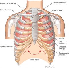
The costal margin is the section at the bottom of the ribcage formed of false ribs and one true rib. Made of cartillage. |
|
|
What are the costal cartillages? What is their function? |
The strands of cartillage connecting the ribs and the sternum. Prolong the ribs and provide elasticity to the walls of the thorax. |
|
|
What is the thoracic inlet? |
The opening at the top of the thorax which the trachea, oesophagus and juggular vessels pass through. |
|
|
Describe the spine with reference to the coccyx and the sacrum. |
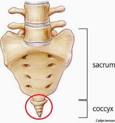
The sacrum is embedded in the pelvis, the tipped end at the bottom is the coccyx. |
|
|
Describe the following terms:
- True rib - False rib - Floating rib |
True rib - Attached to the sternum medially False rib - Attached to the lowest true rib medially Floating rib - Not attached to anything medially |
|
|
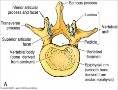
Describe the structures of thoracic vertebrae. |
|
|
|
Describe the anatomy of a single rib. What is the function of the costal groove? |
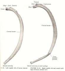
- Identify the head, neck, tubercle and costal grove. - Houses the intercostal nerve, artery and vein |
|
|
What is the fibrous pericardium? |
The layer of fibrous material surrounding the heart muscle. |
|
|
What structure of the diaphragm blends into the pericardium? |
The central tendon |
|
|
What kind of muscle makes up the muscular part of the diaphragm? |
Skeletal Muscle |
|
|
What are the types of muscle found in the body? |
- Skeletal Muscle - Smooth Muscle - Cardiac Muscle |
|
|
What structures are situated below the diaphragm to the right and left? Which side is higher? |
The stomach is below on the left, and the liver is below on the right. The right is higher since there is a large liver underneath. |
|
|
Describe the coeliac artery: |
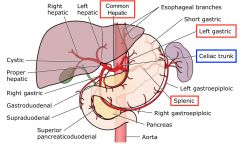
The coeliac artery branches off the aorta. It splits into branches, the splenic, proper hepatic, and gastric arteries. |
|
|
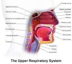
What is the function of the epiglottis? |
Closes over the laryngeal inlet during swallowing to prevent aspiration of food. |
|
|
Describe the components of the upper respiratory tract. |
|
|
|
What is the genioglossus? |
The tongue |