![]()
![]()
![]()
Use LEFT and RIGHT arrow keys to navigate between flashcards;
Use UP and DOWN arrow keys to flip the card;
H to show hint;
A reads text to speech;
41 Cards in this Set
- Front
- Back
|
Functions of the Nervous System |
-Controls the Internal Environment -Controls voluntary movements -Stores memories -Establishes patterns of response based on experience -Contains motor units and neurons |
|
|
CNS vs PNS |
CNS= Brain and spinal cord PNS= Anything outside CNS(Sensory)
-Sensory information from the brain telling the PNS for motor output |
|
|
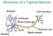
Parts of the Neuron |
Dendrites-Touch receptors Axon-Carries Information Synapse-Connection Transfer |
|
|
Impulse Transmission vs Synaptic Transmission |
Synaptic - diffuse across synaptic cleft Impulse- responds to nerve impulses |
|
|
EPSP vs IPSP |
-protective mechanism to maintain homeostasis until threshold (Control Mechanism) EPSP- closer the threshold IPSP- less likely threshold is reached |
|
|
Temporal Summation vs Spatial Summation |
Temporal- Impulses in a concentrated area
Spatial- Impulses in a spatial area |
|
|
Depolarization vs Repolarization |
-sodium channel opens, some sodium diffuses in -returning to resting potential |
|
|
Threshold and (Resting Potential,Graded Potential,Action Potential) |
-membrane potential level -potential differences between the region inside the membrane to outside -threshold is reached |
|
|
Sodium Potassium Pump |
-movement of sodium and potassium across a cellular membrane |
|
|
What do each joint proprioreceptors provide ? |
Free Nerve Endings (Touch and Pressure) Golgi types receptors (Pressure on joints) Pacnian Corpuscles ( Rate your joint rotation ) |
|
|
How do each Muscle Proprioceptors help us move ? |
Golgi Tendon Organs ( Nerves in tendon) Muscle Spindles (Fibers that wrap around a muscle cell ) |
|
|
Vestibular Apparatus |
-Balance Located in the inner ear Linear and angular acceleration Primary function is to maintain equilibrium |
|
|
How do Motor Unit size impact the movements we make |
Large-Power
Small-Reflexes(Eye Twitch) |
|
|
Movement Control of : Brain Stem Cerebrum/ cerebral cortex Cerebellum Spinal Cord |
- responsible for basic vital life functions such as breathing, heartbeat, and blood pressure. - associated with higher brain function such as thought and action. -structure is associated with regulation and coordination of movement, posture, and balance. - |
|
|
How does exercise positively influence brain function ? |
-Enhances learning and memory -Stimulate Neurogenesus - Improves brains vascular function and blood flow - Levels off mechanisms involved in depression |
|
|
3 types of tissues |
-Skeletal Cardiac Smooth |
|
|
Function of Muscles |
-Producing body movements Stabilizing body positions Regulating organ volumes Movement of substances within the body Producing heat |
|
|
Properties of muscles |
-Excitability Conductivity Contractile Extensibility Elasticity |
|
|
Connective tissues in muscles |
a. Endomysiumb. Perimysiumc. Epimysium |
|
|
Parts of the muscle |
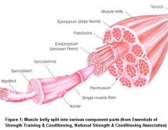
|
|
|
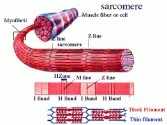
Parts of the muscle fiber – myofibril and sarcoplasm |
Myofibrils- thick and thin filaments (thick is actin and thin is myosin Sarcoplasm- |
|
|
Parts within the sarcoplasm – transverse tubules and sarcoplasmic reticulum
|
Transverse tubules- branch extensively inside the sarcoplasm which allows each sarcomere to be encircles by two T tubules Sarcoplasmic Reticulum-loose network of flattened tubules resembles the smooth endoplasmic reticulum |
|
|
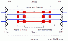
Sarcomeres and parts of striations
|
•TheA band:
•TheI band region: •Sarcomeres = Zline to Z line |
|
|
Actin and myosin
|
•Thick filaments are composed of myosin •Heldin place by the M line proteins
-Actin has troponing and Troppmyons (Connecting signals) |
|
|
I band, A band, H zone, m line, z line, titin
|
- |
|
|
Troponin, tropomyosin
|
-Troponin and tropomyosin regulate contraction via calcium binding Troponin is shown in red (subunits not distinguished). Upon binding calcium, troponin moves tropomyosin away from the myosin-binding sites on actin (bottom), effectively unblocking it. |
|
|
Sliding filament theory and how a contraction begins
|
•Nerveimpulse reaches an axon terminal•AChdiffuses to receptors on the sarcolemma •Amuscle action potential spreads
|
|
|
What happens during relaxation?
|
•Acetylcholinesterase(AChE)breaks down AChwithin the synaptic cleft••Muscleaction potential ceases••Ca+2release channels close
•Activetransport pumps Ca2+ •Calcium-bindingprotein helps hold Ca+2 in SR•Tropomyosin-troponincomplex recovers• |
|
|
Contraction Cycle |
Contraction cycle- •Repeating sequence of events •4 steps to contraction cycle • Cycle keeps repeating as long as there is ATP available & high Ca+2 level near thin filament
|
|
|
Motor unit recruitment
|
•Total strength of a contraction
Motor units in a whole muscle fire asynchronously Produces smooth muscular contraction Precise movements require smaller contractions Large motor units are active when large tension is needed |
|
|
Types of contractions
|
Isotononic Contraction= (Same Tension) a load is moved Isometric Contraction= (Same measure) no movement occurs |
|
|
Types of muscle fibers and the activities they are suited for
|
•Slowoxidative (slow-twitch)
•Fastoxidative-glycolytic (fast-twitch A) •Fastglycolytic (fast-twitch B) |
|
|
Exercise and muscle development |
Atrophy-
hypertrophy- overload- strain - |
|
|
Abnormal Contractions |
Spasm-
Cramp- Tic- Tremor- fasciculation- |
|
|
How does aerobic exercise change the muscle?
|
•Increasesnumber of capillaries •Increasesnumber of mitochondria
•Increasesability to synthesize myoglobin |
|
|
How does anaerobic exercise change the muscle?
|
•Increasesmuscle size
•Increasesmuscle strength •Increasesmuscle power |
|
|
Neuron Diagram (Test question) |
|
|
|
Sarcomere Diagram (Test question) |
|
|
|
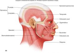
Head Muscles(Test) |
|
|
|
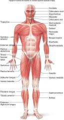
Front view Muscles(Test) |
|
|
|
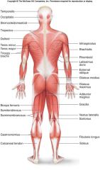
Back view muscles (Test) |
|