Reading...
![]()
Play button
![]()
Play button
![]()
Use LEFT and RIGHT arrow keys to navigate between flashcards;
Use UP and DOWN arrow keys to flip the card;
H to show hint;
A reads text to speech;
25 Cards in this Set
- Front
- Back
|
What is the root word that means muscle?
|
Myo
|
|
|
What are the 3 types of muscles?
|
Skeletal
Cardiac Smooth |
|
|
Skeletal
|
~attach to & cover the bony skeleton.
~Striated ~Voluntary ~Responsible for overall body mobility |
|
|
Cardiac
|
~only in the heart
~Striated ~Involuntary ~Pumps blood in the body |
|
|
Smooth
|
~found in the walls of hollow visceral organs, such as the stomach, urinary bladder, & respiratory passages.
~Not striated ~Involuntary ~Forces fluids & other substances through internal body channels |
|
|
Special Characteristics of Muscle Tissue
|
~Excitability
ability to receive & respond to a stimulus ~Contractility ability to shorten forcibly when adequately stimulated ~Extenibility ability to extend or stretch ~Elasticity ability to recoil & resume its resting length after stretching |
|
|
Muscle Functions
|
~Producing movement
~Maintaining posture & body positioin ~Stabilizing joints ~Generating heat |
|
|
Is muscle an organ?
|
Yes
|
|
|
Epimysium
|
......
|
|
|
Perimysium
|
~ Around the muscle
~ Fascicles ~ Perimysium surrounds each fascicle |
|
|
Endomysium
|
~ Within the muscle
~ Wispy sheath of connective tissue that surrounds each individual muscle fiber ~ Consists of fine areolar tissue |
|
|
A muscle fiber is another name for myofibril.
|
........
|
|
|
What are muscle attachments?
|
~ Direct (fleshy attachment)
the epimysium of the muscle is fused to the periosteum of a bone or perichondrium of a cartilage ~ Indirect the muscle's connective tissue wrapping extend beyond the muscle either as a ropelike tendon or as a sheetlike aponeurosis. |
|
|
What is the difference between origin and insertion?
|
Origin is the attachment point that doesn't move
Insertion is the attachment point that does move |
|
|
What is the difference between a direct or indirect attachment?
|
In direct (fleshy) attachment the epimysium of the muscle is fused to the periosteum of a bone or perichondrium of a cartilage.
Indirect attachments the muscle's connective tissue wrappings extend beyond the muscle either as a ropelike tendon or as a sheetlike aponeurosis. |
|
|
Indirect attachments are much more common because of their durability and small size.
|
...................
|
|
|
What is fascia and aponeurosis?
|
Aponeurosis: fibrous or membranous sheet connecting a muscle and the part it moves
Fascia: layers of fibrous tissue covering and seperating muscle |
|
|
Cross Bridge Cycle
|
1. Cross Bridge Formation
~ Energized myosin head attaches to an actin myofilament, forming a cross bridge. 2. The power (working) stroke. ~ ADP and Pi are released and the myosin head pivots and bends, changing to its bent low-energy state. As a result it pulls the actin lament toward the M line. 3.Cross bridge detachment. ~ After ATP attaches to myosin, the link between myosin and actin weakens, and the myosin head detaches (the cross bridge “breaks”). 4. Cocking of the myosin head ~ As ATP is hydrolyzed to ADP and Pi , the myosin head returns to its prestroke high-energy, or “cocked,” position. * |
|
|
Events at the Neuromuscular Junction
|
1. Action potential arrives at axon terminal of motor neuron
2. Voltage-gated calcium channels open. Calcium enters the axon terminal moving down its electochemical gradient. 3. Ca2+ entry causes ACh (aneurotransmitter) to be released by exocytosis. 4. ACh diffuses across the synaptic cleft and binds to its receptors on the sarcolemma. 5. ACh binding opens ion channels in the receptors that allow simultaneous passage of Na+ into the muscle fiber and K+ out of the muscle fiber. More Na+ ions enter than K+ ions exit, which produces a local change in the membrane potential called the end plate potential. 6.ACh effects are terminated by its breakdown in the synaptic cleft by acetylcholinesterase and diffusion away from the junction. |
|
|
Excitation-Contraction Coupling
|
1. The action potential propogates along the sarcolemma and down the T tubules.
2. Calcium ions are released. Transmission of the AP along the T tubules of the triads causes the voltage-sensitive tubule proteins to change shape. This shape change opens the Ca2+ release channels in the terminal cisterns of the sarcoplasmic reticulum (SR), allowing Ca2+ to flow into the cytosol. 3. Calcium binds to troponin and removes the blocking action of tropomyosin. When Ca2+ binds, troponin changes shape, exposing binding sites for myosin (active sites) on the thin laments. 4. Contraction begins Myosin binding to actin forms cross bridges and contraction (cross bridge cycling) begins. At this point, E-C coupling is over. |
|
|
What are the parts of the muscle cell?
|
Fascicle
muscle fiber sarcolemma nucleus sarcoplasm myofibril filaments |
|
|
glycosome
|
granules of stored glycogen that provide glucose during muscle cell activity
|
|
|
myoglobin
|
a red pigment that stores oxygen
|
|
|
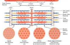
Myofibrils
|
rodlike bundle of contractile filaments found in muscle fibers
they appear striated because of the alternating dark and light bands |
|
|
Sarcomere
|
|