![]()
![]()
![]()
Use LEFT and RIGHT arrow keys to navigate between flashcards;
Use UP and DOWN arrow keys to flip the card;
H to show hint;
A reads text to speech;
150 Cards in this Set
- Front
- Back
|
Sodium Na+ uses |
-Main extracellular cation -Largely determines extracellular fluid volume and influences blood pressure -Important in action potential generation in nerve and muscle tissue -Normal concentration in ECF is about 135-145 mol/L |
|
|
Calcium Ca+ uses |
-Important structural component of bone and teeth -Involved in neurotransmission and muscle contraction -Essential for coagulation (blood clotting) -Regulates enzyme function -Normal total plasma concentration is about 2.2-2.6 mom/L |
|
|
Potassium K+ uses |
-Most abundant intracellular cation -Main determinant of the resting membrane potential (RMP) -Particularly important I'm excitable tissue i.e. nerve and muscle -Normal concentration in ECF is about 3.5-5 mol/L |
|
|
Glucose uses |
-Used by cells (especially neurons) to produce adeonosine triphosphate (ATP) neutrons particularly affected by low glucose -High blood glucose causes other problems both acute and chronic -Normal fasting glucose concentration 3.6-6mmol/L -Non fasting glucose concentration 3.5-8mmol/L |
|
|
Tissues of the Locomotor system |
-Bone -Cartilage -Blood vessels -Muscle -Nerves -Tendons -Ligaments |
|
|
Tissues |
Consist of specialised cells embedded within an extracellular matrix |
|
|
Epithelial |
-Layers/sheets of cells, very little matrix -Covers/protects body surfaces, line cavities -Skin, lining of tracts, glands |
|
|
Connective |
-Sparse cells, lots of matrix containing fibres -Supports structures, transports substances -Bones, cartilage/tendons, fat, blood |
|
|
Muscle |
-Long fibre-like cells, strong fibres capable of pulling loads -Produces movement and heat -Muscles, skeletal, smooth and cardiac |
|
|
Nervous |
-Highly cellular of many types, conducting and supporting -Communication and co-ordination between body parts -Nerves, sensory organs, brain and spinal cord |
|
|
Unicellular organsims immediate external environment |
-Nutrients -Solute concentration -Temperature -pH -Toxins (including own wastes0 -Predators |
|
|
Multicellular organisms |
Can maintain a stable internal environment inside the body |
|
|
Ideas of homeostasis |
1) In our bodies there are mechanisms that act to maintain constancy 2) Any tendency toward change automatically meets with factors that resist change 3) There are co-opertating mechanisms which act simultaneously or successively to maintain homeostasis 4) Homeostasis does not occur by chance, but is the result of organised self-government |
|
|
Core body temperature |
Is generally maintained between 36 to 37 which allows for optimal metabolic and physiological functioning. -oral and axillary temperatures are usually about 0.5 less than rectal core |
|
|
Proteins at high temperatures |
Start to denature |
|
|
Proteins at low temperatures |
Chemical reactions slow down preventing normal cell function |
|
|
Diffusion |
Results from the random movement of individual molecules as a consequence of their thermal energy. |
|
|
Passive diffusion |
Diffusion where the net direction of the movement is 'downhill' and so requires not much energy input form the body. |
|
|
Simple diffusion via membrane channels |
Channels are usually specific and may open/ close spontaneously or in response to various stimuli e.g. chemicals, or change in membrane potential. |
|
|
Carrier mediated passive transport (facilitated diffusion) |
Substance binds to carrier on one side of the membrane which includes the carrier to change shape and release of substance to the other side ("downhill" i.e. down concentration gradient) |
|
|
Primary active transport |
Energy from the hydrolysis of ATP used to move substances against their concentration gradient. Maintains ionic gradients and helps regulate cell volume. |
|
|
Endocytosis and Exocytosis |
Substances transported into or out of the cell membranes (bilayer) vesicles. -Phagocytosis of microbes by neutrophils (endocytosis) -Secretion of insulin by B cells of pancreas (exocytosis) |
|
|
Osmosis |
Is the movement of water across a membrane down its own concentration gradient (or toward the region of higher solute concentration) -The pressure required to just stop this osmosis is the osmotic pressure. -Differences in solute concentration across cell membrane can cause fluid shifts and create pressure that can damage cells. |
|
|
Osmolarity |
Is a measure of the total number of solute particles per litre of solution. |
|
|
Tonicity |
Specifically refers to the effect that a solution has on cell volume |
|
|
Hyper tonic |
Solutions will cause cells to shrink |
|
|
Hypo tonic |
Solutions will cause cells to swell |
|
|
Iso tonic |
Solutions cause no change in cell volume |
|
|
Differences between osmolarity and tonicity |
Osmolarity is a property of a particular solution (independent of any membrane). Tonicity is a property of a solution with reference to a specific membrane. |
|
|
Water crossing cell membranes along osmotic gradients |
If the osmolarity of one compartment changes then water will diffuse by osmosis until equilibrium has been restored |
|
|
Resting membrane potential |
The resting membrane potential is an electrical potential that exists across the cell membrane and is due to different concentrations of ions on each side of the membrane and their respective permeabilities to it. |
|
|
Important points about RMP |
-For most cells the membrane potential remains constant over time- around 70 mV -However for 'excitable' tissues (nerve muscles0 the membrane potential must change in order for them to function -This usually occurs via the opening or closing of specific channels -Because K+ is the major determinant of the RMP it is very important to tissues won't function normally cardiac arrhythmias, muscle weakness. |
|
|
Regulated variable |
The variable that the system senses and tries to keep stable |
|
|
Set point |
The target value for that variable |
|
|
Reference (normal) range |
Values of the regulated variable within acceptable limits |
|
|
Variation |
In regulated variable values within and between 'normal' people. |
|
|
Control systems for homeostasis |
-Homeostasis is achieved by a combination of feedback and feed forward control systems
|
|
|
Negative feedback |
Opposes the change in the regulated variable and moves it back toward the set point. |
|
|
Sensor |
Monitors the actual value of the regulated variable |
|
|
Integrator |
Compares actual and set point values, determines and controls the response (Can be in the same cell as the sensor) |
|
|
Effectors |
Produce the response that restore the regulated variable to its set point |
|
|
Communication pathways |
Carry signals between components |
|
|
Physiological communication pathways |
1) Neuronal- involves action potentials in axions and neurotransmitters releases at synapses, fast, specific. 2) Hormonal- Releases in to the blood or ECF, good for wide spread sustained response. |
|
|
Feed foward |
Establishes a future 'predicted value' for the regulated variable compares this with the 'set-point- and makes anticipatory corrections. |
|
|
Positive feedback |
Response to a stimulus that moves the controlled variable even further away from the 'set-point' it reinforces initial change- not common and often detrimental |
|
|
Anatomical position |
-Upright -Face forwards -Feet together -Palms face forward -Same regardless of movement |
|
|
Posterior |
Close to the back |
|
|
Anterior |
Closer to the front |
|
|
Superior |
Closer to the head |
|
|
Inferior |
Closer to the feet |
|
|
Medial |
Closer to the median line |
|
|
Lateral |
Further away from the median line |
|
|
Median line |
Line right down the middle of the body |
|
|
Proximal |
Is the part of the limn that is closer to the trunk (main part of the body) |
|
|
Distal |
Further away from the trunk (main part of the body) |
|
|
Deep |
Further from the surface within the body. |
|
|
Superficial |
Closer to the surface |
|
|
Sagittal plane |
Divides the body in to left and right portions |
|
|
Coronal plane |
Divides the body in to anterior and posterior portions |
|
|
Transverse plane |
Divides the body in to superior and inferior portions |
|
|
Flexion |
Decreases angle, fleshy parts of limb brought closer together |
|
|
Extension |
Opposite of flexion, angle increases and fleshy parts of limb are further away |
|
|
Dorsiflexion |
Only in ankle, toes brought up towards face |
|
|
Plantar flexion |
Only in ankle, toes pointing towards ground |
|
|
Abduction |
Movement at joint moves limb away from the midline |
|
|
Adduction |
Movement at joint moves limb towards midline |
|
|
Circumduction |
-Combination of the angular movements -Flexion/ Abduction/ Extension/ Adduction -No rotation -Wrist |
|
|
Rotation |
Rotation around the long axis of a joint -Head, shoulder |
|
|
Pronation |
Palm faces posterior |
|
|
Supernation |
Palm faces anterior and forearm bones parallel |
|
|
Inversion |
Sole of foot faces towards midline |
|
|
Eversion |
Sole of foot turns away from midline |
|
|
Functions of the skeleton |
-Support -Movement -Protection -Storage -RBC formation |
|
|
Long bone |
-Longer than they are wide -Shaft or diaphysis -Extremities or epiphyses -Function as levers for movement -Thicker compact bone in diaphysis |
|
|
Short bone |
-Near equal width and length -Weight bearing/ shock absorption -Mostly cancellous bone |
|
|
Flat bones |
-Protection-cranial bones -Muscle attachment- Scapula -Thin plates of compact bone- some cancellous |
|
|
Irregular bones |
-Variable shape and function -Dont fit in to any other class/ can't be classed in to anything else |
|
|
Axial Skeleton |
-Head, ribs, spine -Skull -Cranium (vault) -Facial bone -Mandible -Vertebral column |
|
|
Spine |
-Cervical -Thoracic -Lumbar -Sacrum and coccyx |
|
|
Pelvic gurdle |
-Hip bones -Sacrum -Pelvis |
|
|
Hand bones |
-Carpals -Metacarpals -Phalanges |
|
|
Foot bones |
-Tarsals -Metatarsals -Phalanges |
|
|
Osteoblasts |
Build ECM Increase in periosteum width |
|
|
Osteocytes |
Mature bone cells |
|
|
Osteoclasts |
Breakdown ECM From Enostem mould the bone shape and form the meoularity cavity |
|
|
Cancellous (soft bone) |
Trabeculae- /struts of lamella bone (honeycomb) Marrow- Fills the cavities Osteocytes- Housed in lacuna on surface of trabeculae |
|
|
Compact bone (hard bone) |
Gross level: outer surface seems impenetrable Foramina (holes)-for blood to bring in nutrients Microscopic level: Osteon-Longtutidonal cylinder within compact bone Lamella-Tubes of ECM from series of cylinders logitudionally down shaft (diaphysis) Central Canal- Blood vessels + nerves (communications) Cancaliculi- Chanels for osteocytes through ECM Lacunae- Lakes for osteocytes Periostesum-Protective sheath around the bone covers bone except joint Sub-Periosteum- Below periosteum |
|
|
Ossification |
Process of transforming cartilage bone 1) Primary centre of ossification- Linear growth in Diaphysis 2) Secondary- Epiphysis (begins after diaphysis growth), growth plates/Epiphyseal plates, formed of cartilage, Growth plate-continually replaced by bone Cartilage- continually replaced which results in continuous bone growth |
|
|
Bone pathology |
Imbalance of osteoclasts/blasts activity |
|
|
Osteoporosis |
Where osteoclasts overtake osteoblasts; bone efficiency -Ageing (loss of oestrogen) -Lifestyle (Lack of exercise) -Peak bone mass |
|
|
Fractures |
Step 1: Haematoma - bone broken, bleeding into the site, haemotoma stops the bleeding, capillaries, phagocycytes Step 2: Fibroblasts (cells with differentative chondroblasts) fibro cartilaginous Step 3: Bony callus- cells come in + replace cartilage- osteoblasts (replace soft cell) Step 4: Remodelling |
|
|
Types of fractures |
Closed- Simple Open, compound- Bone breaks through skin, muscle/ nerve damage, risk of infection. Greenstick- Doesn't break bone completely |
|
|
Joint |
Join bone to bone Articulation- Where bone meets, involves bone shapes + soft tissue, allows free/ controlled movement |
|
|
Meniscus |
-Bony congurence -Less BC, more soft tissue support -Deepens concave -Disperses forces |
|
|
Cartilage |
Hyaline/ Articular: Collegen fibres barley visible, high water content in matrix, resists compression, provides smooth frictionless surface Firbrocartilage:Collagen fibres form bundles through matrix, orientation of fibres align with stress, resists compression and tension |
|
|
Ligaments |
DFCT Bone to bone, restrict movement -Movement is restricted away from itself -Lateral restricts adduction -Medial restricts abduction -Holds bones together -Tight and thick where most support is required, loose on sides where movement is allowed -Potential space cavity -Synovial membrane lines the inner surface of the capsule , secretes synovial fluid, lubrication of the joint |
|
|
Tendons |
DFCT Muscle to bone -It facilitates and controls movement, contraction |
|
|
Fibrous Joints |
-Dense fibrous connective tissue DFCT -Ligament bone to bone -Limited movement/ stability -Cranial sutures and tibio fibula joint |
|
|
Cartilaginous joints |
-Some movement allowed -Tissue fibrocartilage (resists tension, associated with joints where some movement is allowed) -Special functions eg invertirai disk (structure), pubis symphysis (joint) |
|
|
Synovial joints |
-Free moving -Most limb joints (except where we require stability) -Complex association of tissues + structures -Facilitation of free movement and control movement -Bone ends determine the range of motion at a joint eg hip, knee |
|
|
Articular (hyaline) cartilage |
-Covers bone ends where they articulate and move over each other -Subcondral bone is smooth |
|
|
Intra capsular |
-In the capsule but not part of the capsule -Restricts movement between bones -Cruciate ligaments (knee) -Arise from tibia inserts into femur -Anterior cruciate restricts posterior displacement of femur -Posterior cruciate restricts anterior displacement of femur |
|
|
Range of movement |
Determined by bone end shape, ligament location and length, body surface contact |
|
|
Hinge joint |
Uniaxial- Flexion, extension ed ankle, elbow, inter phalange joints |
|
|
Pivot Joint |
Uniaxial - Rotation (supernation-pronation) ed radio ulna joints |
|
|
Saddle joint |
Biaxial-Flexion-extension, Abduction-Adduction, cirmduction, obligatory rotation Carpometercapal joint- Base of thumb |
|
|
Ellipsoid joint |
Biaxial-Flexion-extension, Abduction-Adduction, circumdction, no rotation Wrist joint- Radiocarple joint |
|
|
Condylar joint |
Flexion-Extension, rotation Knee, Temperomandibular joint (TMJ) |
|
|
Plane joint |
Multi axil- Sliding anf gliding Flat articular surfaces Can move in any direction Intercarpal and intersal joints |
|
|
Ball and socket joint |
Multi axil-flexion-extension, Abduction, adduction. circumduction, rotation Large range of movement- Stability as well Shoulder, hip |
|
|
Muscles |
1) Movement 2) Heat production (mammals need to generate heat to keep warm) 3) Posture 4) Communion (Sign language, cues, universal cues) |
|
|
Tissue in muscle |
-Fibril 'little muscle' -Myofillaments 'Small proteins' |
|
|
Muscle fibre/ cell |
-Up to 40 cm -Parallel -Cylindrical -Multi-nuclear (as devoplment there are short myositis these join up but don't loose nuclei) -Have striated muscle arrangement |
|
|
Muscle contraction |
-Actin + myosin integrate -Actin + myosin retain their length -Process consumes energy -Calcium ions (Ca2+) essential |
|
|
Muscle form determines function |
1) Length of muscle fibres- Fibre can shorten up to 50% of resting length, large ROM, long muscle fibres that are parallel to the line of pull 2) Number of muscle fibres- Tension is directly proportional to cross- section area (CSA), greater number of fibres, greater tension 3) Arrangement of muscle fibre- Fibres oblique to muscle tendon, more fibres in to same space. |
|
|
Muscle= posture |
You need tension in some muscle fibres to even sit up. Even relaxed muscles are slightly active. Muscle tone: Does not produce movement, keeps muscles trim and healthy, helps trebles and maintain joint, nerve impulses activation fibres. |
|
|
Muscle fibre activation + contraction |
Sends messages to skeletal muscle fibres, trys to get every single sacromere to move at one time |
|
|
Neuromuscular junction |
In the middle of the muscle cell -Sarcolemma, electrical event goes over the sarcolemma in both directions -T-tubules, eletrical event also burrows down the t-tubules, this triggers the sacoplasmic reticulum which is where the calcium is stored to know whern the calcium needs to be released Actin slides over the myosin to get shortening- contracted Relaxed is when the actin is far away from each other |
|
|
A motor unit |
Motor nerve -A motor neuron, axon and branches -Plus all muscle fibres it innervates -Size varies |
|
|
Graded force of concentration |
-Motor unit displays all or none activation of fibres -But the force of contraction of whole muscle is graded |
|
|
Force contraction in a whole muscle |
-Characteristics of muscle fibre -Length -Number -Arrangement -Characteristics of motor units-size, number, rate of timing -Muscle attachment- effort needed for movement, determines muscle tone |
|
|
Anatomical levers |
Bone=levers Joint=pivot/fulcrum Muscle contration= pull Load=External/ internal |
|
|
Lever types |
First- stabilise joint position Second- Effective at overcoming loads Third- Large ROM |
|
|
Concentric |
Muscle is active, decelobs tension (shortening), change in joint position, shortening of muscle |
|
|
Isometric |
Muscle is active, develops tension, no change in joint position |
|
|
Eccentric |
Muscle is active, develops tension, change in joint position, lengthening of muscle |
|
|
Agonist |
Muscle that creates the desired movement acts concentrically |
|
|
Antagonist |
Muscle that try to prevent the movement, acts eccentrically |
|
|
Stabiliser |
When a muscle is active to hold a joint still, action is isometric. |
|
|
Neutraliser |
Muscle eliminates an unwanted movement caused by another movement |
|
|
Concentric actions of muscles |
Anterior Flexion I I Medial--------------------------------------Lateral Adduction I Abduction I Posterior Extension |
|
|
Deltoid |
Greek for triangular Attaches to pectorial girdle proximally, shaft of humorous distally -Middle/ medial= adduction |
|
|
Biceps brachii |
Anterior Both heads attached to the scapula Flexion at the elbow (anterior) Radiulnar joint attaches, when it contracts causes pronation |
|
|
Triceps brachii |
3 heads the muscle Long head comes off proximally to scapula other tho proximal to humorous comes down and attaches ulna at elbow joint= extension |
|
|
Illipsoas |
Anterior= hip flexion Attaches:vertebrae and anterior surface of pelvis Proximal to the thoracic vertebra and all 5 lumbar vertebra Distally attaches to the femur |
|
|
Gluteus maximum |
posterior=extension Attaches, sacrum and pelvis to the iliotibial band, distal tendon and femur |
|
|
Quadriceps femoris |
-anterior to the knee=extension no flexion -4 separate muscles, straight muscle of the femur -Rectus femerus- straight muscle of the anterior side of the hip, knee joint Lateralis femerus Intermedius femerus Medials femerus |
|
|
Tibalias anterior |
Crosses ankle on anterior sideAnkle=dorsiflexion Attaches tibia, medial tarsal |
|
|
Triceps surae |
Gastroanemius and soles Support Plantarflexion Attaches femur, tibia to canlcaneus via tendon |
|
|
Hamstrings |
Looking pastorally, hip= extension Knee joint= flexion and rotation attaches pelvis and femur goes to tibia and fibula |
|
|
General functions of muscle |
-Heat production -Posture -Movement -Communication |
|
|
Gravity |
Action as an agonist or antagonist. Sometimes gravity helps the movement, sometimes it opposes the movement |
|
|
Bipedal standing |
-Relatively small area of contact with ground -Plantar surface of feet -Energy efficient |
|
|
Quadrupedal standing |
-Base of support -Legs flexed at several joints -needs lots of energy -Bigger area base of support-more stable -Muscles have to contract as the muscles are flexed |
|
|
Bipedal standing- Hip |
-Posterior to joint, joint pushed in to extension -Extension= ligaments are tight |
|
|
Bipedal standing- knee |
-Anterior to joint -Joint pushed in to extension=ligaments are tight |
|
|
Bipedal standing- Ankle |
-Anterior to the joint -Falls in to dorsiflexion, not locked |
|
|
Walking- bipedal |
-Learnt -Gait is characterised -Basic pattern is gait cycle |
|
|
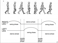
Gait cycle |
|
|
|
Swing and stance phase |

|