![]()
![]()
![]()
Use LEFT and RIGHT arrow keys to navigate between flashcards;
Use UP and DOWN arrow keys to flip the card;
H to show hint;
A reads text to speech;
10 Cards in this Set
- Front
- Back
|
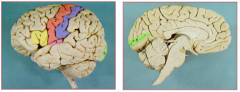
5 major functional areas of the brain |
|
|
|
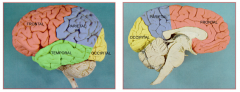
lobes of the brain |
|
|
|
position and surrounding bones of the middle meningeal artery |
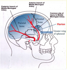
the branches of the middle meningeal artery lies in grooves of bones between the skull and the dura mater (unlike the venous sinuses which lie between the layers of the dura mater)
|
|
|
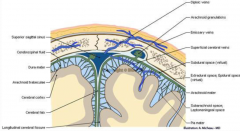
picture showing venous drainage and meninges |
|
|
|
what is the clinical significance of the pterion? |
pterion is a common site of injury to middle meningeal artery
where frontal, parietal, temporal and sphenoid bones meet
anterior division of middle meningeal artery runs underneath, a traumatic blow to the pterion may cause an epidural haemorrhage
a blow to the top or back of the head may also fracture the pterion |
|
|
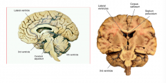
sagittal and coronal view of ventricles |
|
|
|
production and flow of CSF in the brain |
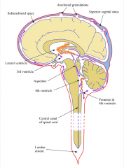
CSF is made in choroid plexuses (orange)
escapes through foramen in 4th ventricle to enter subarachnoid space which surrounds the brain and spinal cord
CSF is removed from the subarachnoid space through arachnoid granulations which project into the superior sagittal venous sinus, re-entering the bloodstream |
|
|
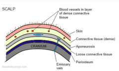
layers of the scalp |
|
|
|
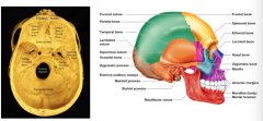
bones of the skull |
|
|
|
signs of increasing ICP |
decreasing GCS
diminished pupil response to light
lateralising signs |