Reading...
![]()
Play button
![]()
Play button
![]()
Use LEFT and RIGHT arrow keys to navigate between flashcards;
Use UP and DOWN arrow keys to flip the card;
H to show hint;
A reads text to speech;
166 Cards in this Set
- Front
- Back

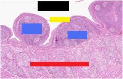
What are the arrows branching off the black box pointing to?
|
Developing Alveoli
|
|

What is this a picture of?
|
Fetal lung
|
|

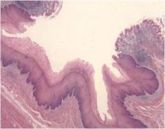
What is this a picture of?
|
Nasal Cavity
|
|

What type of cell is the red box pointing to?
|
Ciliated cell
|
|

What is the blue box on top of? What is it used for?
|
A goblet cell
to secrete mucus |
|

What type of tissue is the black box on top of?
|
Connective tissue
|
|

What is the green box pointing to?
|
olfactory epithelium
|
|

What is the orange box pointing to?
|
Olfactory glands
|
|

What is the yellow box pointing to?
|
Olfactory nerves
|
|

What are the white boxes referring to?
|
Ducts
|
|

What is this a histological section of?
|
Olfactory mucosa
|
|

What is the red box referring to?
|
epiglottis
|
|

What is the sky blue box referring to?
|
lingual tonsil
|
|

What is the royal blue box 'hole' referring to?
|
Foramen cecum
|
|

What is the black box referring to?
|
palatine tonsil
|
|

What is the yellow box referring to?
|
Foliate papillae
|
|
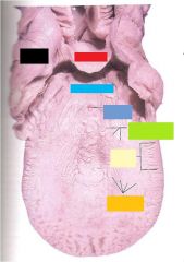
What is the orange box referring to?
|
fungiform papillae
|
|

What is this histological section depicting?
|
The inferior concha of the nasal cavity
|
|

What is the orange box pointing to?
|
Respiratory epithelium
|
|

What are the lavender and purple boxes referring to?
|
Lavender - lamina propria
Purple - capillaries |
|

What is the red box referring to?
|
Goblet cells
|
|

What is the function of what the black box is pointing to?
|
To protect from dust particles and move liquid over the surface
|
|

What is this a histological section of?
|
Olfactory region of the nasal cavity
|
|

What kind of stain is performed in this histological section?
|
Alcian blue & van Gieson
|
|

What is the yellow box, green box, and orange, box referring to?
|
Yellow = olfactory cells & sustentacular cells
Green box = Bowman's capsule Orange box = Bone |
|

What is this a histological section of?
|
Tonsil
|
|

What is the pink, sky blue, and green box referring to?
|
Pink = epithelium
Sky Blue = tonsillar crypt Green = Lymph follicle |
|

What is the histological section of?
What type of tissue lines the section? |
Pharynx
stratified squamous epithelium |
|

What is this a histological section of?
|
pharynx
|
|

What is this a histological section of? What is its function?
|
Larynx
Conduit for air, speech organ |
|

What is this a histological section of? What kind of cartilage does it contain and why?
What are the "VF" lined with and what do they contain? |
Larynx
Elastic cartilage allows us to vary the pitch of our voice (Also contains hyaline) Lined with stratified squamous and contained striated skeletal muscle as well as ligaments |
|

What are the "VF" and what are they used for?
|
The vocal folds are to control the flow of air through the larynx and vibrate to produce sound
|
|

What is this a histological section of?
|
larynx
|
|

What is this a histological section of?
|
The trachea and the esophagus
|
|

What do the red and pink box refer to?
|
Red = Esophagus
Pink = Trachea |
|

What do the sky blue and green box refer to? What kind of membrane is the orange box pointing to?
|
Sky blue = Cartilage
Green = Bone tissue Orange = Fibroelastic |
|

What is the function of what the red box is pointing to?
|
The red box is the hyaline cartilage ring and it is used to keep the trachea from collapsing on itself during breathing.
This is the trachea |
|

What is this a histological section of? What are the different layers?
|
Trachea
1. Mucosa 2. Submucosa 3. Cartilaginous 4. Adventitia |
|

What layers do the Green box (smiley facey :P), Other green box, and light green box at the bottom refer to?
|
Smiley Face = Mucosa
Other green box = Submucosa Light green box at the bottom = Cartilaginous |
|

What do the lavender and orange box referring to?
|
Lavender = lamina propria
Orange = respiratory epithelium |
|

What type of tissue is within each layer of this histological section?
|
1. mucosa - ciliated pseudostratified epithelium & elastic, fiber-rich lamina propria
2. submucosa - slightly denser connective tissue than lamina propria 3. Cartilaginous - c-shaped hyaline cartilage 4. adventitia - connective tissue that binds trachea to adjacent structures |
|

What is this a histological section of? What type of cells are present and what are their functions?
|
Trachea
Cells: 1. Cilia - short, hairlike 2. Goblet cells - secrete mucus and increase during infection 3. Basal cells - reserve cell populations (most nuclei belong to basal cells) |
|
What is this a histological section of?
|
Trachea
|
|
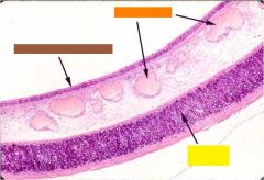
What do the brown, orange, and yellow boxes refer to?
|
Brown = Respiratory Epithelium
Orange = Blood Vessels Yellow = Hyaline Cartilage |
|

What lumens are the orange and green box referring to?
|
Orange = Bronchiole
Green = Arteriole |
|

What is the lavender box referring to?
|
Alveolar air space
|
|

What is this a histological section of?
|
Alveoli
|
|

What is this a histological section of?
|
Alveoli
|
|

What are black and yellow boxes referring to?
|
The respiratory duct
|
|

What are the blue and green boxes referring to?
|
Blood vessels and bronchiole
|
|

What is this a histological section of?
|
Bronchi
|
|

What do the red boxes refer to?
|
Pseudostratified respiratory epithelium, cilia, and goblet cells
|
|

What type of tissue is the orange box? the yellow box?
|
Orange = connective tissue
Yellow = smooth muscle tissue |
|

What does the brown box refer to?
|
Glands
|
|

What is this a histological section of?
|
Bronchi
|
|

What do the green, red, and brown boxes represent?
|
Red = blood vessel
Green = Lumen of the alveolar Brown = capillaries |
|

What do the yellow and blue boxes represent?
|
Yellow = smooth muscle tissue
Blue = Cartilage |
|

What is this a histological section of?
|
alveoli
|
|

What are the pink, green, sky blue, and red boxes referring to?
|
Pink = Alveoli
Green = Respiratory duct Blue = Bronchiole Red = Artery |
|

What is this a histological section of?
|
Lung elastin
|
|

What do the orange and sky blue boxes refer to?
|
Orange = Elastic Fibers
Sky blue = alveolus |
|

What is this a histological section of?
|
Lung
|
|

What does the sky blue, purple, yellow, and green refer to?
|
Sky Blue = alveolus
Purple = Capillaries Yellow = Clara Type II Green = Nucleolus of Clara Type II |
|

What is this a histological section of?
|
the lung
|
|

What are the yellow, orange, and blue boxes representing?
|
Yellow = Smooth muscle
Blue = Bronchiole Orange = Connective Tissue |
|

What is this a histological section of?
|
Bronchiole
|
|

What do the sky blue, red, and black boxes represent?
|
Sky blue = Terminal Bronchiole
Red = muscle Black = Alveolus |
|
What is this a histological section of?
|
diaphragm
|
|

dx
|
Acute broncopneumonia
|
|

dx
|
Bronchial asthma
|
|

dx
|
Tuberculosis
|
|

dx
|
Asthma
|
|

dx
|
pneumocystis carnii pneumonia
|
|

What type of specialized mucosa is this histological section?
|
foliate papillae
|
|

What type of mucosa is depicted in this section?
|
Filiform papillae
|
|

What type of mucosa is depicted in this section?
|
Fungiform papillae
|
|

What is this a histological section of?
|
Taste buds
|
|

What are the black and grey boxes pointing to?
|
Black = Taste pore
Grey = Microvilli |
|

What are the green and blue boxes pointing to?
|
Green = Supporting cells
Blue = Sensory cells |
|

What are the red and yellow boxes pointing to?
|
Red = basal cells
Yellow = nerve |
|

What is this a histological section of?
|
Tonsil
|
|

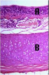
What type of layer is at the top? What type of tissue is at the bottom?
|
Top - stratified squamous
Bottom - lymphoid tissue |
|
What is this a histological section of?
|
Oral cavity
|
|

What are the blue and red boxes referring to?
|
Blue = Lymphatic tissue
Red = Mucus-type salivary glands |
|

What are the blue and red boxes referring to?
|
Blue = Lymphatic tissue
Red = Mucus-type salivary glands |
|

What are the black box, purple box, and red box pointing to?
|
Black = Ameloblasts
Purple = Enamel Red = Dentin |
|

What are the green and yellow boxes pointing to?
|
Green = odontoblasts
Yellow = Outer enamel epithelium |
|

What are the sky blue and orange boxes pointing to?
|
Sky blue = Stellate reticulum
Orange = Dental papillae |
|

What type of duct is depicted?
|
Intercalcalated duct
|
|

What type of duct is depicted?
|
Striated duct
|
|

What type of duct is depicted?
|
Excretory
|
|

What is this a histological section of?
|
Parotid gland
|
|

What is this a histological section of? What are the red dotted lines for?
|
Submandicular gland
Red dotted lines - serous and mucus acini striated duct |
|

dx
|
Salivary gland tumor
|
|

What do the different roman numerals stand for?
|
I. Mucosa
a.Lamina propria b. Muscularis mucosae II. Submucosa III. Muscularis Externa a.Longitudinal b. Circular IV. Serosa (or adventitia) |
|

Tell me about this figure.
|
The blastula (1) undergoes gastrulation forming the endoderm (red). The archenteron (green arrow) becomes the gastrointestinal lumen. The gastrula (2) has all three germ layers.
|
|

What is this a histological section of and what kind of tissue is at the surface?
|
Esophagus
Stratified Squamous epithelium |
|

What do the purple, yellow, blue, and red boxes represent?
|
Purple = lamina propria
Yellow = Smooth muscle Sky Blue = esophageal glands Red = Skeletal muscle |
|

What is this a histological section of?
|
Esophagus
|
|

What do the white, red, and lavender boxes represent?
|
White = respiratory epithelium
Red = Lymph Nodule Lavender = lamina propria |
|

What layer of the esophagus is this a histological section of? What types of tissue does it have indicated by the red, purple, and green boxes?
|
Esophagus mucosa
Red = Epithelium lining Purple = lamina propria (connective tissue) Green = Muscularis mucosae (smooth muscle) |
|

What is this a histological section of? What do the black, red, and blue boxes represent?
|
Esophagus
Black - Submucosa Red = Muscular mucosae Blue = Circular muscle layer |
|

What do the grey and pink boxes represent?
|
Grey = keratinized stratified squamous epithelium
Pink = longtudinal muscle layer |
|
What is this a histological section of?
|
The gastroesophageal junction
|
|

What is this a histological section of?
|
the gastroesophageal junction
|
|

What is this sheet of connective tissue that binds together the loops of the GI tract?
|
mesentary
|
|

What layer is this histological section and what do the different color boxes represent?
|
Muscularis externae
Orange = Outer longitudinal smooth muscle Red = inner circular longitudinal muscle Green = collagen |
|

What layer is this a histological section of? What are the three layers as represented by the pink, red, and brown boxes?
|
Submucosa layer of the esophagus
Pink = muscularis externae Red = Submucosa Brown = Mucosa |
|

What do the yellow and green boxes represent?
|
Yellow = Gland
Green = Duct |
|

What do the purple, red, and white boxes represent?
|
Purple = lamina propria
Red = Lymph nodule White = esophageal epithelium |
|

What is this a histological section of? What do the red and yellow boxes represent?
|
Stomach
Red = Gastric pits Yellow = Cardiac glands |
|

dx dx dx
|
Barrett's Esophagus without dysplasia
Barrett's Esophagus with low-grade dysplasia Barrett's Esophagus with high-grade dysplasia |
|

dx
|
Barrett's Esophagus
|
|

What is this a histological section of and what do the red and blue boxes represent?
|
Loop of bowel
Red = Artery Blue = Vein |
|

What do the yellow, green, brown, and grey boxes stand for?
|
Yellow = Mesothelium
Grey = Mesenary Green = Muscularis Externae Brown = Serosa |
|

What is this a histological section of?
|
Fundic glands of gastric mucosa
|
|

What do the various letters represent?
|
MSC: surface mucous cells
PC: parietal cells MNC: mucous neck cells CC: chief cells PC: parietal cells |
|

What is this a histological section of?
|
Gastric glands
N= mucous neck cells? P = parietal? |
|

What is this a histological section of?
|
Gastric glands
C = Chief cells? P = Parietal cells? |
|

What is this a histological section of and what do the C and L stand for?
|
GI
C = circular muscle layer L = Longitudinal muscle layer |
|

What is this a histological section of? What do the different letters stand for?
|
The Pyloric-Duodenal Junction
G = Brunner's glands CM = circular muscle layer LM = longitudinal muscle layer PS = pyloric sphincter |
|

What is this a histological section of and what do the red and pink boxes stand for?
|
Pyloric-Duodenal junction
Red = Duodenum Pink = pylorus |
|

What do the yellow, grey, sky blue, and green boxes stand for?
|
Yellow = Brunner's glands
Grey = Pyloric Glands Sky blue = muscularis mucosae Green = submucosa |
|

dx
|
Fundic Gland Polyposis
|
|

dx
|
gastric ulcer
|
|

What is this a histological section of?
|
Liver lobule
|
|

What is this a histological section of?
|
Central vein
|
|

What is this a histological section of?
|
Portal Triad
|
|

What is this a histological section of?
|
Left Liver lobe
|
|

What are the different zones of this histological section?
|
Zone 1
Closest to the short axis and blood supply Corresponds to the periphery of the classic lobules 1st to receive oxygen, nutrients, and toxins from sinusoidal blood 1st to show morphological changes Zone 3 Farthest from the short axis and closest to the central vein Corresponds to the most central part of the classic lobule that surrounds the central vein 1st to show necrosis Zone 2 Between zones 1 & 3 with no sharp boundaries Intermediate responses to zones 1 & 3 |
|

What do the sky blue, brown, and green boxes represent?
|
Sky blue = Kupffer cells
Brown = Endothelial cells Green = Hepatocytes |
|

What types of cells are in this histological section? What are their functions?
|
Stellate = primary storage for hepatic vitamin A
|
|

What is this a histological section of? What do the brown, orange, and purple boxes represent?
|
Liver
Brown = Branch of portal vein Orange = Branch of bile duct Purple = Branch of hepatic artery |
|

dx
|
Hepatitis B
|
|

dx
|
Sclerosing Cholangitis
|
|

dx
|
Several Central Fibrosis
|
|

dx
|
Fibrous scarring due to cirrhosis
|
|

dx
|
Hepatic adenoma
|
|
What is this a histological section of? What is its function?
|
Canaliculi
Site for hepatocyte bile secretion |
|

What is this a histological section of?
|
Bile duct
|
|

What is this a histological section of?
|
ampulla of vater
|
|

What is this a histological section of? What do the purple, red, and grey boxes represent?
|
Gall Bladder
Purple = lamina propria Red = Epithelium Grey = microvilli |
|

What is this a histological section of? What do the blue, green, and yellow boxes represent?
|
Gall bladder
Blue = Mucosa Green = Muscularis Yellow = adventitia |
|

dx
|
Papillary Adecnocarcinoma of Gallbladder
|
|

dx
|
Papillary Adecnocarcinoma of Gallbladder
|
|

dx
|
Gallbladder cholestasis
|
|

dx
|
Gallbladder cholestasis
|
|

dx
|
Ectopic pancreatic
acinar tissue |
|

dx
|
Ectopic pancreatic
acinar tissue |
|

What do the red, blue, yellow, and purple boxes represent?
|
Red = Crypts
Blue = Interstitial space of the submucosa Yellow = Smooth muscle fibers of the muscularis mucosa Purple = Lamina propria |
|

What part of the small intestine is this a histological section of? What do the red and yellow boxes represent?
|
Red = crypt epithelium
Yellow = crypt lumen |
|

dx
|
Acute cholecystitis
|
|

dx
|
Acute cholecystitis
|
|

dx
|
acute cholecystitis
|
|

What is this a histological section of? What do the black, red, orange, and green boxes represent?
|
Large Intestines
Red = Mucosa Orange = Blood Vessels Green = Submucosa Black = Crypts |
|
dx
|
Chronic Diarrhea
|
|

What is this a histological section of?
|
Colon
|
|

dx
|
Cystic Fibrosis
|
|

dx
|
Acute pancreatitis
|
|

dx
|
Adenocarcinoma
|
|

Dx
|
Adenocarcinoma
|
|

dx
|
Type II Diabetes Mellitus
|
|

dx
|
Type I diabetes mellitus
|
|

What is this a histological section of?
|
Islets of langerhans
|
|

What is this a histological section of? What do the black, red, yellow, and green boxes stand for?
|
Exocrine Pancreas
Black = aciner cells Green = Centroaciner cells Red = Blood Vessels Yellow = intercalated duct |
|

What is this a histological section of?
|
Islets of langerhans
|