Reading...
![]()
Play button
![]()
Play button
![]()
Use LEFT and RIGHT arrow keys to navigate between flashcards;
Use UP and DOWN arrow keys to flip the card;
H to show hint;
A reads text to speech;
52 Cards in this Set
- Front
- Back
|
Connective Tissue Proper
|
Has 2 subclasses:
1. Loose c.t. (areolar, adipose, and reticular) 2. Dense Connective Tissue (dense regular, dense irregular, and elastic) |
|
|
Functions of Areolar Connective Tissue
|
1. Supporting and binding other tissues(the job of the fibers)
2. holding body fluids (the ground substance role) 3. defending against infection (via the cavity of white blood cells and macrophages) 4. storing nutrients as fat (in fat cells) |
|
|
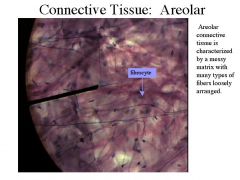
Areolar Connective Tissue
|

A type of loose connective tissue.
|
|
|
Fibroblasts
|
Young, actively mitotic cell that forms the fibers of connective tissue.
|
|
|
Adipose Tissue
|

-Areolar connective tissue modified to store nutrients;
- A connective tissue consisting chiefly of fat cells. |
|
|
Adipocytes
|
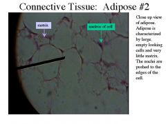
An adipose, or fat, cell.
|
|
|
Brown Adipose Tissue
|
-Contain abundant mitochondra, which use the lipid fuels to heat the bloodstream to warm the body rather then to produce ATP molecules.
-Occurs only in babies who lack the ability to produce body heat |
|
|
Reticular Connective Tissue
|
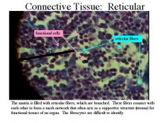
Connective tissue with a fine network of reticular fibers that form the internal supporting framework of lymphoid organs.
|
|
|
Dense Regular Connective Tissue
|
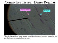
-One variety of the dense connective tissues, all of which have fibers as there predominant element.
|
|
|
Fibrous Connective Tissues
|
Another term for Dense Regular C.T.
|
|
|
Dense Irregular Connective Tissue
|
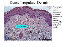
Has the same structural elements as the regular variety, howevever the bundles of collagen fibers are much thicker and are arranged irregularly.
|
|
|
Cartilage
|
White, semiopaque connective tissue
|
|
|
Chondroblasts
|
Actively mitotic cell of cartilage.
|
|
|
Hyaline Cartilage
|
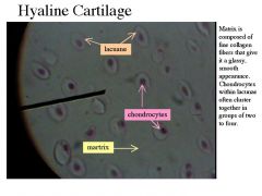
The most abundant cartilage type in the body; provides firm support with some pliability.
|
|
|
Articular Cartilage
|
Hyaline cartilage covering bone ends at movable joints
|
|
|
Epiphyseal Plates
|
Plate of hyaline cartilage at the junction of the diaphysis and epiphysis that provides for growth in length of a long bone
|
|
|
Elastic Cartilage
|
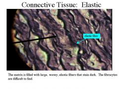
Cartilage with abundant elastic fibers; more flexible than hyaline cartilage.
|
|
|
Fibrocartilage
|
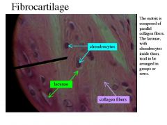
The most compressible type of cartilage; resistant to stretch. Forms vertebral discs and knee joint cartilages.
|
|
|
Bone (Osseous Tissue)
|

A connective tissue that forms the bony skeleton.
|
|
|
Osteoblasts
|
Bone-forming cells
|
|
|
Osteocytes
|
Mature bone cell.
|
|
|
Osteons
|
System of interconnecting canals in the microscopic structure of adult compact bone; unit of bone; also called Haversian system.
|
|
|
Blood
|
-The fluid within blood vessels
-The most atypical connective tissue. |
|
|
Nervous Tissue
|
The main component of the nervous sytsem.
|
|
|
Neurons
|
Cell of the nervous system specialized to generate and transmit electrical signals (action potentials and graded potentials).
|
|
|
Dendrites
|
Branching neuron process that serves as a receptive, or input, region; transmits an electrical signal toward the cell body.
|
|
|
Axons
|
Neuron process that carries impulses away from the nerve cell body; efferent process; the conducting portion of a nerve cell.
|
|
|
Muscle Tissue
|
Highly cellular, well vascularized tissues that are responsible for most types of body movement.
|
|
|
Myofilaments
|
Filament that constitutes myofibrils. Of two types: actin and myosin.
|
|
|
Actin
|
A contractile protein of muscle.
|
|
|
Myosin
|
One of the principal contractile proteins found in muscle.
|
|
|
Skeletal Muscle Tissues
|
Packaged by C.T. sheets into organs called skeletal muscles that are attached to the bones of the skeleton.
|
|
|
Muscle Fibers
|
A muscle cell.
|
|
|
Striated
|
-Banded;
-Having parallel lines or grooves on the surface |
|
|
Cardiac Muscle
|
Found only in the wall of the heart. Its Contractions help propel blood through blood vessels to all parts of the body.
|
|
|
Intercalated Disks
|
Specialized connections between myocardial cells containing gap junctions and desmosomes.
|
|
|
Smooth Muscle
|
Spindle-shaped cells with one centrally located nucleus and no externally visible striations (bands). Found mainly in the walls of hollow organs.
|
|
|
Voluntary Muscle
|
Muscle under strict nervous control; skeletal muscle.
|
|
|
Involuntary Muscle
|
Muscle that cannot ordinarily be controlled voluntarily (e.g., smooth and cardiac muscle)
|
|
|
Cutaneous Membranes
|
Pertaining to the skin;
An organ system consisting of keratinized stratified squamous epithelium(epidermis) firmly attached to a thick layer of dense irregular c.t. (dermis) |
|
|
Mucous Membranes
|
Membranes that form the linings of body cavities open to the exterior (digestive, respiratory, urinary, and reproductive tracts).
|
|
|
Serous Membranes
|
-aka serosa
-The moist membrane found in closed ventral body cavities. |
|
|
Pleura
|
Membranes that form the linings of body cavities open to the exterior (digestive, respiratory, urinary, and reproductive tracts).
|
|
|
Pericardium
|
Double-layered sac enclosing the heart and forming its superficial layer; has fibrous and serous layers.
|
|
|
Peritoneums
|
Serous membrane lining the interior of the abdominal cavity and covering the surfaces of abdominal organs.
|
|
|
Steps of Tissue Repair
|
1. Inflammation sets the stage
2. Organization restores the blood supply. 3. Regeneration and fibrosis effect permanent repair. |
|
|
Primary Germ Layer
|
-One of the 1st events of the embryonic development.
-Lie one atop the next like a 3 layered cellular pancake -From superficial to deep: Ectoderm, mesoderm, and endoderm. |
|
|
Ectoderm
|
Embryonic germ layer; forms the epidermis of the skin and its derivatives, and nervous tissues.
|
|
|
Mesoderm
|
Primary germ layer that forms the skeleton and muscles of the body.
|
|
|
Endoderm
|
Embryonic germ layer; forms the lining of the digestive tube and its associated structures.
|
|
|
_________ is always found in Cartilage.
|
Chondryocytes
|
|
|
Chondrocytes lie in the ________ of Hyaline Cartilage tissue.
|
Lacunaue
|