![]()
![]()
![]()
Use LEFT and RIGHT arrow keys to navigate between flashcards;
Use UP and DOWN arrow keys to flip the card;
H to show hint;
A reads text to speech;
274 Cards in this Set
- Front
- Back
- 3rd side (hint)
|
Membranous Organelle |
Classification of intracellular organelles with plasma membranes (separating the internal env of the organelle from the cytoplasm) |
|
|
|
Nonmembranous Organelles |
Classi of organelles without plasma membranes |
|
|
|
Intracellular microcompartment |
Spaces enclosed by organelle's membranes constitute the _______ |
|
|
|
Inclusions |
◾not always enclosed by PM ◾cont's crystals, pigment granules, lipids, glycogen and stored waste prod's |
|
|
|
Plasma membrane |
Membranous Organelles (MO) lipid bilayer serves as cell boundary |
|
|
|
rER |
A region of the endoplasmic reticulum assoc with ribosomes: the site of CHON synthesis |
|
|
|
sER |
MO site for lipid and sterol synthesis. Glycogen metabolism, membrane formation & recylcing Not assoc with ribosomes |
|
|
|
Golgi Apparatus |
MO composed of flattened cisternae responsible for modifying sorting packaging CHONS and lipids for intra/extracellular transpo |
|
|
|
Endosomes |
MO interposed with edocytotic pathways that have the major function of sorting CHONs delivered to them via endocytotic vesicles and redirecting them to different cellular compartments for the final destination |
|
|
|
Lysosomes |
Small organelles containing digestive enzymes that are formed from endosomes |
|
|
|
➰pinocytotic vesicles ➰endocytotic vesicles ➰coated vesicles |
(3) transport vesicles |
|
|
|
Transport Vesicles |
mo involved in both endocytosis and exocytosis and vary in shape and material that they transpo |
|
|
|
Mitochondria |
Mo that provide most of the energu to cell by producing ATP through oxidative phosphorylation |
|
|
|
Peroxisomes |
Small organelles involved in the production and degradation of H2O2 and FA's |
|
|
|
Microtubules |
NMO which together with actin and intermediatw filaments form elements of the cytoskeleton and continuously elongate ( by ➕tubulin dimers) and shorten (by ➖ tubilin dimers) a property refered to as dynamic instability |
|
|
|
Centrioles |
◾NMO short paired cylindrical structures found in center of MTOC or centrosome ◾whose derivatives give rise to the basal bodies of cilia |
|
|
|
Ribosomes |
NMO structures essential for CHON synthesis and composed of ribosomal RNA (rRNA) and (rCHON) attached to the rER. |
|
|
|
Nucleus |
LMx: Largest organelle within the cell with distinct boundary |
|
|
|
Nucleus |
EMx: surrounded by 2 membranes containing nuclear pore complexes and perinuclear cisternal space regions with condensed and diffused chromatin pattern (heterochroma and euchroma) |
|
|
|
Nucleolus |
LMx: roughly circular, basophilic, visible in living cells with interference Mx |
|
|
|
Nucleolus |
EMx: dense non membranous structure cont fibrillar and granular material |
|
|
|
PM sER Endosomes Ribosomes |
Not visible under LMx (4) |
|
|
|
rER |
LMx: basophilic, aka ergastoplasm |
|
|
|
rER |
EMx: flattened sheets, sacs and tubes with attached ribosomes |
|
|
|
sER |
LMx: not visible, may exhibit eosinophilia |
|
|
|
sER |
EMx: flattened sheets, sacs, tubes without attached ribosomes |
|
|
|
Golgi apparatus |
LMx: observed as negative staining in heavy metail stained preps. Visible in living cells with interference Mx :: exhibit a clear area partially surrounded by ergastoplasm |
|
|
|
Golgi Apparatus |
EMx: flattened membrane sheets often adjacent to one side of the nucleus ::embedded in a network of microtubules near the Mtoc |
|
|
|
Secretory Vesicles |
LMx: only visible when such are very large |
Eg. Zymogen granules in pancreas |
|
|
Secretory Vesicles |
EMx: they are of uniform diameter often polarized on one side of cell |
|
|
|
Mitochondria |
LMx: miniscule, dark dots, visible in living cells using Vital dyes |
|
|
|
Mitochondria |
EMx: two membrane system, arranged in numerous folds called cristae. In steroid producing cells, the inner membrane arranged in tubular cristae |
|
|
|
Endosomes |
EMx: tubulovesicular structures with subdivided lumen containing electron lucent material or other smaller vesicles |
|
|
|
Lysosomes and Peroxisomes |
LMx: visible only after special enzyme histochemical staining (2) |
|
|
|
Lysosomes |
EMx: membrane bounded, electron densed |
|
|
|
Peroxisomes |
EMx: membrane bounded often with electron dense crystalloid inclusions |
|
|
|
Cytoskeletal elements |
LM: only observedbwhen organized into large structures |
Eg muscle fibrils |
|
|
Cytoskeletal elements |
EMx: long linear staining pattern |
|
|
|
Ribosomes |
EMx: minute dark dots, assoc with rER |
|
|
|
Glycogen |
LMx: purple haze region in cytoplasm, metachromasia with toluidine blue stained specimen |
|
|
|
Glycogen |
EMx: non membranous, dense grape-like inclusions ➿ |
|
|
|
Lipid droplets |
LMx: readily visible when extremely large. Appear as large empty holes in section (removed by embedding solvents) |
Eg in adipocytes |
|
|
Lipid droplets |
EMx: Nonmembranous inclusions generally appear as void in the section |
|
|
|
➰ microtubules ➰ filaments ➰Centrioles ➰Ribosomes |
Nonmembranous organelles (4) |
|
|
|
Actin filaments |
Flexible chains of actin molecules |
|
|
|
Intermediate filaments |
Ropelike fibers formed fr variety of CHONs |
|
|
|
Filaments |
Groups providing tensile strength to withstand tension and confer resistance to shearing forces |
|
|
|
➰phospholipid ➰cholesterol ➰protein |
The membrane primarily consists of _______ molecules (3) |
|
|
|
Nucleus |
Storage and use of genome |
|
|
|
Nucleus |
EAP (eg of assoc pathology) Inherited genetic disease, induced mutations |
|
|
|
Nucleolus |
Synthesis of rRNA, involved in regulation of the cell cycle |
|
|
|
Nucleolus |
EAP: Werner syndrome, cancerogenesis |
|
|
|
Plasmamembrane |
EAP: Cystic fibrosis, intestinal malabsorption, lactose intolerance |
|
|
|
Golgi apparatus |
EAP I-cell disease, polycystic kidney disease |
|
|
|
Secretory vesicles |
EAP: Lewy bodies of parkinson's Dsse, proinsulin diabetes |
|
|
|
Endosomes |
EAP: M6P receptor deficiency |
|
|
|
Lysosomes |
Fx: Digestion of macromolecus |
|
|
|
Glycogen |
Fx: short term storage of glucose |
|
|
|
Peroxisomes |
EAP: zellweger's syndrome |
|
|
Fx and EAP |
Organelles and cytoplasmic inclusions: Fx and EAP |
|
|
|
Lipid rafts |
Control the movt and distribution of CHONs within the lipid bilayer, Also contain memb chon involved in cell signaling, aka signaling platforms |
|
|
|
High conc of Cholesterol and GSL |
Lipid rafts are localized regions within the PM containing ⬆⬆ conc of _________ |
|
|
|
Freeze fracture |
Technique how integral CHONs can be confirmed |
|
|
|
P Face |
E Face or P face? Displays more particles? |
E Face = extracellular phase P Face = protoplasm phase |
|
|
ATP synthase |
Major CHON of the inner mitochondrial membrane. For ion pumping |
|
|
|
Linker CHONs |
Anchor the intracellular cystoskeleton to the extracellular matrix |
|
|
|
Structural CHONs |
Membrane CHON visualized by freeze fracture method |
|
|
|
Blebbing |
It is caused by the detachment of the plasma membrane from underlying actin filaments of the cell cytoskeleton |
|
|
|
Plasma Membrane blebs |
Are dynamic protrusions of the PM that are commonly observed in acute cell injury |
|
|
|
Cell injury |
Often manifests as morphologic changes in cell's PM which results in formation of PM-blebs |
|
|
|
Phalloidin, Cytochalasin-B |
Cytoskeletal poisons that act on actin filaments and cause extensive membrane blebbing (2) |
|
|
|
➰fat soluble ➰small ➰ uncharged |
(3) characteristics of substances that traverse the PM via simple diffusion |
|
|
|
➰transfer small & water soluble subs ➰some require energy; ATP (Na K) ➰some are just passive (glucose) ➰highly selective, transfers 1 type of molecule ➰undergoes conformational change |
Characteristic/Fx of a carrier CHON |
|
|
|
Channel Protein |
Also transfer small water soluble subs, Made of transmembrane CHONs with several membrane spanning domain that create hydrophillic channels |
|
|
|
Pore domain |
What serves as the ion selectivity filter of the channel protein which partially penetrates the membrane bilayer |
|
|
|
Yes |
Are ion channels ion selective or not? |
|
|
|
Membrane potential in neurons |
Eg of Voltage gated channel |
|
|
|
Neurotransmitter such as ACH receptors in muscle cells |
Eg of ligand gated ion channel |
|
|
|
Vesicular Transport |
A type of transport that undergoes conformational changes in PM at localized sites and subsequent formation of vesicles/fusion of vesicles with the membrane |
|
|
|
Vesicle Budding |
Major mechanism by which large molecules enter leave and move within the cell |
|
|
|
Endocytosis, exocytosis |
2 types of vesicular transport |
|
|
|
A. Endocytosis B. Exocytosis |
Endo/exocytosis A. Assoc w/ the formation & budding of vesicles from the PM B. Assoc with the fusion of veeicles originating from intracellular organelles with the pm |
|
|
|
Clathrin |
Chon that interacts with PM in vesicle formation for endocytotic mechanism |
|
|
|
Non specific |
Is pinocytosis specific or non specific? |
|
|
|
True |
Pinocytosis is constitutive |
It involves a continuous dynamic formation of small vesicles at the cell surface |
|
|
➰Blood vessels ➰smooth mucle cells |
(2) areas where pinocytotic vesicles are numerous |
|
|
|
Clathrin-independent |
Is pinocytosis clathrin depended or independed? |
|
|
|
Yes |
Is phagocytosis non selective? |
|
|
|
Phagosomes |
The large vesicle containing the engulfed phagocytosed particles |
|
|
|
Pathogen-associated molecular patterns |
Pagocytosis is triggered by fhe recognition of ___________ that are commonly expressed on pathogen surfaces by Toll-like receptors |
|
|
|
Nuclear factor kappa B |
Activated by PAMP. It regulates genese that control cell responses for phagocytosis |
|
|
|
Phagocytosis |
Clathrin independeny but also actin depended endocytosis |
|
|
|
➰Pinocytosis ➰phagocytosis ➰ receptor mediated endocytosis |
Three mechanisms for Endocytosis |
|
|
|
A. Specific B. Non C. Non |
Specific/nonspecific: A. Receptor mediated endo B. Phagocytosis C. Pinocytosis |
|
|
|
Cargo receptor |
Term for the receptors in receptor mediated endocytosis in regions of the PM where such regions eventually become coated pits |
|
|
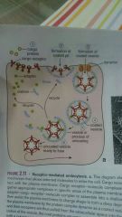
Adaptin |
A chon that helps select and gather appropriate complexes in specific areas of the plasma membrane for transport into cells, They recognize cargo receptor molecule complexes |
|
|
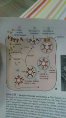
Dynamin |
Chon that pinches off the fully formed coated from the plasma membrane |
|
|
|
True |
T/F Coated pits and clathrin coated vesicles are formed in areas devoid of actin filaments |
|
|
|
Dynamin |
A large mechanoenzyme GTPase mediates the liberation of forming clathrin coated vesicles from PM during receptor mediated endo |
|
|
|
Receptor mediated endocytosis |
A clathrin depended endocytosis |
|
|
|
Receptor mediated endocytosis |
Coated vesicles are found in what mechanism of endocytosis? |
|
|
|
Clathrin coated vesicles |
They are also involved in movt of cargo mtrl from PM-Golgi app to early endosomes, early-late endosomes |
|
|
|
Coatomers COP-I, COP-II |
Mediates intracellular traffic during exocytosis |
|
|
|
➰Constitutive pathway ➰regulated secretory pathway |
2 general pathways for exocytosis |
|
|
|
Constitutive pathway |
Exocytosis pathway wherein chon that leave the cell are secreted immediately after their synthesis and and exit from golgi. |
|
|
|
Constitutive pathway |
Type of exocytosis pathway present to some degree in all cells |
|
|
|
Regulated secretory pathway |
A regulated event (hormonal or neural stimulus) must be activated for secretion to occur. |
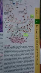
Diagram |
|
|
Calcium Ions |
The signaling stimulus in the process of regulated secretory pathway causes a transient influx of ________ |
|
|
|
➰chief cells of gastric mucosa ➰acinar cells of the pancreas |
Eg of cells that exhibit regulated secretory pathway (2) |
|
|
|
Rab-GTPase |
Interacts with the tethering proteins located on the target membrane. Provides recognition |
|
|
|
Docking complex between rabGTPase and receptor |
Immobilizes the vesicle near the target membrane |
|
|
|
SNARE (specific membrane CHON) |
Fam of transmembrane chon's, they guarantee the specificity of interaction between a particular vesicle and its target membrane and also promote membrane fusion |
|
|
|
Nsf/-snap chon complex |
Dismantle the snare complexed after they are formed |
|
|
|
Restricted to a a portion near the cell membrane where vesicles originating from the cell membrane fuse |
Early endosomes are termed such bc? |
|
|
|
Lysosomes |
Mature form of late endosomes? |
|
|
|
➰cellular localization ➰morphology ➰state of acidification ➰function |
Differences between early and late endosomes |
|
|
|
A. Near cell membrane B. Near golgi apparatus & nucleus |
Location: A. Early endosomes is to _______ B. Late endosomes is to ______ |
|
|
|
A. Tubolevesicular structure B. Onion like internal membrane |
Structure: Early endosome is to ___a____ Late endosome is to ____b____ |
|
|
|
Early: less acidic (6.2-6.5) Late: more acidic (ave 5.5) |
(Acidicity level) Early/late endosome More acidic? Less acidic? |
|
|
|
MultiVesicular Bodies (MVB) |
These are specific vesicles that transport substances between early and late endosomes. Highly selective transporters |
|
|
|
Prelysosomes |
Other name for late endosomes |
|
|
|
Sort and recycle CHONs internalized by endocytotic pathways |
Major function of early lysosomes |
|
|
|
Endosomes |
Transport endocytosed material. And biogenesis of lysosomes |
|
|
|
➰Morphologic shape ➰geometry of tubules & Vesicles |
Basis for the sorting mechanism in early endosomes |
|
|
|
➰receptor is recycled, ligand is degraded ➰ both recycled ➰both degraded ➰both transported through the cell |
Pathways for processing internalized receptor complexes of early endosomes |
|
|
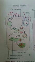
➰LDL receptor complex ➰Insulin glucose transporter (GLUT) recep comp ➰peptide hormones and their recep |
Eg of receptor complexes that "receptor is recycled, ligand is degraded" |
|
|
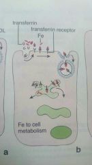
Iron-Transferrin receptor complex MHC 1 and 2 molecules |
Eg of showing "both receptor and ligands recycled" in endosomes |
|
|
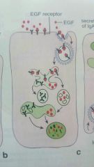
Egf (epiderman growth factor) and receptors |
Eg of "both receptor and ligand are degraded" pathway in endosomes |
|
|
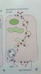
Iga-receptor complex |
Eg of "both receptor and ligand are transported through the cell" pathway in endosomes |
|
|
|
Transport of a substance (ligand receptor complexes) through the cell to be released to a different site of the cell surface |
What is transcytosis? |
|
|
|
Proteases, nucleases, glycosidases, lipases, phospholipases |
Lysosomes are rich in hydrolytic enzymes such as: |
|
|
|
Autophagy |
The process of Removal of cytoplasmic components particularly membrane bounded organelles by digesting them within lysosomes |
|
|
|
a. rER (synthesis) b. Golgi (sorting) c. M6P receptor (binding ability) |
Lysosomal enzymes are synthesized in the _____(a)______ and sorted in the ____(b)_____ based on their binding ability to ____(c)____ |
|
|
|
➰cholesterol in the "unusual phospholipid structure" ➰ lyso-bis-phosphatidic acid (lipid) |
2 unique components of the lysosomal membrane that makes ot unique and resistant to hydrolytic digestion |
|
|
|
Lysobiphosphatidic acid |
May play an impt role in restricting the activity of hydrolytic enzymes directed against the PM |
|
|
|
True |
Lysosomes and late endosomes contain proton pumps that transports H into lysosomal lumen to maintain low ph. The lysosomal membrane also contains transport CHON that transpo final products of digestion to the cytoplasm used in synthetic processes or exocytosed
|
|
|
|
Chloroquine (tx for malaria) |
Eg of drugs that effect the lysosomal Fx; it raises PH level inactivating the enz's |
|
|
|
C terminus domain recognized by adaptin protein complexes |
Lysosomal membrane proteins are synthesized in rER but have specific lysosomal targeting signal. Instead of M6P, they have _________ |
|
|
|
➰lamp's (lysosomal assoc membrane CHON) ➰lgp's (Lysosomal membrane glycoCHON) ➰limp's (Lysosomal integral membrane CHON) |
Classifications of lysosomal membrane CHONs |
|
|
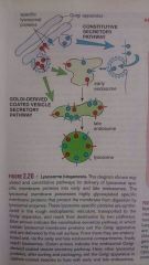
Golgi derived coated vesicle secretory pathway |
LIMP's after sorting and packaging, exit the golgi in clathrin coated vesic's, they travel and fuse with late endosomes |
|
|
|
Autophagy |
Represents major cellular pathway in which a number of cytoplasmic CHONs, organelles, and other cellular structures are degraded in lysosomal compartments |
|
|
|
A. Phagocytosis- bacteria, cell debris B. Pinocytosis & receptor mediated endocytosis- CHONs, ligand receptor complexes C. Autophagy- entire organelles, cytoplasmic CHONs |
Intracellular digestion in lysosomes Three pathways: ➰extracellular large particles via (a) ➰extracellular small particles via (b) ➰intracellular particles via (c) |
|
|
|
True, Bc of number, size or contents |
Lysosomes in some cells are recognizable under LMx? T/F Why? |
|
|
|
Residual body |
Formed from hydrolytic breakdown of the contents of lysosomes, a debri filled vacuole |
|
|
|
Age pigment or lipofuscin granules |
Residual bodies in neurons are called: ________ |
|
|
|
A. Normal B.1 slower growth B.2 slpw changes in facial features B.3 bone and joint deformities |
Children born with LSD usually appear ___(a)___ at birth Soon, they show (b.1) (b.2) (b.3) |
|
|
|
Glucocerebrosidase. Sphingomyelinase |
LSD Protein deficiency in: Gaucher Disease? Niemann Pick disease A1B? |
|
|
|
Nutrient starvation, hypoxia, high temperature |
⬆mTOR (mammalian target of rapamycin) = inhibits autophagy What causes ⬇⬇ mtor activity (3) |
|
|
|
Macroautophagy Microautophagy Chaperon mediated autophagy |
(3) pathways for autophagy |
|
|
|
Atg12, Atg5, Atg16L attaches to ER, and localizes the isolation membrane and Atg8- recruited after Together they change the shape of the isolation membrane |
Autophagy proteins |
|
|
|
First stages of starvation |
Macroautophagy occurs in the liver during _______ |
|
|
|
Microautophagy |
Small cytoplasmic solube chon's ae internalized into the lysosomes. Degraded in a slow and continuous process under normal physiologic conditions |
|
|
|
A. Chaperon mediated autophagy B. Hsc73 (heat-shock chaper-one protein) |
The only selective process of CHON degradation ___(a)_____ It requires assistance from specific cytosolic chaperones such as ___(b)___ |
|
|
|
True |
Starvation activates autophagy |
|
|
|
Lysosomes and Proteosomes |
Oranelles that can or are involved in Protein Degradation in cells |
|
|
|
➰starvation ➰cell death ➰cell aging ➰cellular differentiation |
Autophagy plays an essential role during what situations (4) |
|
|
|
Proteosome-mediated degradation |
Used by cells to destroy abnormal proteins that are misfolded, denatured , or contain abnormal AA's |
|
|
|
Polyubiquitin chain |
CHON's destined for proteasome-mediated degradation need to be recognizedband specifically tagged by the _________ |
|
|
|
Ubiquitin |
Polyubiquitination in which CHONs targeted for destruction are repeatedly tagged by covalent attachments of a small protein called ______ |
|
|
|
Four in a form of a polyubiquitin chain |
A CHON targeted for destruction within the proteosome must be labeled with at least how many ubiquitin molecules? |
|
|
|
➰polyubiquitination ➰by 26S proteasome complex |
2 mechanisms on protein degradation in proteasomes |
|
|
|
(1) ergastoplasm, rER (2) presence of RNA |
What portion of the cell cytoplasm stains with basic dye? (1) The basophilic staining is caused by? (2) |
|
|
|
Transcription |
Process in which the genetic code for a CHON is transcribed from DNA to pre-mRNA |
|
|
|
rER |
Serves as a checkpoint in the process of CHON production |
|
|
|
Nissl Bodies |
Large basophillic bodies of the nerve cells are called & consist both rER and large number of free ribosomes |
|
|
|
A. Tubular |
sER is _______ A. Tubular B. Sheetlike |
|
|
|
Ribosomal docking CHON |
How are ribosomes attached to the membrame of the rER |
|
|
|
rRNA |
Each subunit of ribosome contains_____ of diff length as well as numerous of other CHON's |
|
|
|
Polyribosomes and polysomes |
Other name for group of ribosomes |
|
|
|
Transcription and translation Transcription and translation |
Chon synthesis involved two processes |
|
|
|
Transcription |
Process by which the genetic cpde for a CHON is transcribed from DNA to pre MRNA |
|
|
|
(1)Aminoglycosides (streptomycin) Macrolides (erythromycin) Lincosamides (clindamycin) Tetracycline Chloramphenicol (2) binding to diff portions of bacterial ribosomes |
(1)Types of antibiotics that inhibit CHON synthesis. (2)And the manner of inhibition |
|
|
|
(1)Signal sequences (2) 15-60 AA on amino terminal |
(1)Sorting signals that direct CHONs to their correct destinations (2) often found at? |
|
|
|
Signal recognition particle (SRP) |
Arrests further growth of the polypeptide chain of a nascent (beginning) peptide |
|
|
|
1. Signal peptidase 2. Signal peptide peptidase |
After CHON synthesis is resumed, the signal sequence iscleaved from ths poly peptide by ___(1)___ and is susequently digested by ___(2)___ |
|
|
|
(1)true (2)golgi apparatus within minutes |
(1)Few proteins remaind permanent residents of rER (T/F). (2)The newly synthesized CHONs are delivered where? |
|
|
|
1-antitrypsin deficiency |
Inability of rER to export a mutated protein to golgi, which leads to decreased activity of A1AT in blood and lungs |
|
|
|
rER |
Serves as quality checkpoint for CHON synthesis |
|
|
|
Active secretory cells (glndular cellS, odontoblast, ameloblast, osteoblasts) and in neurons (cells with large plasma membrane) |
The rER is most highly developed in what type of celles |
|
|
|
Coatomer |
They mediate bidirectional traffic between the rER and Golgi appatatus |
|
|
|
Clathrins |
Mediate bidirectional transport from and to the plasma membrane |
|
|
|
(1)anterogade transport or COP-II (2)retrogade transport or COP1 [COP-I COP-II are the two classes of CHONS] |
(1)Type of transport where CHONs is transpo from rER to Cis Golgi Network (2) " from cis golgi network back to rER |
|
|
|
COP-I |
Class of CHON thats responsible for maintaining retrograde transport between the Golgi Cisternae |
|
|
|
True |
In the absence of a signal sequence proteins that are synthesized on free ribosomes remain in the cytosol |
|
|
|
True |
Cells with large amounts of sER may exhibit distinct cytoplasmic eosinophilia |
|
|
|
Adrenocortical Cells Testicular Leydig Cells |
sER is well developed in cells that synthesize and secrete steroids such as..(2) |
|
|
|
sER |
It sequesters/holds/keeps Calcium ions essential for contractile process |
|
|
|
sER |
Principal organelle for the detoxification and conjugatiob of npxious substances |
|
|
|
Cytochrome P450 |
The sER contains Detoxifying enzymes related to ______ that is anchored directly into sER PM |
|
|
|
Golgi apparatus |
Organelle well developed in secretory cells (similar with rER) and does not stain with EOSIN or hematoxylin |
|
|
|
cis golgi network (1) Trans golgi network (2) |
The flattened cisternae located closest to the rER represent the forming face (1) The cisternae located away from the rER represent the maturing face |
|
|
|
Remodelling of N-linked oligosaccharides previous added in rER |
Post translational modification in golgi apparatus involves what process? |
|
|
|
:: apical PM - uses non clathrin coated vesicles :: basolateral PM - uses vesicles coated with an unidentified protein associated with an epithelium specific adaptor protein :: endosomes or lysosomes - Bear specific signal sequences. Take extended route :: apical cytoplasm - uses clathrin coated vesicles
|
Four major pathways of CHON secretion from Golgi to various cell destinations |
|
|
|
B. Trans GN |
Sorting and packaging of CHONs into transpo vesicles occurs in A. Cis-GN B. Trans-GN |
|
|
|
Sorting signals Physical Properties |
The sorting and packaging of CHON in TGN is based on (2) factors |
|
|
|
RBC and Terminal Keratinocytes |
Mitochondria is present in all cells except (2) |
|
|
|
Acidophilia |
Mitochondria contribute to the Acidophilia or basophilia of cytoplasm? |
|
|
|
The phospholipid cardiolipin |
Inner membrane of mitochondria is rich in _____ |
|
|
|
ck, cytochrome C, ak |
Intermembrane space contains specific enzymes |
|
|
|
calcium and other divalent and trivalent cations. |
Mitochondria contain dense matriz granules that store |
|
|
|
Bcl-2 |
The mitochondria initiating apoptosis via relase of cytochrome c is regulated by |
|
|
|
Oxidative enzymes: catalase, peroxidase (which are impt in liver cells, performs detoxification process) |
Lysosomes is to hydrolytic enzymes such as proteases, nucleases glycosidses lipases and phosphatases.. While peroxisomes is to.. |
|
|
|
Regulated the cellular H2O2 content by breaking down H2O2 , thus protecting the cell |
Function of catalase in peroxisomes |
|
|
|
Crystalloid Inclusion (nucleoid) |
In most animals, peroxisomes contain urate oxidase which gives a characteristic appearance of... |
|
|
|
Non functional peroxisomes Bc of lack of necessary enzymes. Caused by mutation in the gene encoding the receptor for the peroxisome targeting signal that does not recognize the signal ser-lys-leu |
Zellweggers syndrome |
|
|
|
1. Mitochondria 2. Peroxisome |
Organelle defect: Merff is to (1) Zellweggers is to (2) |
|
|
|
True |
Microtubules are elongated conoosed of equal parts of a-tubulin, and b-tubulin |
|
|
|
Guanosine triphosphate (gtp) Mg |
Polymerization of tubulin dimers requires the presence of |
|
|
|
Plus growing end |
Alpha tubulin is to minus (non growing end) Beta tubulin is to |
|
|
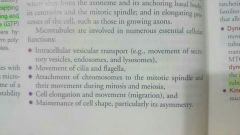
Fx |
Fx of microtubules |
|
|
|
Molecular motor proteins: Dynein (1) Kinesin (2)
|
They move along the microtubules toward the minus end. Capable of transpo organelles from periphery to MTOC (1)
Toward the plus ends, slash cell periphery (2) |
|
|
|
Axonemal dynein |
Responsible for the sliding of one microtubule against an adjacent microtubule of the axoneme |
|
|
|
Both |
Dynein and kinesin involved in mitosis or meiosis? |
|
|
|
Actin is thinner shorter more flexible |
Actin vs microtubules |
|
|
|
G actin (globular) f actin (filamentous) |
Free actin molecules in cytoplasm (1) Polymerized actin filament (2) |
|
|
|
Controlling rate of polymerization of actin filaments(1) Filament organization (2) |
Actin binding proteins are responsible for |
|
|
|
A. Gelsolin B. Tropomodulin C. Spectrin, Adductin, protein 4.1, protein 4.9 D. Myofilaments, thin thick filaments |
Actin filament severing CHON eg (a) Actin capping CHON (b) Actin cross linking CHON (c) Actin motor CHON (d) |
|
|
Fx actin filaments |
Fx of Actin Filaments |
|
|
|
True |
Intermediate do not possess enzymatic activity and form from non polar filaments |
|
|
|
Class 1 and Class 2 - Keratin (acidic) (basic) Class 3 -vimentin Class 4 - neurofilaments Class 5 - lamin Class 6 - beaded filaments |
6 classes of Intermediate Filaments |
|
|
|
Vimentin |
Most widely distributed intermediate filament |
|
|
|
A. Fibroblast B. Muscle cells C. Glial cells D. Peripheral nerve cells |
Locations of class 3 IF: Vimentin: (a) Desmin: (b) GFAP: (c) Peripherin: (d) |
|
|
|
Axons of nerve cells [neurofilaments] |
Class 4 IF is expressed mostly where? |
|
|
|
Lamin class 5 |
IF thats located in the nucleoplasm |
|
|
|
Beaded filaments: 1. Phakinin 2. Filensin |
2 proteins of class 6 IF |
|
|
|
Gamma-tubulin ring |
Serves as a starting point (nucleation site) for growth of microtubule in MTOC |
|
|
|
Centriole (both) |
Centriole/ centrosome Provide the basal bodies for cilia and flagella? Align mitotic spindle? |
|
|
|
Basal Bodies |
What acts as organizing center for cilium? |
|
|

Table for cytoskeleton part II |
Table for cytoskeleton part II |
|
|
|
Kartageners |
Disease of microtubules. Affects sperm motility |
|
|
|
1. Alzheimers 2 liver cirrhosis |
1. Neurofibriallary tangles (Intermediate filaments) 2. Mallory bodies |
|
|
|
Alexanders disease- rosenthal fibers (cytoplasmic inclusions in astrocytes) |
Mutations in the coding region of the GFAP gene. And pathologic feature? |
|
|
|
to position the mitotic spindle |
Primary role of centrioles in mitosis is |
|
|
|
G1 PHASE |
What phase in the cell cycle does cilia is assembled? |
|
|
|
Lipofuscin; Wear and tear pigment |
Brownish gold pigment visible in routine H&E prep; Other name |
|
|
|
Dna folded forms the chromatin. Chromatim folded forms the chromosome. Paired chromosome by centromere is chromatid |
Level of organization in nucleus |
|
|
|
46 (diploid 2n)- 23 homolpgous pairs. 22 autosome, 23rd is sex chromosome |
Each human cell contains ___ chromosome |
|
|
|
Histones and non histones |
Chromatin contains 5 basic CHONs called |
|
|
|
Marginal Chromatin- periphery of nucleus Karyosomes-discrete bodies throughout the nucleus Nuclear associated chromatin- found in assoc with the nucleolus |
Heterochromatin is disposed in 3 locations |
|
|
|
H&E Vital dyes: hoescht dyes, propidium iodode |
Stains heterochromatin |
|
|
|
Nucleosomes- foumd both in eurochroma and hetero. |
Smallest unit of chromatin. Made up of complexes of dna and histone molecules (octamer) |
|
|
|
Nucleosomal substructure of chromatin |
Beads of string appearance under EMx |
|
|
|
Telomerase |
Enz present in malignant cells that adds repeated nucleotide sequences to telomere ends. Expression of this enzyme has been shown to extend lifespan of cells (telomere length indicate lifespan of cell, as it shortens after every cell division) |
|
|
|
rRNA synthesis and initial ribosomal prod & assembly |
Nucleolus is the site of |
|
|
|
Fibrillar centers- DNA loops of 5 diff chromosomes (13,14,15,21,22) Fibrillar material (pars fibrosa)- ribosomal genes actively undergoing transcription, ⬆⬆amnts of rRNA Granular material (pars granulosa)- site of initial ribosomal assembly, densely packed preribosomal particles |
Nucleolus cont 3 distinct regions |
|
|
|
True |
Nucleolus: rRNA is present in both granular and fibrillar material |
|
|
|
Nucleostemin- regulates cell cycle and differentiation |
Newly ID'd CHON within the nucleolus |
|
|
|
Hematoxylin and basic dyes. Metachromatically with thionine dyes |
Nucleolus stains with |
|
|
|
True |
Dna is present in nucleolus altho no staining rxn with feulgen due to low conc |
|
|
|
Emery dreifuss muscular dystrophy Dominant form- mutation in lamin a/c Recessive form- mutation in emerin |
Mutation in lamin or lamin receptors |
|
|
|
Late anaphase |
Re assemly of the nuclear envelope begins at what phase in mitosis |
|
|
|
Static population |
Consist of cell that no longer divide (CNS cells, skeletal or cardiac muscle cells) |
|
|
|
Stable cell populations |
Cells that divide episodically and slowly to maintain normal tissue and organ (perichondrial cells, periosteal cells, smooth muscle cells,endothelial cells of BV, fibroblast of Connective tissue) |
|
|
|
Renewing cell populations |
May be slowly or rapidly but display regular mitotic activity |
|
|
|
1. Smooth muscle cells, fibroblast of uterine wall, epithelial cells of the lens of the eye, 2. Blood cells, epithelial cells, dermal fibroblasts, epith and ubepith fibroblasts of mucosal lining of alimentary tract |
Slowly renewing cells eg (1) Rapidly renewing eg (2) |
|
|
|
To produce 2 daughter cells that has identical chromosomes of the parent cell |
Goal of cell cycle |
|
|
|
Interphase- continuous growth of cell, 3 phases G1, S, G2 subdivide interphase Mitosia- cell division |
2 phases of the cell cycle |
|
|
|
G1- cell gathers nutrients and synthesizes RNA and chons for DNA synthesis |
Longest phase. And events in such phase |
|
|
|
Terminal differentiation after Leaving the cell cycle at G1 |
What is the GO phase |
|
|
|
1. Restriction point- checks size of cell, state of physio process, interactions with extra cellular matrix 2. G1 DNA damage checkpoint- monitors the integrity of new replicated dna |
Checkpoints of G1 |
|
|
|
1.Dna is replicated 2. S DNA damage checkpoint- monitors quality of the replicating DNA |
Event in S Phase. And checkpoint |
|
|
|
"Cells prepares for cell division" A. G2 dna damage checkpoint B. Unreplicated DNA checkpoint - prevents progression to Mphase before DNA synthesis is complete |
Even in G2 phase. And checkpoints |
|
|
|
pRb- retinoblastoma susceptibility CHON E2F- essential transcription factors |
Restriction point is mediated by what? |
|
|
|
1. Spindle assembly checkpoint 2. Chromosome segregation checkpoint |
MPhase checkpoints |
|
|
|
Associated with mesothelioma (pleural cavities in thorax caner) osteosarcoma, ependydoma (childhood brain tumor) |
T Antigen Simian Virus (Sv40) |
|
|
|
Maturation Promoting Factor- control the initation of mitosis. Contains Cdc2, Cyclin B. |
Helps power the cells through the checkpoints of cell cycle divisions |
|
|
|
2n, 2d ➡ 2n 4d |
Mitosis chromosome number (n) and dna number (d) |
|
|
|
Cohesins and the centromere |
During prophase: The sister chromatids are held together by a ring of CHONs called |
|
|
|
Kinetochore- attached to specific repetitive DNA sequences known as satellite DNA |
A highly specialized protein attaches on each chromatid side oppositr to the centromete |
|
|
|
Metaphase 1. Astral microtubules- nucleated from the gamma-tubulin rings in a starlike fashion around each MTOC. 2. Polar microtubules- grow away from MTOC 3. Kinetochore microtubules- to probe the cytoplasm in search of kinetochore |
The phase of cell division that consist of three type of Microtubules, becomes organized at opposite poles of the cell |
|
|
|
Metaphase. Equatorial plate or metaphase plate |
Chromosomes move to the plane in the middle of the cell. what region and what phase of cell division |
|
|
|
Anaphase. Dyneins |
Phase when Initial separation of sister chromatids and Cohesins breakdown? And they are pulled by? |
|
|
|
2d 2n |
Mitosis effect of dna and chromosome number |
|
|
|
1n 2d after 1meiotic division 1n 2n after 2nd |
Result of meiosis |
|