Reading...
![]()
Play button
![]()
Play button
![]()
Use LEFT and RIGHT arrow keys to navigate between flashcards;
Use UP and DOWN arrow keys to flip the card;
H to show hint;
A reads text to speech;
60 Cards in this Set
- Front
- Back
|
What embryonic layer is connective tissue derived from?
|
Embryonic mesoderm
|
|
|
What are the three components of CT?
|
Ground substance, fibers, Cells
|
|
|
What are some characteristics of ground substance?
|
Amorphous, colorless, homogenous
|
|
|
What are the three main components of ground substance?
|
1. Glycoaminoglycans (GAGs) - most abundant, negativly charged- hydrated
2. Proteoglycans - secreted by resident cells, have a protein backbone bound to GAGS. Hydrophyllic- surrounded by water . Multiadhesive glycoproteins - Laminin (900 kD) is a main component of BL, and fibronectin. |
|
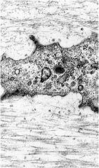
What type of CT surrounds this cell? What is it composed of?
|
Hyaline cartilage - composed of type 2 collagen
|
|
|
What is the term for accumulating tissue fluid in CT?
|
Edema (swelling)
|
|
|
What are the three main types of fibers?
|
Collagen, Elastic, Reticular
|
|
|
How many types of collagen are there? What color does it stain in H&E?
|
There are 19 types, and it stains pink (eosin) in H&E due to it being acidophilic.
|
|
|
What are the common sites to find type 1 collagen? What is its function?
|
1. Found in bone, CT of skin, tnedon, ligaments, dentin, sclera, fascia, and organ capsules (makes up 90% of collagen found in body).
2. It provides resistance to force, tension, and stretch |
|
|
What the the common sites to find type 2 collagen? What is its function?
|
1. Cartilage (hyaline and elastic), notochord, and intervertebral disks
2. Provides resistance to intermittent pressure. |
|
|
What are common sites to find type 3 collagen? what are its functions?
|
1. Found in loose connective tissue and organs (uterus, liver, spleen, kidney, lungs...), smooth muscle; endoneurium, blood vessels, and fetal skin
2. Forms reticular fibers, arranged as a loose network of fibers, provides a supportive scaffolding for specialized cells of organs and blood vessels. |
|
|
What are some common sites for type 4 collagen? What are its functions?
|
1. Basal Laminae of epithelia, kidney glomeruli, and lens capsule.
2. Provides support and filtration barrier |
|
|
What is Ehlers-Danlos syndrom (EDS)?
|
A name given to a group of more than 10 types of collagen disorders.
|
|
|
What two AA's are unique to Elastic Fibers?
|
Desmosine and isodesmosine
|
|
|
What special protein is an integral part of elastic fibers? What disease is characterized by a defect in this protein?
|
Fibrillin - marfans syndrome
|
|
|
What kins of stains must be used to visualize elastic fibers? What colors does it stain?
|
Orecin or resorcin-fuchin to stain the fibers purple, dark blue, or blue black.
|
|
|
Where are elastic fibers commonly found?
|
The walls of blood vessels, usually forming fenestrated membranes in larger vessels (aorta)
|
|
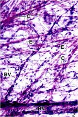
Identify the tissues abbreviated in the picture
|
BV- Blood vessel
E - elastic fibers (orecin stained) C - collagen |
|
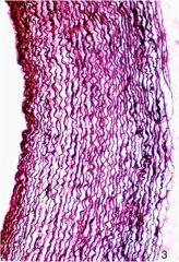
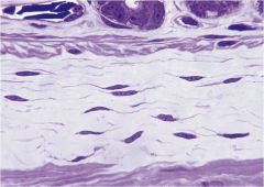
Identify the tissue, and location.
|
Elastic Laminae of the aorta that allow it to recoil in response to changes in systolic pressure.
|
|
|
What are some characteristics of Marfans Syndrome?
|
Patients are tall, with long extremities. Mitral valve prolapse, dilation of the root of aorta, aortic dissection. Caused by a deficiency in the protein fibrillin 1.
|
|
|
What type of collagen fibers make up reticular fibers?
|
Type 3
|
|
|
What kind of stain is used to visualize reticular fibers?
|
silver
|
|
|
Where is the reticular laminae located?
|
Beneath the basal Laminae
|
|
|
What kind of fibers form the framework for the lymph nodes and the spleen?
|
Reticular fibers
|
|
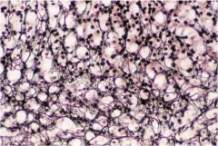
What type of tissue is displayed? what kind of stain was used?
|
Reticular fibers in the adrenal cortex, stained with silver
|
|
|
What are the two types of connective tissue cells? Name examples of each.
|
1. Resident (permanent)
- Fibroblasts - adipose - macrophage - mast cells - specialized -e.g. osteoblasts 2. Immigrant (wandering) - plasma cells - leukocytes |
|
|
What is the most abundant CT cell type? What is its function?
|
Fibroblasts - synthesizes extracellular matrix (ground substance and fibers)
|
|
|
What is a specialized form of the fibroblast that has a role in wound healing?
|
Myofibroblast - contains bundles of actin microfilaments and dense bodies.
|
|
|
Name for a quiescent fibroblast
|
Fibrocyte
|
|
Name the cell type of the CT
|
Fibroblast
|
|
|
What are the two types of adipose tissue?
|
White fat (unilocular) and Brown Fat (Multilocular)
|
|
|
What is the role of brown fat?
|
Found in infants and is heat generating when mobilized by symp. nervous system. Found only in kidneys, adrenal glands, aorta, neck and mediastinum of adults
|
|
|
Where are adipose cells derived from?
|
undifferentiated mesenchymal cells
|
|
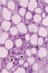
Identify the tissue
|
White fat (unilocular)
|
|
|
What is Prader-Willi syndrome?
|
Overproduction of Ghrelin, making the patient hungry all of the time.
|
|
|
What is peptide YY?
|
A gatrointestinal peptide signaling satiety through the hypothalamus
|
|
|
What is leptin?
|
Hormone secreted by fat cells that works on the hypothalamus to suppress appetite. Obese people are resistant.
|
|
|
What is the origin of the mast cell?
|
Hemopoietic in bone marrow, differentiate in CT
|
|
|
What substances do mast cells secrete?
|
- Histamine and slow reacting substances of anaphylaxis (SRS-A)
- Eosiophil and neutrophil chemotactic factors (ECF and NCF) - Heparin - anticoagulent |
|
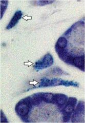
Identify the CT type
|
Mast cells
|
|
|
Where are plasma cells derived from? What do they produce?
|
B-lymphocytes, they produce antibodies.
|
|
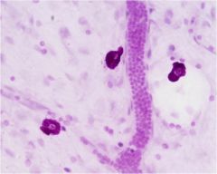
Identify the CT
|
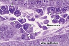
Plasma cells
|
|
|
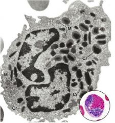
What immunoglobulin is produced by plasma cells that interacts with mast cell receptors?
|
IgE - causes mast cells to degranulate, which causes a release of its chemical factors.
|
|
|
Where are tissue macrophages derived from?
|
Blood borne monocytes
|
|
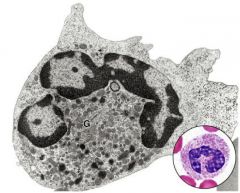
Identify the cells
|
Macrohphage
|
|
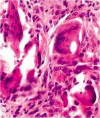
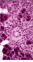
Identify the arrowed cells. What is this a response to?
|
Macrohpages that have fused into multinucleated cells in response to a foreign body (i.e. hait in an incision or a suture.)
|
|
|
Name the 3 types of lymphocytes and their functions.
|
1. B Lymphocytes give rise to plasma which make immunoglobulin
2. T Lymphocytes are involved in cell-mediated immunity 3. Natural killer cells (NK) destroy virus infected cells and some tumors. |
|
What are the functions of neutrophils?
|
First to arrive at the site of inflammation; leave by a process called diapedesis. Active phagocytes (microphages) that can phagocytose some bacteria.
|
|
|
What is pus?
|
An accumulation of dead neutrophils
|
|
|
What processes are eisonphils associated with?
|
Allergic reactions, parasitic infections, and chronic inflammatory processes. Found in lamina propria of guy.
|
|
|
What morphological characteristics make eosinophils distinct?
|
Large red granules, and a bilobed nucleus
|
|
|
What is edema?
|
Accumulation of fluid in the CT comparment.
|
|
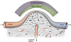
1. What happens if hydrostatic pressure is too high, as in congestive heart failure?
2. What happens if colloid osmotic pressure is too low, as in starvation? |
1. Fluid accumulates in the ECM
2. Fluid accumulates in ECM |
|
|
What is the difference between loose and dense CT?
|
Loose CT has a lot of cells, but few fibers. Dense CT has the opposite.
Dense CT is broken up into two groups, Irregular and Regular. |
|
|
Where are Dense irregular CT found?
|
Dermis - reticular layer
|
|
|
Where would one find regular CT?
|
Tendons, ligaments
|
|
|
Where is loose CT found?
|
Found around glands. Cells include Mast cells, plasma, macrophages, lymphocytes, and fibroblasts
|
|
Identify the CT type
|
Loose CT
|
|
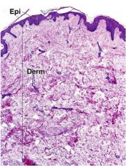
Identify the CT type
|
Dense irregular CT containing mainly fibroblasts
|
|
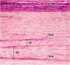
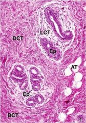
Identify the CT type
|
Dense Regular CT - fibroblasts predominate. EPT - epitendineum, TF - tendon fscicles, Ent - endotendineum
|