Reading...
![]()
Play button
![]()
Play button
![]()
Use LEFT and RIGHT arrow keys to navigate between flashcards;
Use UP and DOWN arrow keys to flip the card;
H to show hint;
A reads text to speech;
117 Cards in this Set
- Front
- Back
- 3rd side (hint)
|
|

x
|
|
|
|
x
|
|
|
|
x
|
|
|
|
x
|
|
|
|
x
|
|
|
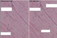
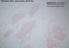
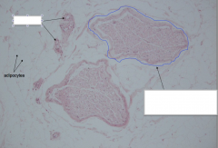
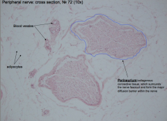
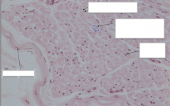
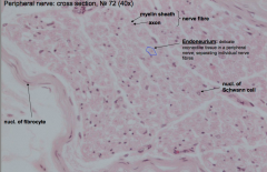
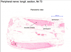
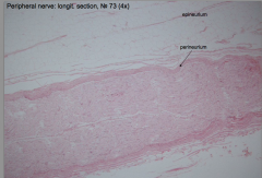
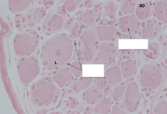
peripheral nerve cross section
|
x
|
|
|
|
x
|
|
|
|
x
|
|
|
|
x
|
|
|
|
x
|
|
|
|
|
|
|
|
x
|
|
|
|
x
|
|
|

|
x
|
|
|
|
x
|
|
|
|
x
|
|
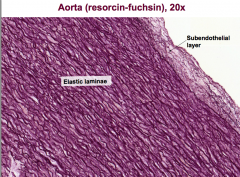
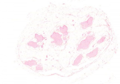
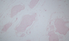
other is aorta x 20
|
|
x
|
|
|
|
x
|
|
|

|
xx
|
|
|

|
x
|
|

|
|
x
|
|
|
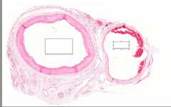
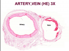
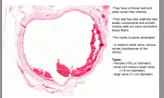
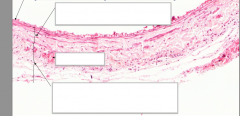
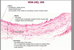
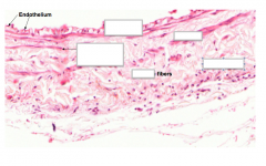
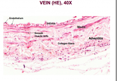
Vein features
|
|
x
|
|
|
|
x
|
|
|
|
x
|
|
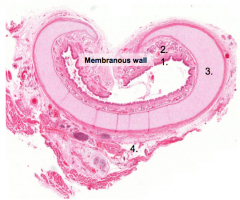
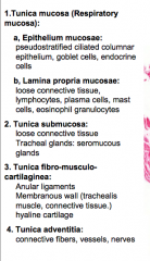
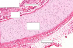
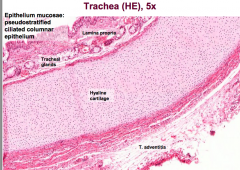
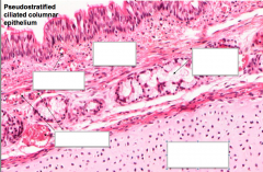
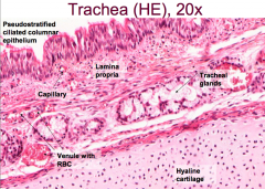
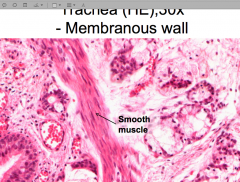
what is this slide? define it layers
|
|
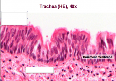
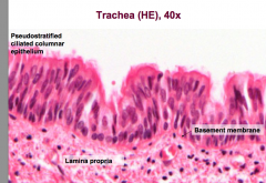
the trachea
|
|
|
|
x
|
|
|
|
x
|
|
|
|
x
|
|
|
|
x
|
|
|

|
x
|
|
|
|
x
|
|
|
|
x
|
|
|
|
|
|
|
|
|
|
|
|
|
|
|
|
|
|
|
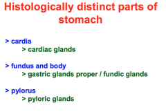
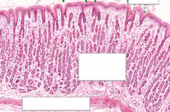
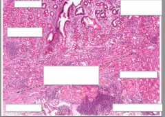
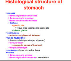
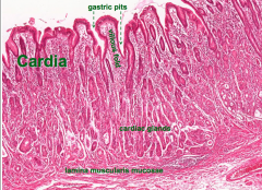
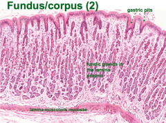
stomach glands
|
|
|
|
|
|
|
|
|
|
|
|
|
|
|
|

|
|
|
|
|
|
|
|
|
|
|
|
|
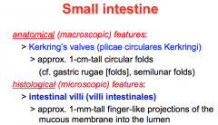
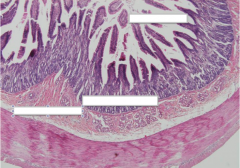
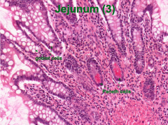
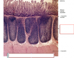
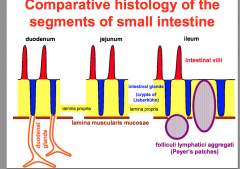
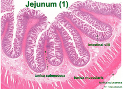
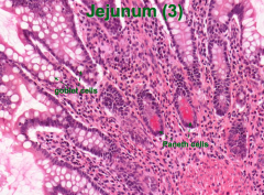
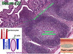
HISTOLOGICAL features of SI
|
|
but microvilli NOT a specific feature bc theyre found in other parts
|
|
|
|
|
|
|
|
|
|
|
|
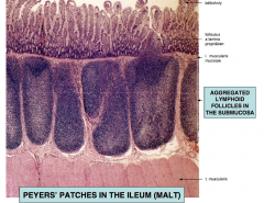
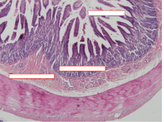
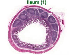
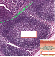
ileum HE
|
|
|
|
|
|
|
|
|
|
|
|
|
|
|
|
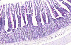
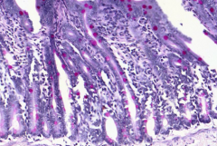
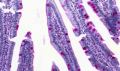
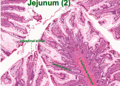
Jejunum: PAS&H Staining!
|
|
|
|
|
|
|
|
|
|
|
|
|
|
|
|
|
|

|
|
|
|
|
|
|
|
|
|
|
|
|
|
|
|
|
|
|
|
|
|
|
|
|
|
|
|
|
|
|
|
|
|
|
|
|
|
|
|
|
|
|
|
|
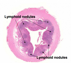
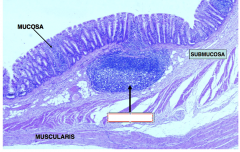
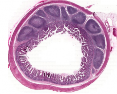
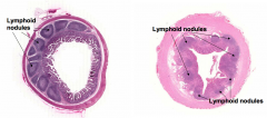
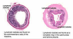
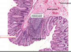
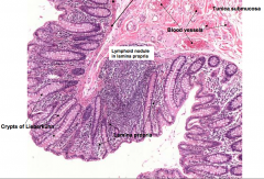
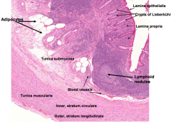
vermiform process
|
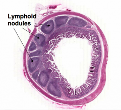
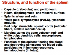
Lymphoid nodules are found as a complete ring in the submucosa and lamina propria.
|
|
|
|
|
|
|
|
|
|
|

|
|
|
|
|
|
|
|
|
|
|
|
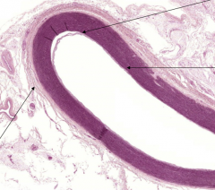
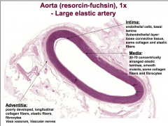
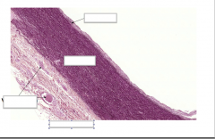
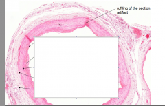
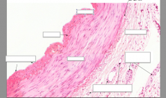
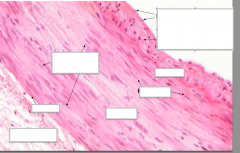
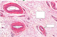
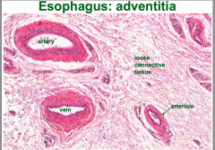
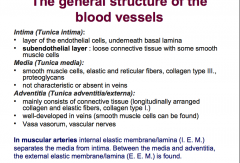
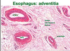
the general structure of blood vessels
|
|
|
|
|
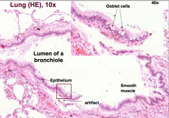
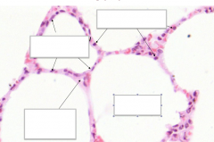
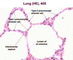
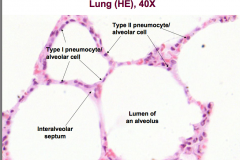
lung to know
|
|
|
|
|
blood smear 100 ml contents
|

|
|
|
|
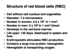
rbc structure
|
|
|
|
|
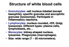
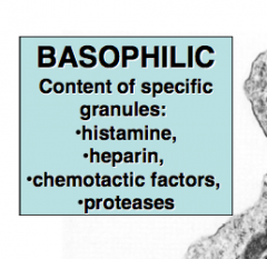
wbc structure
|
|
|
|
|
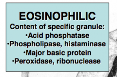
define nucleophilic, then basophilic
|
|
|
|
|
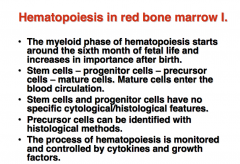
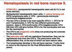
hematopoiesis in red bone marrow I and II
|
|
|
|
|
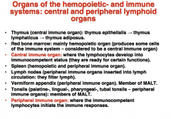
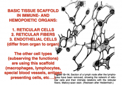
name 8 Organs of the hemopoietic- and immune
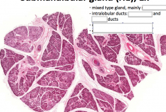
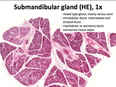
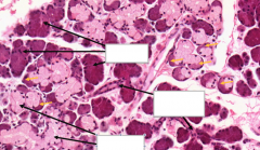
Organs of the hemopoietic- and immune systems: central and peripheral lymphoid systems: central and peripheral lymphoid organs ALSO their general organization |
|
|
|
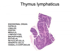
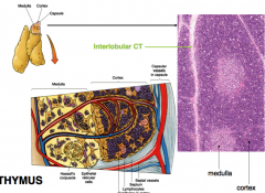
this+structure
|
|
|
|
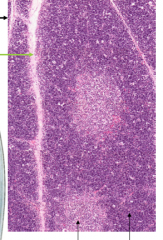
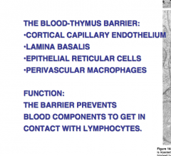
name the arrows AND then!! define the thymus blood barrier and its function.
|
|
|
|
|
|
|
|
|
|
|
|
|
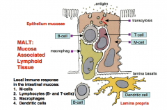
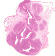
what is MALT? what are its components?
|
|
|
|
|
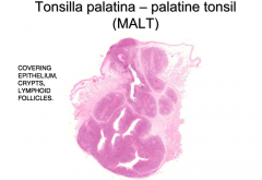
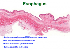
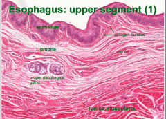
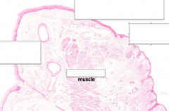
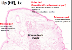
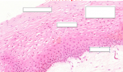
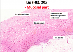
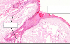
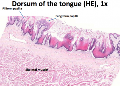
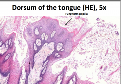
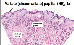
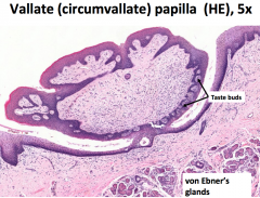
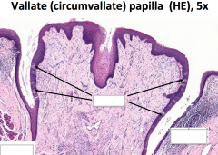
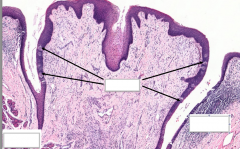
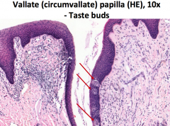
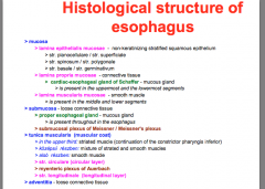
stratified squamous epithelium
lingual glands, lymphoid nodules beneath epithelium |
|
|
|
|
|
|
|
|
|
|
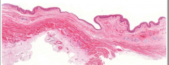
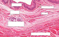
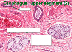
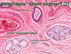
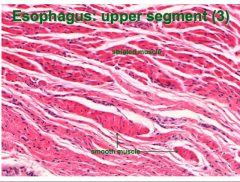
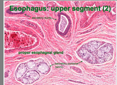
histo of eso
|
|
|
|
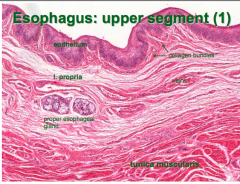
look at it!
|
|
|
|
|
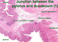
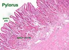
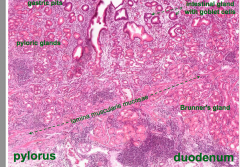
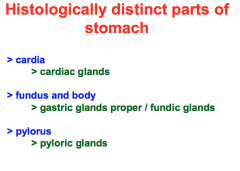
stomach histo and distinct parts
|
|
|
|
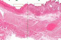
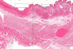
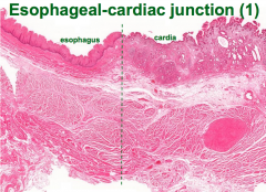
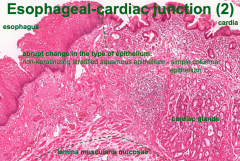
what is this? how can you tell?
|
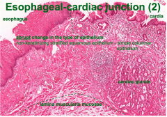
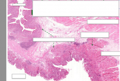
esophageal (cardiac junction)... eso is thick, cardia thinner epi
|
|
|
|
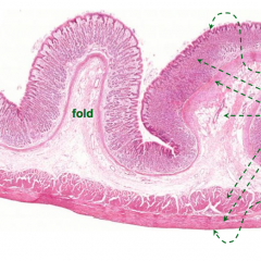
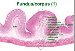
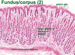
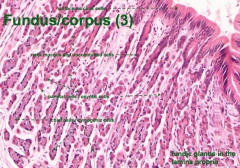
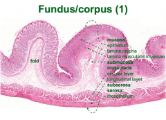
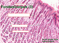
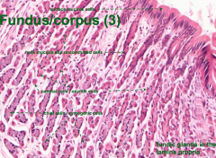
fundus/corpus IDing parts
|
|
|
|
|
|
|
|
|
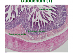
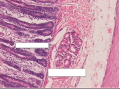
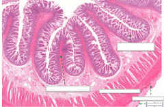
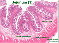
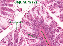
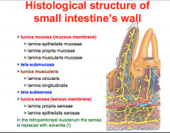
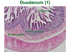
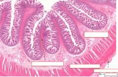
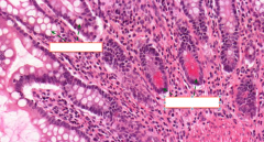
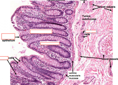
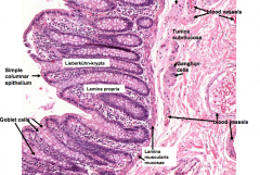
SI histology and special part explained
|
|
|
|
|
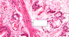
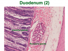
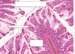
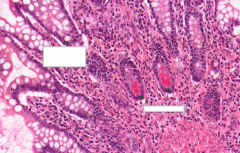
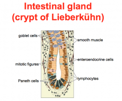
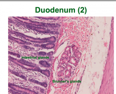
intestinal gland (lieberk...wdkjnwkjn) parts
|
|
|
|
|
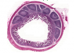
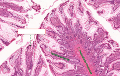
D-J-I pictures.. draw these!
|
|
|
|
|
|
|
|
|
|
|
|
|
|
|
|
|
|
|
|
|
|
|
|
|
|
|
|
|
|
|
|
|
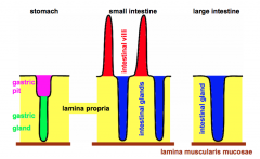
compare mucosa of stomach, LI, SI... via drawing YAY!<3
|
|
|
|
|
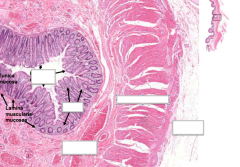
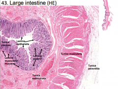
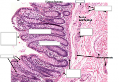
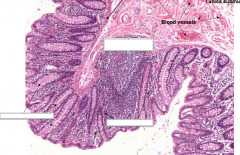
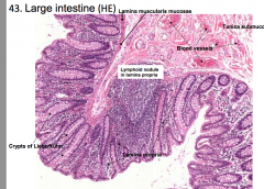
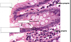
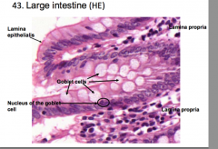
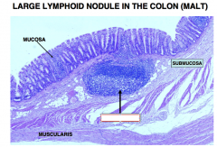
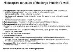
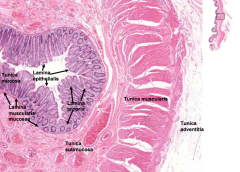
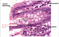
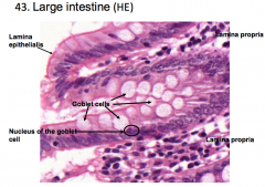
LI histology
|
|
|
|
|
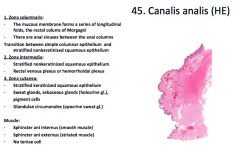
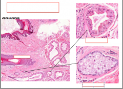
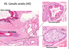
anal canal histology
|
|
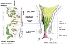
see change in epi?!?
|
|
|
vermi vs ileum
|
|
|
|
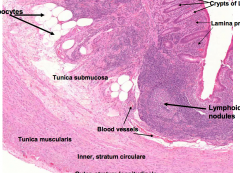
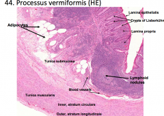
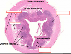
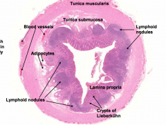
verminformisss... now name histologically important features layers
|
|
|
name
|

LI
|
|
|
|
large intestine
|
|
|
|
LI
|
|
|
|
|
|
|
|
vermiform process
|
|
|
|
|
|
|
|
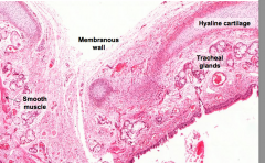
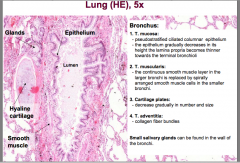
trachea vs bronchi
|
Bronchi differ from the trachea in having plates rather than rings of cartilage, and in having a layer of smooth muscle between the lamina propria and submucosa. In smaller branches, the amount of cartilage decreases, whereas the amount of smooth muscle increases. Also, the number of glands and goblet cells decreases.
|
|
|
|
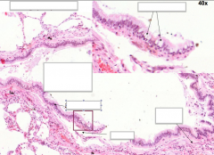
bronchioles
|
Bronchioles are smaller branches of the bronchi, and are distinguished from them by the absence of cartilage and glands. In larger bronchioles, the epithelium is still ciliated, but is now usually simple columnar, whereas in the smallest bronchioles, the epithelium will be simple cuboidal (mostly Clara cells) and lack cilia altogether. the smallest conducting bronchioles consist of a simple cuboidal epithelium of mostly Clara cells, a few ciliated cells, and NO goblet cells, and are called terminal bronchioles
|
|