Reading...
![]()
Play button
![]()
Play button
![]()
Use LEFT and RIGHT arrow keys to navigate between flashcards;
Use UP and DOWN arrow keys to flip the card;
H to show hint;
A reads text to speech;
31 Cards in this Set
- Front
- Back

|
Skeletal Muscle (voluntary)
-striated and contain cross striations in transverse section and are long multi nucleated cells. -Contains myofibrils extending entire length Heterogenous mixture of fibers (3) |
|
|
What is the organization of skeletal muscle?
|
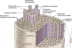
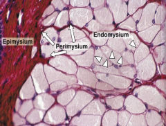
Epimysium - surround sentire muscle
Perimysium - invests each fascicle Endomysium -envelopes each fiber Sarcolemma - cell membrane of muscle cell- called myofibrils |
|
|
Epimysium
Peimysium Endomysium |
|

|
Contain large amount of myoglobin pigment, many mitochondria and rich in oxidative enzymes.
-called slow fibers - slow conduction |
|

|
Contain less myoglobin and few mitochondria than fibers. Poor in oxidative enzymes but rich in phosphorylases.
-Fast twitch fibers, rapid conduction -high staining with ATPase |
|
|
Intermediate Fibers
|
Features are midway between red and white fibers.
Nerve innervation will constitute fiber type. |
|
|
Sarcoplasm
|
Is the cytoplasm of the muscle fiber. Which is enveloped by plasma membrane called sarcolemma. Contains multinuclei in periphery.
|
|
|
Sarcoplasmic Reticulum (SR) and Terminal cisternae
|
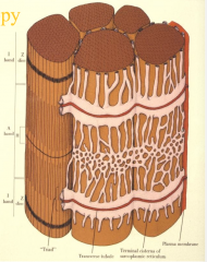
Surrounds myofilaments and forms a meshwork around each fibril. At junction of A and I bands, formes a pair of dilated terminal cistern, which pass around myofibrils.
Cisternae - regulated muscle contraction by sequestering calcium ions - relaxation |
|
|
Traids
|
Located at A-I junction. Consists of a narrow transverse tubule (T), between two cisternae. T-tubule is deep invagination of sarcolemma into the muscle cell. They conduct impulses from exterior of fiber to trigger release of calcium from terminal cisternae.
|
|
|
Myofibrils
|
Longitudinally arranged cylindrical bundles of thick and thin filaments. Lie parallel to axis. Held by intermediate filaments - DESMIN and VIMENTIN
Surrounded by SR |
|
|
Myofilaments
|
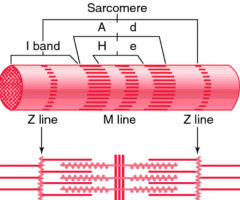
Arranged in myofibril. Functional unit - sarcomere
I band contains only thin filaments - anchor at Z-line A band - consist of both thick and think M line - formed by connections of thick myosin filaments After contraction, A remains constant. I and H bands both decrease as Z draws closer to ends of A band. H band - consists of thick myosin filaments only |
|
|
Myosin
|
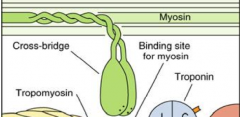
double headed golf club, with heads that posses ATP binding site
-two identical heavy and light chains -heavy chains are cleaved to form two fragments, light and heavy meromyosin -Light meomyosin make rod -like portion where heavy meromyosin represent globular head |
|
|
Muscle contraction mechanism
|
1) Initiated by binding o Ca2 to TnC (troponin), which exposes myosin binding site on actin
2) Myosin head binds to actin, and ATP breaks into ADP, yielding energy that contracts (pulls head of myosin) 3) Thin filaments slide over thick filaments (complete overlapping of action and myosin) |
|
|
Actin
|
Is the thin filament that consists of F-action. Consists of two strands to form double helix. Troponin is distributed along filaments.
|
|
|
Troponin complex
|
TnC - which bonds calcium ions
TnT - binds tropomyosin TnL - prevents interaction between actin and myosin Ca2+ binds to TnC, which exposes myosin binding site. |
|
|
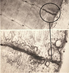
Myoneural Junction
|
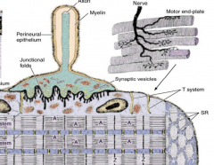
Region on cell where nerve terminates. As axon approaches muscle, loses myelin sheath. But schwann cell continues to cover nonsynaptic surface of nerve terminal.
Nerve terminal lies in indentation (primary synaptic cleft) Additionally invaginates to the sarcolemma to form junctional folds (secondary synaptic clefts) -terminal contains many mitochondria and small storage vesicles for transmitter acetycholine |
|
|
Neural Stimulation
|
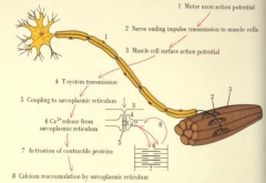
At presynaptic terminate stimulates an influx of Ca2+ ions, which cause synaptic vesicles to release acertylcholine - receptors located on sarcolemma.
Causes increase in Na+ ions, depolarizes muscle membrane causing action potential - propagates over sarcolemma and into T-tubules, activating release of calcium, triggering contraction. Basal lamina lined with secondary clefts of the motor end plate is acetylcholinesterase which limits response by breaking down acetylcholine. |
|
|
Muscle Spindle
|
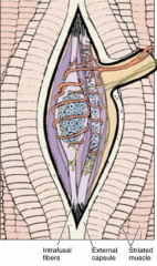
Sensory organ in skeletal muscle that functions a stretch receptor. Large afferent sensory nerve fibers end at spindle - several endings - intrafusual fibers:
- nuclear bag filters - thin nuclear chain fibers |
|

|
Cardiac Muscel
-striated and centralized nucleus -attached end to end by intercalated disk |
|

|
Transverse shows muscle fibers are irregularly shaped and nuclei are infrequent but are large and central.
Myofibrils are clamped as Cohnheims field -T tubules are larger, located at Z-lines rather than A-I junctions -SR poorly developed contributes to diads |
|
|
Mitochondria in cardiac muscle
|
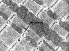
Lie parallel to I bands and glycogen is common in the cell
|
|

|
This is atrial cardiac muscle, which contains atrial granules which contain two hormones
1) Cardiodilatin - vasodilator 2) Cardionatrin - diuretic hormone |
|
|
These are intercalated disks, which form the end-to-end stepwise junction attachments of muscle cells.
Contain: fasica adherens and desmosomes in transverse Longitudinal - gap junctions Transverse, shown at Z-line |
|

|
Purkinje fibers
-are conducting cells of atrioventricular bundle -large and filled with glycogen and contain mitochondria -few myofibrils -make contact with cardiac through gap, desmo and fascia adherents |
|

|
Smooth Muscle - BV and Iris
-tightly packed, staggered, only widest profiles contain nucleus. Nuclei not observed in every cell in cross section. Contracted state will show nucleus with indention. -innervated by sympathetic and parasympthatic -contain alpha-actin -in non-vascular - filaments are desmin but in vascular are vimentin |
|

|
Transverse section, impregnated with silver to stain for reticular fibers that surround muscle.
|
|
|
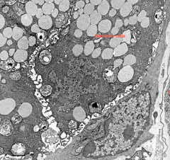
Organelles of Smooth muscle
|
Cell contains, mitochondria, RER, golgi near nucleus
Organelles are involved in synthesis of type II collagen, elastin, GAGs, glycoproteins and lamina growth |
|

|
N- nucleus
DB- dense body C- capillary |
|
|
What kinds of junctions are between smooth muscle?
|
Gap junctions (nexus) which facilitate spread of excitation.
|
|
|
How does contraction vary in vascular and visceral smooth muscle?
|
Vascular - initiated by nerve impulse - little spread
Visceral - myogenic (stretch) and signal spreads via gap In pregnancy smooth muscle contracts due to oxytocin. |
|
|
Myoepithelial cells
|
Very similar to smooth cells. Originate from ectoderm.
-basket like - located between epithelium and glands -contain hemidesosomes -contain action, myosin and cytokertain -contraction forces secretory material |