Reading...
![]()
Play button
![]()
Play button
![]()
Use LEFT and RIGHT arrow keys to navigate between flashcards;
Use UP and DOWN arrow keys to flip the card;
H to show hint;
A reads text to speech;
37 Cards in this Set
- Front
- Back
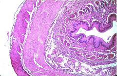
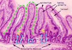
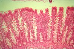
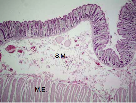
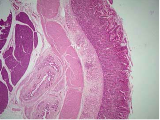
Identify the structure and layers. 7 layers.
|
Esophagus.
Mucosa: Stratified Squamous Epithelium, lamina propria, muscularis mucosa. Submucosa: CT Muscularis Externa: inner circular later, outer longitudinal layer Adventitia: CT (no mesothelium) |
|
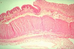
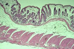
identify structures and epithelial types. how far do these epithelial types go in each direction?
|
esophagus: strat. squamous from oral cavity to esophagus
stomach: simple columnar from stomach to recto-anal junction |
|
identify structures and epithelial types
|
esophagus on left; strat. squamous
stomach on right; simple columnar |
|
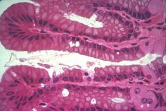
identify this structure
|
stomach
|
|
identify
|
surface mucous cells in the stomach
|
|
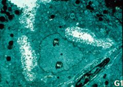
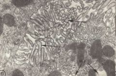
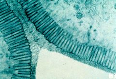
identify
|
EM of a parietal cell.
|
|
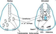
ID
|
parietal cell
|
|
identify
|
parietal cell
|
|
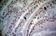
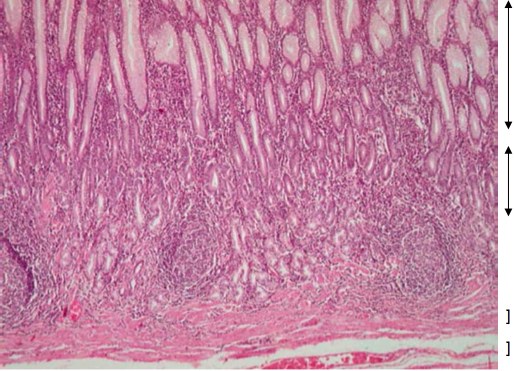
identify the cells and name their function. what kind of stain has to be used to distinguish these?
|
enteroendocrine cells; they secrete hormones.
silver stain must be used, called "argentaffin cells." |
|
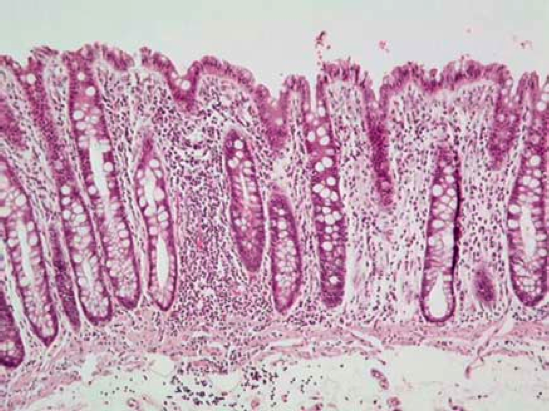
what is this and what helps it increase surface area?
|
intestine. plicae increase SA.
|
|
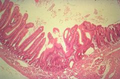
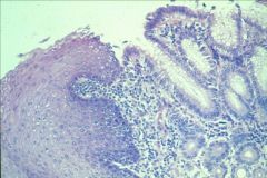
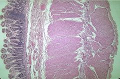
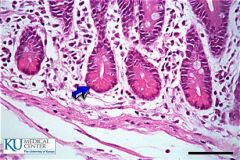
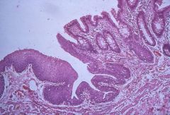
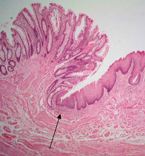
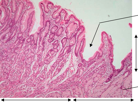
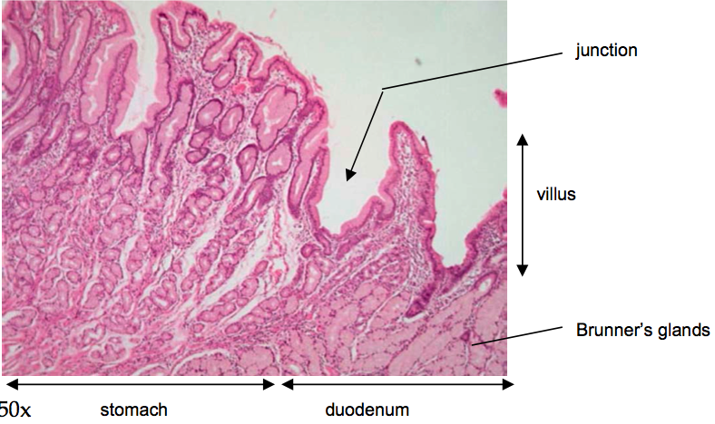
identify the structure on either side of this junction. how can you tell?
|
duodenum on the left; stomach on the right. duodenum has villi.
|
|
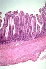
where is this? what projections are these?
|
small intestine; villi.
|
|
what two structures are most obvious?
|
villi and microvilli
|
|
where is this?
|
jejunum
|
|
where is this?
|
ileum.
|
|
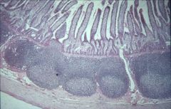
Where is this? What structures are at the bottom?
|
Ileum; peyer's patches.
|
|
what structure is pointed out? what does it do?
|
a paneth cell; secretes lysozyme.
|
|
where is this?
|
colon
|
|
where is this?
|
colon
|
|
where is this and what particular portion is being shown?
|
mucosa of the colon.
|
|
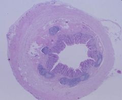
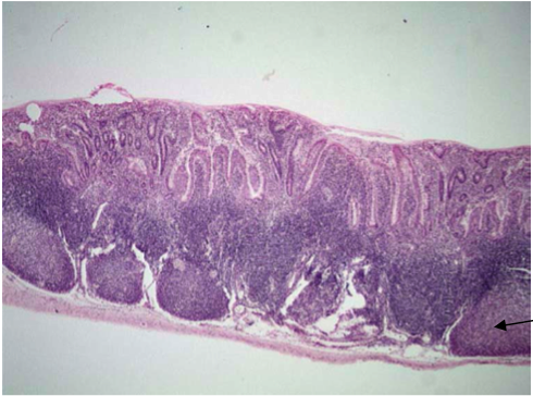
where is this?
|
appendix
|
|
where is this?
|
appendix
|
|
identify this junction and changes in epithelium
|
Rectoanal junction; strat. squamous on left and simple columnar on right.
|
|
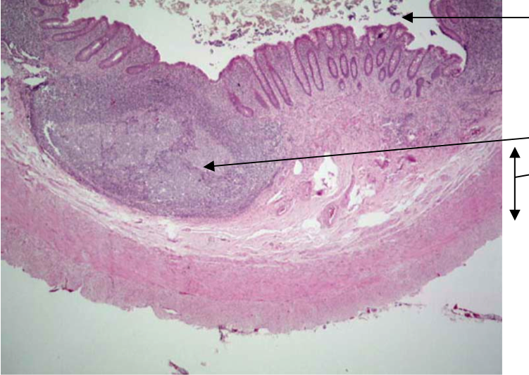
|
Appendix
top: lumen left: lymphoid tissue right: muscle layers |
|
what's this?
|
colon
|
|
what's this?
|
colon
|
|
identify
|
colon
|
|
|
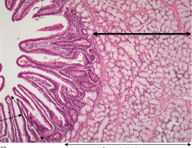
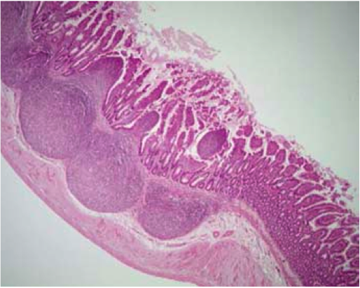
Duodenum
left: villi right: Brunner's glands |
|
|
duodenum
|
|
|
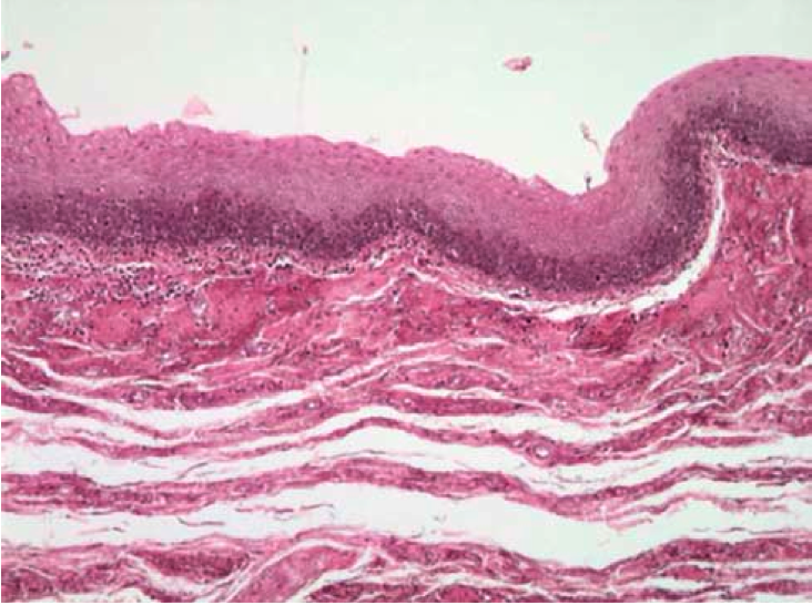
esophagus
|
|
|
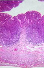
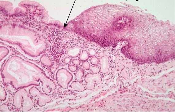
esophageal/cardia junction
|
|
|
ileum
|
|
|
ileum; peyer's patch
|
|
|
Anorectal junction
|
|
|
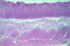
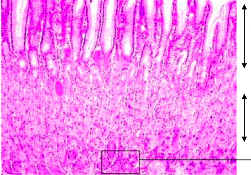
Stomach
Top to bottom: Long pits with mucus glands Short glands Muscularis mucosa Submucosa |
|
|
top: surface and mucus neck cells
bottom: parietal cells square: chief cells |
|
|
|