Reading...
![]()
Play button
![]()
Play button
![]()
Use LEFT and RIGHT arrow keys to navigate between flashcards;
Use UP and DOWN arrow keys to flip the card;
H to show hint;
A reads text to speech;
91 Cards in this Set
- Front
- Back
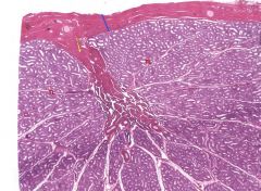
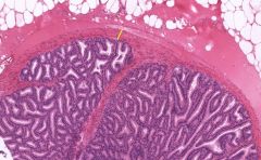
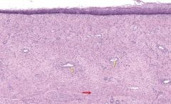
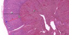
Identify the regions and structures.
|
Blue line - Tunica Albuginea
Yellow Arrow - mediastinum testis TL - Testicular Lobules |
|
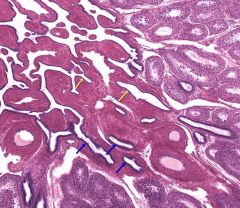
Identify the indicated structures or regions.
|
Blue Arrows - Straight tubules (tubuli recti)
Yellow Arrows - Reti testes |
|
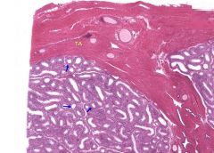
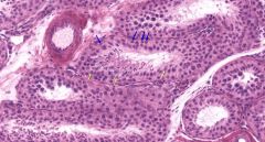
Identify the indicated structures or regions
|
Blue arrows - Seminiferous tubules
TA - Tunia albuginea |
|
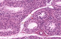
Identify the indicated structures or regions.
|
Leydig Cells
|
|
Identify the indicated structures or regions
|
Myeloid Cells
|
|
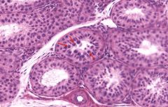
Identify the indicated structures or regions
|
Red Arrows - Spermatazoa
Blue Arrows - Spermatids |
|
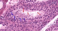
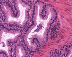
Identify the indicated structures or regions
|
Red Arrows - Spermatocytes
Green Arrows - Spermatids |
|
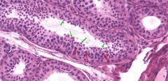
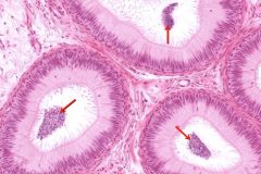
Identify the indicated structures or regions
|
Red Arrows - Primary Spermatocytes
|
|
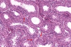
Identify the indicated structures or regions
|
Yellow Arrows - Sertoli Cells
Blue Arrows - Spermatogonia |
|
|
Where are the vasa recta?
|
Blood vessel In the medulla of the Kidney.
|
|
|
What is the major histological difference between the anterior and posterior pituitary?
|
Anterior is darker than the pituitary because the pituitary is nervous tissue.
|
|
|
Pituicyte
|
Glial cells of the posterior pituitary.
|
|
|
What are chief cells and where are they located?
|
The predominant cells in the parathyroid that produce PTH.
|
|
|
What are the major layers of the uterus?
|
Stratum functionalis, endometrium, stratum basale, and the myometrium.
|
|
|
What are chromaffin cells?
|
The most common cells in the medulla adrenal gland.
|
|
|
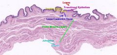
What is the distinguishing features of the bladder?
|
Transitional epithelium and lots of smooth muscle.
|
|
|
What's the difference between early and late spermatids?
|
Early spermatids look like spermatocytes near the lumen while late spermatids look more like spermatozoa (mature sperm).
|
|
|
What are the main functions of Sertoli cells?
|
1 - Nutrition for developing spermatozoa
2 - Produce androgen binding protein (ABP) and inhibin 3 - Produce anti-mullerian hormone 4 - Phagocytize residual bodies shed during spermeogenesis |
|
|
What function does the blood-testes barrier serve for sperm production?
|
The blood-testes barrier protects the sperm from the immune system and blood borne toxins.
|
|
|
What is the main function of Leydig cells?
|
1 - Produce testosterone under the influence of LH
|
|
Identify the indicated structures or cells.
|

Smooth muscle cells (epididymis).
|
|
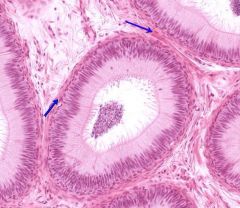
Identify the indicated structures or cells.
|
Stereociliated pseudostratefied columnar epithelium
|
|
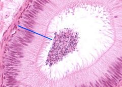
Identify the indicated structures or cells.
|
Stereocilia
|
|
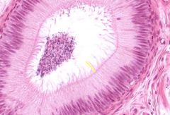
Identify the indicated structures or cells.
|
Spermatozoa
|
|
What organ is this? Identify the cell type of the layer indicated using the yellow line.
|
Seminal Vesicle - Smooth Muscle
|
|
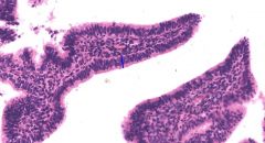
Identify the organ and the indicated structures or cells.
|
Pseudostratified columnar epithelium and sperm.
|
|

Identify the organ and indicated layer, cells, or structures.
|
- The Vas Deferens
- The inner circular, median longitudinal, and outer longitudinal smooth muscle layers |
|
Identify the organ and indicated layer, cells, or structures.
|
- The Vas Deferens
- The inner circular, median longitudinal, and outer longitudinal smooth muscle layers |
|
|
What do the seminal vesicles produce?
|
A yellow alkaline fluid that includes fructose, citrate, prostaglandins, and proteins.
|
|
|
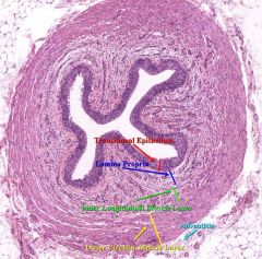
The layers of the Vas Deferns.
|
L - The Lumen
EP - Stereociliated pseudostratified columnar epithelium LP - Lamina Propria IL - Inner longitudinal muscle layer MC - Middle circular muscle layer OL - Outer longitudinal muscle layer |
|
Identify the organ and its function.
|
The prostate gland surrounds the urethra and produces postatic fluid hat includes citric acid, acid phosphates, amylase, fibrolysin, and lipids.
|
|
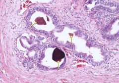
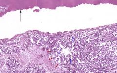
What are these and where are they found and under what conditions?
|
Postatic concretions (corpora amylacea) found in an aged prostate.
|
|
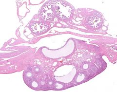
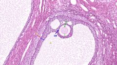
What organ(s) is/are shown here?
|
The ovary and oviduct/fallopian tube.
|
|
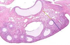
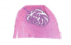
Identify the organ and the noted structures or layers.
|
Ovary - Blue is cortex and Yellow is medulla
|
|
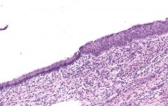
Identify the organ and the layer or structure indicated.
|
Ovary - Germinal epithelium (cuboidal)
|
|
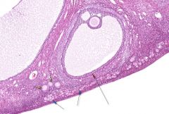
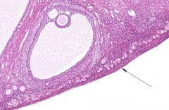
Identify the organ and the major structures indicated.
|
Ovary
Blue Arrows - Primordial Follicles Yellow Arrows - Primary (Unilaminar) Follicles Red Arrow - Secondary Follicle |
|
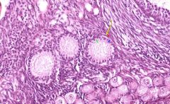
Identify the organ and structures/layers/cells indicated.
|
Ovary
Yellow arrow - Unilaminar primary follice Green line - Zona pellucida Blue line - Granulosa cell layer Red line - Theca interna |
|
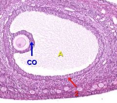
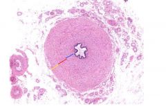
Identify the organ and structures/layers/cells indicated.
|
Ovarian secondary follice:
A - Antrum CO - Cumulus oophorus (specifically the corona radiata) |
|
Identify the organ and the indicated structures/layers/cells.
|
Ovarian secondary follicle:
Yellow line - Theca externa Red line - Theca interna Blue line - Granulosa Green line - Zona Pellucidaa A - Antrum O - Oocyte CR - Corona radiata |
|

Identify the organ and the indicated structures/layers/cells.
|
Atretic follices
|
|
|
Which two layers of an ovarian follice become the corpus luteum?
|
The theca interna and the granulosa
|
|
|
What happens to the corpus luteum when fertilization does not occur.
|
It degrades and regresses into the corpus albicans.
|
|
|
When does the first meiotic division of an oocyte take place and what hormone induces it?
|
During ovulation under the influence of LH.
|
|
|
What maintains the corpus luteum after pregnancy?
|
Human chorionic gonadotropin.
|
|
Identify the organ and the indicated layer. What type of cells are found here?
|
Oviduct
The ampulla, under the blue line, is composed of three smooth muscle layers. |
|
Identify the organ and the type of cells that are indicated.
|
Oviduct
These are ciliated columnar epithelial cells. Peg cells (non ciliated) are also found here. |
|
Identify the organ and the noted structure
|
Cervix
Junction of Oss |
|
Identify the organ and the indicated structures.
|
Vagina
Red Arrows - Smooth muscle Yellow Arrows - Blood vessels |
|
Identify the organ and indicated structure.
|
Mammary Gland
Intralobular duct |
|
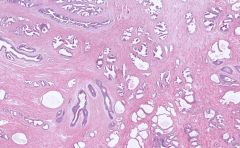
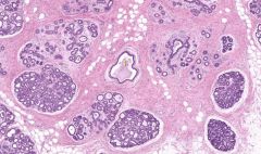
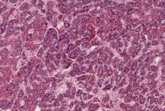
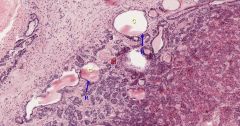
Identify the organ and the structures indicated. What cells are found here and what do they produce?
|
Pancreas
Islets of Langerhaans contain alpha, beta, and gamma cells which produce glucagon, insulin, and somatostatin respectively. |
|

Identify the organ and indicated regions.
|
Pituitary
The lower (darker) portion is the anterior pituitary which is divided into the Pars Tuberalis (PT), Pars Intermedia (PI), and the Pars Distalis (PD). |
|
Identify the organ and indicated regions.
|
Pituitary
The lower (darker) portion is the anterior pituitary which is divided into the Pars Tuberalis (PT), Pars Intermedia (PI), and the Pars Distalis (PD). |
|
Identify the organ and the indicated structures, cells, or layers.
|
Anterior Pituitary
C - Chromaphobes B - Basophils A - Acidophils (Somatotrophs & Mammotrophs) PD - Pars Distalis |
|
Identify the organ and the indicated structures, cells, or layers.
|
Anterior Pituitary
C - Colloids PI - Pars Intermedia |
|
Identify the organ and the indicated structures, layers, or cells.
|
Posterior Pituitary
PN - Pars Nervoa P - Pituicytes |
|
Identify the organ and the indicated structures, layers, or cells.
|
Anterior/Posterior Pituitary
I - Infundibulum PT - Pars Tuberalis Yellow Arrows - Venous channels |
|
|
Name the major cell types of the anterior pituitary and their respective functions.
|
Basophils
1. Gonadotrops - Produce LH and FSH 2. Thyrotrops - Produce TSH 3. Corticotrops - Produce ACTH Acidophils 1. Somatotrophs - Produce HGH 2. Mammotrophs - Produce PRL |
|
|
Name the major cell types of the posterior pituitary and their respective functions.
|
Pituicytes - The glial cells of pars nervoa
|
|
|
What are herring bodies?
|
These are the posterior pituitary's stores of ADH and Oxytocin produced in the hypothalamus.
|
|
|
Name the major cell types of the anterior pituitary and their respective functions.
|
Basophils
1. Gonadotrops - Produce LH and FSH 2. Thyrotrops - Produce TSH 3. Corticotrops - Produce ACTH Acidophils 1. Somatotrophs - Produce HGH 2. Mammotrophs - Produce PRL |
|
|
Name the major cell types of the posterior pituitary and their respective functions.
|
Pituicytes - The glial cells of pars nervoa
|
|
|
What are herring bodies?
|
These are the posterior pituitary's stores of ADH and Oxytocin produced in the hypothalamus.
|
|
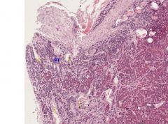
Identify the organ and the indicated cells. What do these cells produce?
|
Parathyroid
C - Chief cells produce PTH O - Oxyphils have no known function |
|
|
What is the major function of parathyroid hormone?
|
It increases calcium levels in the blood by reducing renal calcium excretion and increasing calcium absorption from bones.
|
|
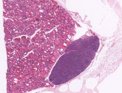
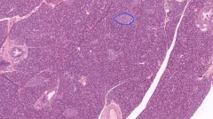
Identify the organs shown.
|
The thyroid and parathyroid.
|
|
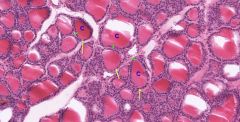
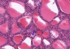
Identify the organ and the indicated structures/cells/layers.
|
Thyroid
C - Colloid Green Line - Simple cuboidal epithelium Yellow Arrows - Follicular cells |
|
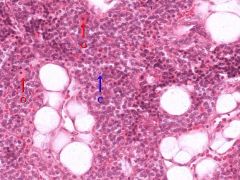
Identify the organ and the indicated cells.
|
Thyroid
Parafollicular cells |
|
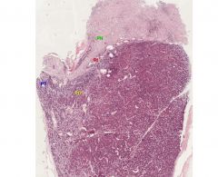
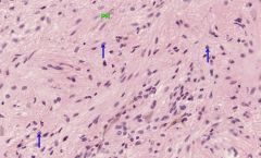
Identify the organ and the indicated layers. What cells are found in each layer and what are their functions?
|
Organ: Adrenal Gland
C - Cortex M - Medulla Chromaffin cells here produce epinephrine and noepinephrine (catecholamines). G - Zona Glomerulosa The cells here produce the mineralocorticoid aldosterone. F - Zona Fasiculata Produce the glucocorticoid cortisol and some androgens R - Zona Reticularis Cells produce androgens. |
|
|
Where are chromaffin cells found, what is their function, and where are they derived from?
|
They are found in the Adrenal Medulla and derive from Neural crest cells. They produce catecholamines such as epinephrine and norepinepherine.
|
|
Identify the organ and the indicated structures/layers/cells.
|
Pineal Gland
Blue Arrows - Pinealocyte (Produce maltonin) |
|
|
Where are medullary rays found and what structures constitute them?
|
They are found in the center of a lobule in the Medulla of the kindey and extend into the cortex. They are made up of the straight portions of the proximal tubule (thick descending), distal tubule (thick ascending), and the collecting duct.
|
|
|
Where are vasa recta found and what are they associated with?
|
These are blood vesseles found in the medulla of the kidneys associated with Juxtamedullary nephrons.
|
|
|
What's special about the glomerular capillaries?
|
They are fenestrated and without a diaphram.
|
|
|
What type of collagen is found in the glomerular basement membrane?
|
Collagen Type IV
|
|
|
What is the role of heparin sulfate in the glomerular basement membrane?
|
It makes the BM of the glomerulus act as a charge barrier.
|
|
|
What are podocytes and where are they found?
|
They are epitherlial cells that are found at the periphery of the glomerulus.
|
|
|
What are mesangial cells and where are they found?
|
Mesangial cells are found in the kidney. They are the phagocytic cells of the glomerular BM and respond to angiotensin II.
|
|
|
Identify the organ and the indicated structures, cells, or layers.
|
|
|
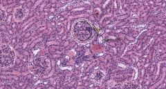
Identify the organ and the indicated structures, cells, or layers.
|
|
|
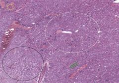
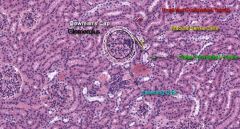
Identify the organ and the indicated structures.
What makes up the structure that the green lines demarcate? |
Kidney
White Circle - Cortex Black Circle - Medulla Green Lines - Medullary rays made up of proximal + distal tubules and collecting ducts Yellow Circle - Arctuate artery |
|
|
What is the function of the proximal convoluted tubule?
|
It resorbs glucose, amino acids, and small proteins. It's junctional complexes are leaky and it contains many mitochondria for active transport across the epithelium.
|
|
|
What is the function of the distal convoluted tubule?
|
Responds to Aldosterone by resorbing Na+ and secreting K+. This increases blood pressure and volume. It also maintains the acid-base balance in blood by secreting hydrogen and ammonium ions into the urine.
|
|
|
What is the function of ADH in the kidneys?
|
ADH permeabilizes the tight epithelium of the collecting ducts allowing water to leak out. This produce hypertonic urine (useful when fluid intake is low).
|
|
|
What are the three cellular components of the juxtaglomerular apparatus?
|
The three cellular components of the apparatus are the macula densa, extraglomerular mesangial cells, and juxtaglomerular cells.
|
|
|
What is the function of juxtaglomerular cells?
|
They produce renin in response to:
- Beta1 adrenergic stimulation - Decrease in renal perfusion pressure (detected directly by the granular cells) - Decrease in NaCl absorption in the Macula Densa (often due to a decrease in glomerular filtration rate, or GFR). |
|
|
What is the function of macula densa cells and where are they found?
|
The macula densa senses NaCl concentration in the distal tubule of the kidney and secretes a vasopressor which acts on the adjacent afferent arteriole to decrease glomerular filtration rate (GFR), as part of the tubuloglomerular feedback loop
|
|
|
What is the function of extramesangial cells and where are they found?
|
They are associated with secretion of erythropoietin and are found near the JGA of the kidney nephron.
|
|
|
Where is transition epithelium found in the body?
|
Bladder, ureters, superior urethra and gland ducts of the prostate.
|
|
|
What are the two distinguishing histological characteristics of the ureter vs the fallopian tubes and the vas deferens?
|
Transitional epithelium and two muscle layer
|
|
Identify the organ.
|
Ureter
|
|
Identify this organ.
|
Urinary Bladder
|