Reading...
![]()
Play button
![]()
Play button
![]()
Use LEFT and RIGHT arrow keys to navigate between flashcards;
Use UP and DOWN arrow keys to flip the card;
H to show hint;
A reads text to speech;
187 Cards in this Set
- Front
- Back
|
Tissue sections are preserved in ___ buffered formalin
|
10%
|
|
|
Effects of fixation on tissues
|
denature & cross link proteins
|
|
|
3 important features of a marking agent
|
Insoluble to fixative; stays external to tissue; no rxn with stains
|
|
|
Why do we dissolve calcium in tissues?
|
Calcium deposits will not section properly
|
|
|
Two most common stains
|
hematoxylin and eosin
|
|
|
What does hematoxylin do?
|
Turns acidic structures blue
|
|
|
What does eosin do?
|
Turns basic structures pink
|
|
|
3 causes of artifacts from fixation procedures
|
freezing, inadequate fixation, bacterial contamination
|
|
|
2 causes of artifacts from necropsy procedures
|
plant/hair fragments and bone pieces
|
|
|
Cytology uses ___ instead of tissue samples
|
air-dried smears
|
|
|
Benefit of cytology
|
faster results
|
|
|
Downside of cytology
|
less-specific results
|
|
|
An example of a cytology stain
|
Romanovsky
|
|
|
This stain turns fungi and spirochetes black
|
GMS (Gomoris' Methenamine Silver)
|
|
|
Silver stains highlight 3 things
|
goblets, reticular fibers, plant tissue
|
|
|
Most common stain used to find collagen, turns collagen blue
|
Masson's trichrome
|
|
|
Trichrome stains that aren't Masson's turn collagen what color?
|
green
|
|
|
Stain used to identify hemosiderin in tissue
|
Prussian blue
|
|
|
Prussian blue turns what blue?
|
iron
|
|
|
Stain that turns acid fast bacteria fuschia
|
Ziehl-Neelsen acid fast
|
|
|
A strongly basic dye
|
Toluidene blue
|
|
|
Name of state of polyanions in tissue causing a color shift from blue to reddish-purple
|
Metachromasia
|
|
|
Two structures that will exhibit metachromasia
|
Ionized sulfate groups & ionized phosphate groups
|
|
|
Toluidene blue is very good for identifying what?
|
Mast cells that are showing metachromasia
|
|
|
A stain that turns carbohydrates pink
|
Periodic Acid-Schiff (PAS)
|
|
|
Immunohistochemistry allows for the identification of ...
|
specific antigenic substances
|
|
|
A major subunit protein of intermediate filaments of mesenchymal cells
|
Vimentin
|
|
|
A group comprising of 29 different proteins that is characteristic of epithelial cells
|
Cytokeratin
|
|
|
Definition of resolution
|
The shortest distance between 2 points on a specimen that can still be distinguished as 2 separate entitites
|
|
|
Electron microscopes can visualize items as small as
|
1nm
|
|
|
Transmission Electron Microscopy allows us to see
|
inner structure & contours of tissue components
|
|
|
TEM requires what sort of samples?
|
very thin
|
|
|
In TEM, electrons interact with what?
|
heavy metal atoms (appear black)
|
|
|
SEM is used to visualize
|
the surface of tissues
|
|
|
For SEM, the tissue has to be coated with
|
metal (eg gold)
|
|
|
Which type of microscopy gives you a 3D image?
|
SEM
|
|
|
Fixation stabilizes 2 types of reactions
|
protein-protein, protein-nucleic acid
|
|
|
Fixation immobilizes
|
fats and carbohydrates
|
|
|
The time between death and sampling/fixation should be as short as possible because
|
autolysis begins right after death/disruption of blood flow and can degrade the sample
|
|
|
Four things whose presence can greatly slow fixation
|
skin, blood, fat, fibrous capsules
|
|
|
Size rules of samples for fixation
|
No thicker than 3-5mm and no bigger than a postage stamp (2cm)
|
|
|
A commonly used black pigment
|
India ink
|
|
|
A downside to India ink
|
it takes a long time to dry
|
|
|
Two strong mineral acids that will decalcify bone
|
nitric acid, hydrochloric acid
|
|
|
Downside of using strong decalcifying acids
|
they will harm cell morphology
|
|
|
Two slower, less harsh acids for decalcification
|
acetic acid, formic acid
|
|
|
What is used to dehydrate tissues?
|
ethanol solutions
|
|
|
Most common embedding media
|
paraffin
|
|
|
Why is methacrylate sometimes used instead of paraffin?
|
It allows thinner sections
|
|
|
Clearing is the transition between
|
dehydration and infiltration with paraffin or plastic
|
|
|
Most common chemical for removing the dehydrant from tissues
|
xylene (or a safer xylene alternative)
|
|
|
Dehydration and clearing takes a minimum amount of time of
|
7 hours
|
|
|
How do we remove wrinkles from sections?
|
float in water
|
|
|
Basic dyes carry a net __ charge
|
positive
|
|
|
Hematoxylin needs what to bind to tissues?
|
a mordant (often a metal cation) to link it to the tissue as it does not bind directly
|
|
|
The only problem with H&E is when...
|
eosin overstains decalcified sections
|
|
|
Freezing does this more than formalin
|
destroys cell structures
|
|
|
Tissues are frozen inside these
|
cryostates, which are myotomes inside a refrigerated compartment
|
|
|
Romanovsky's stain is a combination of two things
|
basic dye and eosin
|
|
|
Methylene blue will bind to two cellular structures
|
ribosomes, nucleic acids
|
|
|
Eosin binds to
|
positively charged cytoplasmic elements
|
|
|
The stain used to see tuberculosis cells is
|
Ziehl-Neelsen Acid Fast
|
|
|
Immunohistochemistry: the process
|
A substance is injected into an animal to elicit an immune response involving the production of antibodies. This immune-response antibody is targeted by a second antibody, which is then labeled.
|
|
|
Two most common labels for antibodies in immunohistochemistry
|
horseradish peroxidase and alkaline phosphatase
|
|
|
3 determining factors of resolution
|
light, lens aperture, and refractive index of mounting media
|
|
|
Eukaryotic cells all use the same things to accomplish these two important functions
|
contraction and duplication of DNA
|
|
|
The glycalyx is the
|
coating around a eukaryotic cell
|
|
|
The glycalyx is formed by three things
|
glycoproteins, glycolipids, proteoglycans
|
|
|
The glycalyx is used for 2 functions
|
cell signaling and houses hormone receptors
|
|
|
In the phospholipid bilayer of cell coats, the hydrophobic head is where?
|
face out of the cell
|
|
|
Intergral membrane components are where?
|
embedded in the membrane
|
|
|
Peripheral membrane components are where?
|
attached to inner or outer surfaces of membrane
|
|
|
The name of sphingolipid rich areas with cholesterol packed against hydrocarbon chains
|
lipid rafts
|
|
|
Lipid rafts are ____ and rise above other areas
|
rigid
|
|
|
Lipid rafts serve as
|
signaling platforms
|
|
|
What controls the distribution of specific proteins?
|
lipid rafts
|
|
|
5 cell structures that are visible with light microscopy
|
cell membrane; nucleus; golgi; nucleolus; centriole
|
|
|
Membrane-bound organelles have ___ membranes
|
double
|
|
|
Two membrane bound organelles
|
mitochondria and nucleus
|
|
|
What moves things from one membrane bound compartment of a cell to another?
|
vesicles
|
|
|
A Wright stain is a type of ____ stain
|
Romanovsky
|
|
|
Nuclei will stain what colors?
|
blue to purple
|
|
|
Three purposes of chromatin
|
Strengthen DNA (allows division and prevents damage), package it to fit into cell, control gene expression/replication
|
|
|
Chromatin is a combination of
|
DNA and proteins
|
|
|
What maintain the structure of chromatin?
|
histones
|
|
|
Three levels of chromatin organization
|
Euchromatin (DNA wraps around histones to form nucleosomes), heterochromatin (compact nucleosome arrays); chromosomes (highest level of DNA packaging)
|
|
|
The type of chromatin that is actively transcribed
|
euchromatin
|
|
|
Amount of chromatin is correlated to
|
cell activity level
|
|
|
Is euchromatin visible under a light microscope?
|
no
|
|
|
Nuceosomes each have __ histones
|
8
|
|
|
Disruption of balance between heterochromatin and euchromatin can lead to
|
uncontrolled cell growth (cancer)
|
|
|
3 locations for heterochromatin
|
ends of euchromatin, nucleus, karyosomes
|
|
|
In typical mammals, __% of the genome is packaged in heterochromatin
|
10%
|
|
|
Nuclear-associated heterochromatin typically forms a
|
ring around the nucleus
|
|
|
What is a karyosome?
|
a discreet body inside heterochromatin, irregular in size and shape
|
|
|
What is responsible for the nucleus staining blue?
|
heterochromatin
|
|
|
The name of an X chromosome in women that exists only as tightly packed, inactive heterochromatin
|
Barr body
|
|
|
Barr bodies are most easily seen in
|
smears
|
|
|
The Barr Body is normally found where in the cell?
|
adjacent to nuclear envelope
|
|
|
The Barr Body was used to identify what?
|
males trying to pass as females at the Olympics
|
|
|
Chromosomes can be said to resemble
|
spiders
|
|
|
Mitotic activity: static
|
no longer capable of division
|
|
|
Mitotic activity: stable
|
undergo periodic division to maintain normal function
|
|
|
Mitotic activity: continuously dividing (3 examples)
|
skin, intestinal epithelium, bone marrow
|
|
|
Main purpose of nucleolus
|
synthesis of ribosome components from rRNA
|
|
|
Nucleoli are found around NOR's, which are
|
nucleolar organizing regions
|
|
|
Three components of the nucleolus
|
rRNA, proteins, and a small amount of DNA
|
|
|
The name of partially assembled ribosomal subunits
|
preribosomes
|
|
|
Preribosomes are sent from ___ to the ___ for completion
|
nucleus via nuclear pores to the cytoplasm
|
|
|
The protein inside the nucleolus that seems to regulate cell cycling and cell differentiation
|
nucleostemin
|
|
|
Nucleostemin has been found in this type of cells
|
cancer, and may contribute to their uncontrolled growth
|
|
|
The nuceolus is prominent in cells that are
|
metabolically active or involved in protein secretion
|
|
|
In H&E, the nucleolus will stain
|
purple
|
|
|
In Romanowsky stains, the nucleolus will stain
|
blue
|
|
|
In Romanowsky stains, chromatin will stain
|
purple
|
|
|
Sometimes the nucloelus can be hidden by
|
heterochromatin
|
|
|
Heterochromatin vs. Euchromatin: appearance on slide
|
Heterochromatin looks clumped, course, and granular; euchromatin looks finer and diffuse
|
|
|
The outer nuclear membrane is continuous with the
|
endoplastic reticulum
|
|
|
The perinuclear cisternal space is
|
the space between inner and outer nuclear membranes
|
|
|
The perinuclear cisternal space is continuous with
|
the rough ER's cisternal space
|
|
|
The purposes of a ribosome's 2 subunits
|
smaller strand binds mRNA; larger strand hold tRNA, holds growing peptide chain, and binds ER
|
|
|
Polysomes are
|
chains of ribosomes on an mRNA strand
|
|
|
Polysomes are ____ in stains
|
basophilic
|
|
|
What stain is better for seeing ribosomes than H&E?
|
Romanowsky
|
|
|
The membrane-limited, flattened, interconnecting spaces of the ER are called
|
cisternae
|
|
|
The rough ER's main purpose is
|
protein synthesis
|
|
|
Where do integral membrane proteins embed themselves?
|
rough ER
|
|
|
Process of exporting proteins made by rough ER
|
Condense them, package into vesicles, vesicles bud off the ER and go to the golgi or plasma membrane
|
|
|
These cells have well developed rough ER
|
secretory cells
|
|
|
Rough ER gives a ____ color tinge to cytoplasm
|
blue
|
|
|
Darker blue in a Romanowsky stain means
|
more ribosomes
|
|
|
The type of tubules on smooth ER
|
short anastomosing tubules
|
|
|
Does the smooth ER have ribosomes?
|
no
|
|
|
Five things the smooth ER is involved in
|
Fatty acid/phospholipid synthesis; drug detoxification; steroid synthesis; sequestering Ca for muscle contraction; membrane formation and recycling
|
|
|
The smooth ER is usually associated with many
|
enzymes, depending on its cell's function
|
|
|
Large amounts of smooth ER will make the cell stain more
|
pink
|
|
|
The name of smooth ER in cardiac and skeletal muscle
|
sarcoplasmic reticulum
|
|
|
An organ with very well developed smooth ER
|
liver
|
|
|
What has enzymes anchored in the smooth ER of the liver?
|
Cytochrome P450, which conjugate carcinogens and drugs
|
|
|
Golgi apparatus' function
|
post-translational protein sortage and packaging
|
|
|
The Cis-Golgi Network consists of
|
the Golgi cisternae that are closest to the ER (the cisternae that are farthest from the ER are the Trans-Golgi Network)
|
|
|
The golgi is best seen using
|
an electron microscope
|
|
|
The golgi won't stain with these stains
|
H&E or Romanowsky stains
|
|
|
These cells are best for viewing golgi with a light microscope
|
plasma cells
|
|
|
3 destinations of golgi vesicles
|
regulated secretory pathway (eg, dense core sensory vesicles); constitutive secretory pathway (eg, vesicles carrying membranes and proteins); inclusion in lysosomes via the lysosomal pathway
|
|
|
Constitutive secretion
|
proteins are released immediately after synthesis and this occurs in all cells with no need for stimulation from outside factors
|
|
|
Three cells that use constitutive secretion to send out their products
|
Osteoblasts, chondrocytes, fibroblasts
|
|
|
Regulated secretion
|
proteins are released as the release is triggered by hormones, etc.
|
|
|
Two glands that participate in regulated secretion
|
exocrine pancreas and thyroid gland
|
|
|
The movement of vesicles out of the cell
|
exocytosis
|
|
|
Exocytosis involves fusion of
|
the vesicle membrane to the cell membrane
|
|
|
Three types of endocytosis
|
Phagocytosis (ingestion of large particles); pinocytosis (nonspecific ingestion of fluid and small particles); receptor mediated endocytosis (entry of specific molecules)
|
|
|
Receptor mediated endocytosis
|
Receptors for hormones accumulate in lipid rafts; when a hormone arrives, it is enclosed by clathrin molecules into a coated vesicle. It will lose its coat and become an endosome and its internalized proteins are sorted and sent to different parts of the cell.
|
|
|
Three things about phagocytosis
|
Can be receptor mediated; results in a phagosome; involves fusion of lysosome to form a phagolysosome or secondary lysosome
|
|
|
Phagocytosis is usually performed by
|
specialized cells such as macrophages and histiocytes
|
|
|
Macrophage receptors recognize
|
antibody molecules on outside of invading microorganism
|
|
|
Macrophages live in
|
tissues
|
|
|
Monocytes and neutrophils live in
|
blood
|
|
|
5 commonly phagocytosed materials
|
RBC's; cell debris; carbon particles; asbestos fibers; bacteria/yeast
|
|
|
When you inhale smoke, your lung cells will
|
phagocytose too many carbon molecules
|
|
|
Heterophagy
|
ingestion of outside materials; phagosomes will fuse with a primary lysosome to form a secondary lysosome (or phagolysosome), inside which digestion will occur
|
|
|
Autophages
|
ingestion of old cell organelles within the smooth ER
|
|
|
Lysosomes are
|
membrane bound organelles that have hydrolytic enzymes
|
|
|
Lysosomes maintain pH with
|
proton pumps to keep pH low
|
|
|
Lysosome formation
|
start as vesicles from the golgi or rough ER
|
|
|
The lysosome is protected from its own enzymes by
|
a unique lipid on its membrane that protects against hydrolytic enzymes
|
|
|
Main purpose of lysosomes
|
digestive system function, especially to recycle old/damaged cell parts; commonly found in phagocytes
|
|
|
What cells do not have mitochondria?
|
erythrocytes; mature keratinocytes (skin cells)
|
|
|
Describe the inner layer of mitochondria
|
It is folded into cristae to increase surface area available for chemical reactions
|
|
|
Three main jobs of mitochondria
|
Generate ATP (ox. phosphorylation, B oxidation of fatty acids, TCA), synthesize steroid hormones, initiate apoptosis
|
|
|
Mitochondria are _____philic
|
eosinophilic
|
|
|
Microfilaments are made of
|
actin monomers
|
|
|
Two parts of a microtubule
|
a-tubulin monomers and tubulin dimers
|
|
|
Actin: diameter, shape, traits, polarity, location and function
|
6-8nm; double stranded linear array; thin & flexible; - end is slow growing, + end is fast growing; microvilli & terminal web, beneath plasma membrane, muscle contractile elements; provide structural support and are part of contractile elements in muscle and motile cells
|
|
|
Intermediate filament: diameter, shape, traits, polarity, location and function
|
8-10nm; rope-like; strong & stable; nonpolar; desmosomes, hemidesmosomes, beneath inner nuclear membrane; add mechanical strength and resistance to shearing forces
|
|
|
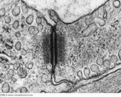
Microtubule: diameter, shape, traits, polarity, location and function
|
20-25nm; nonbranching hollow cylinders; dynamic instability; - end is nongrowing and embedded in MTOC, + end grows; cilia & flagella, basal bodies, centrioles, mitotic spindle; network for organelle mov't, mov't of cilia and chromosomes during mitosis
|
|
What are these?
|
Actin filaments
|
|
What are these?
|
Intermediate filaments
|
|
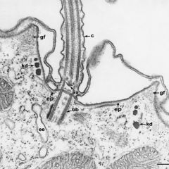
What are these?
|
microtubules
|
|
|
Intermediate filaments: tissue specificifity
|
Epithelial cells (cytokeratins, class I); mesenchymal cells like connective tissue and muscle (vimentin and desmin, class II); nerve cells (neurofilaments, class III); nuclei in all cells (lamins, class IV)
|
|
|
How can intermediate filaments be used diagnostically?
|
Tumors will maintain intermediate filaments of the same type as the site of the cancer's origin, so by analyzing the intermediate filaments inside a tumor the original site and type of cancer can be determined and used to narrow down a treatment.
|
|
|
Lipofuscin: what is it and where does it come from?
|
"wear and tear pigment;" a brown/yellow pigment seen in neurons of muscle cells that is the result of oxidative degradation of mitochondria and lysosomal digestion
|
|
|
Melanin: what is it and where is it?
|
Brown pigment to provide protection from solar damage; in skin, gums, eyelids, brain; provides color to skin, hair, iris
|
|
|
Hemosiderin: what does it look like and where is it?
|
Chunky brown/yellow pigment; it is usually found in macrophages after hemoglobin breakdown, in spleen and bone marrow, and in tissues following hemorrhage. It will stain blue with Prussian Blue stain bc of iron content, and thus can be differentiated from lipofuscin and melanin
|
|
|
Glycogen can be seen with these stains
|
PAS (makes glycogen magenta) and Carmin (for seeing it in hepatic samples). Glycogen is water soluble and thus will disappear during most fixation with 10% buffered formalin.
|
|
|
Lipid can be seen with what stain?
|
Oil red-O (stain lipid red), and the sample must first be frozen
|
|
|
Necrosis vs. Apoptosis
|
Necrosis: cell murder; caused by hypothermia, overheating, low pH, pathogens, radiation; always results in secondary inflammation response; usually bad for organism
Apoptosis: cell suicide; happens to cells that are irreperably damaged or just no longer needed; triggered by intrinsic or extrinsic factors (TNF-a); no inflammatory response; normal part of life |