Reading...
![]()
Play button
![]()
Play button
![]()
Use LEFT and RIGHT arrow keys to navigate between flashcards;
Use UP and DOWN arrow keys to flip the card;
H to show hint;
A reads text to speech;
49 Cards in this Set
- Front
- Back
|
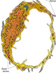
What are the three layers of an artery?
|
1. Tunica intima
2. Tunica media 3. Tunica adventitia |
|
|
What is in the Tunica Intima?
|
1. Endothelium
2. Basal lamina 3. Subendothelial connective tissue 4. Internal elastic lamina |
|
|
What is in the Tunica Media?
|
1. Smooth muscle layer
2. External elastic lamina |
|
|
What is in the Tunica Adventitia?
|
1. Loose connective tissue
|
|
|
What vessels feed blood vessels?
|
Vasa vasorum
|
|
|
What cell type forms the elastic fibers in the tunica media?
|
Smooth muscle fibers (not fibroblasts!)
|
|
|
On a histological slide, what are some visual differences between arteries and veins?
|
Artery is thicker and will appear round.
Vein will appear irregular. Blood will more often be found in the vein rather than the artery. |
|
|
What are the layers of a capillary?
|
Only endothelium.
|
|
|
What type of epithelium is the endothelium of capillaries, arteries, and veins?
|
Simple squamous epithelium.
|
|
|
What is the diameter of a capillary?
|
One cell thick, roughly 10uM.
|
|
|
Name the types of arteries
|
1. Elastic arteries
2. Muscular arteries 3. Arterioles |
|
|
Name an elastic artery
|
1. Aorta and major branches
2. Iliacs |
|
|
What are the largest arteries in the body?
|
Elastic arteries
|
|
|
What is the most prominent feature of elastic arteries?
|
Thick elastic tunica media that does not show internal or external elastic lamina layers.
|
|
|
What propels blood through an elastic artery?
|
Contraction action after expansion.
|
|
|
What is Marfan's syndrome?
|
A defect in the fibrillin gene. Prone to aneurism and dissection of elastic arteries.
|
|
|
What are some prominent features of muscular arteries?
|
1. Thick tunica media (Smooth muscle layer)
2. Distinct internal and external elastic lamina |
|
|
Describe an arteriole
|
1. If you can see the whole structure on a histological slide, it is an arteriole.
2. Tunica adventitia is only a few layers thick. |
|
|
Name the artery that feeds into the capillary bed
|
Metarteriole
|
|
|
Name the different types of capillaries
|
1. Continuous
2. Fenestrated 3. Sinusoidal |
|
|
Name the characteristic of a continuous capillary
|
Tight junctions between cells
|
|
|
Where are continuous capillaries found?
|
1. nerves
2. muscles 3. connective tissue |
|
|
Name the characteristic of a fenestrated capillary
|
Have small windows with diaphragms.
|
|
|
Where are fenestrated capillaries found?
|
In the pancreas and endocrine glands, intestines, and glomerulus
|
|
|
Name the identifying characteristic of sinusoidal capillaries and where they are found.
|
They have large holes and are the largest capillaries (30uM) and are found in the spleen, liver, and bone marrow.
|
|
|
Describe the innervation of blood vessels.
|
Parasympathetic and sympathetic innervation. Nerves stimulate the outmost layer and the gap junctions allow communication of the signal throughout the vessel (tunica media). Epinephrine is the NT.
|
|
|
What is arteriovenous anastomosis?
|
Artery goes directly to the vein, or through a venous plexus to the vein. Capillaries are bypassed. Heat conserved.
|
|
|
What do metarterioles do?
|
Regulates amount of blood going into the true capillaries, where heat is lost.
|
|
|
How does blood get to veins?
|
1. Through capillaries
2. Through AV anastomosis |
|
|
Name the layers of a vein
|
1. Tunica intima
2. Tunica media 3. Tunica adventitia |
|
|
What is in the tunica intima layer in a vein?
|
1. Endothelium
2. Basal lamina 3. Subendothelial connective tissue 4. Internal elastic lamina |
|
|
What is in in the tunica media layer of a vein?
|
1. smooth muscle
2. fibroelastic connective tissue This is a very thin layer. |
|
|
What is in the tunica advantitia layer of a vein?
|
1. Collagenous, connective tissue
2. Fibroblasts 3. Elastic fibers 4. Smooth muscle 5. Vasa vasorum. |
|
|
What are the layers of the heart?
|
1. Endocardium
2. Myocardium 3. Epicardium (or visceral pericardium) |
|
|
What is the function of lymphatic ducts in peripheral tissue?
|
Cells and membranes are leaky. Fluid goes into the interstitial fluid. Lymphatic ducts have a blind cap with open valves (flaplike).
|
|
|
What is lymphadema?
|
Blockage of lymph ducts that prevent drainage. Causes swelling
|
|
|
What are the characteristics of lymph vessels?
|
1. Thin walled
2. Will have white blood cells 3. Larger than a capillary 4. Only have a single cell layer |
|
|
What is a pericyte?
|
AKA Rouget cell or mural cell.
Is a mesenchymal-like cell, associated with the walls of small blood vessels. Undifferentiated cell, it serves to support these vessels, but it can differentiate into fibroblasts, smooth muscle cells, or macrophages. |
|
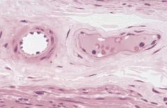
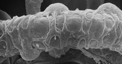
Identify the structures in this image
|
Vein and Arteriole
|
|
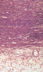
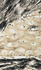
Identify the structure
|
Muscular artery
|
|
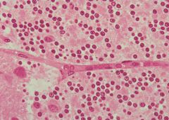
Identify the structure
|
Capillary
|
|
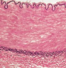
Identify the vessel
|
Elastic artery
|
|
Identify the structure
|
Lymph Vessel
|
|
Identify the cell type and where they would be found
|
Pericyte, associated with the walls of small vessels
|
|
Identify the cell type and tissue where it can be found
|
Purkinje fibers/cell, found in the myocardium of the heart.
|
|
|
Where Weibel-Palade bodies are found?
|
Weibel-Palade bodies are organelles in the endothelial cells.
|
|
|
What do Weibel-Palade bodies do?
|
1. von Willebrand factor (vWF), a multimeric protein involved in blood coagulation.
2. P-selectin, which binds to passing leukocytes. This allows leukocytes to bind to the cells lining the blood vessels in a series of steps called the leukocyte adhesion cascade. Leukocytes transmigrate across the endothelium and enter the surrounding tissue where they can migrate to the site of infection. |
|
|
Explain why the tunica media of large arteries is nourished by the vasa vasorum only during diastole.
|
During systole, the walls of the vessel are under pressure, not allowing blood flow.
|
|
|
Explain why the vasa vasorum is more prolific in large veins.
|
Partial oxygen pressure and osmotic pressure is lower than in arteries. More vasa vasorum needed to supply the vessels sufficiently.
|