![]()
![]()
![]()
Use LEFT and RIGHT arrow keys to navigate between flashcards;
Use UP and DOWN arrow keys to flip the card;
H to show hint;
A reads text to speech;
138 Cards in this Set
- Front
- Back
|
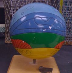
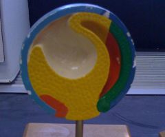
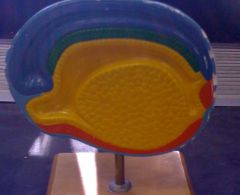
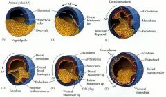
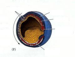
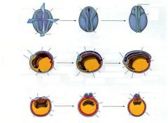
Blastula ectoderm - light blue ectoderm (nervous tissue) - dark blue mesoderm - red mesoderm (notochord) - green endoderm - yellow |
|
|
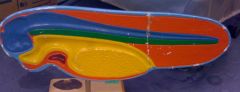
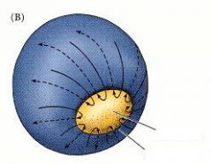
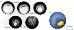
Blastula cross-section -Cleavage has created a blastula with a blastocoel (white space inside). Notice the external surface of the model, which illustrates the animal (less yolk) and vegetal poles (more yolk). |
|
|
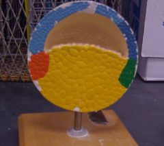
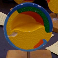
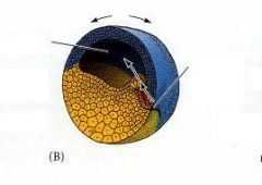
Early gastrulation -The movement of cells to produce the three primary germ layers has begun. The green end of mesoderm represents the dorsal lip of the blastopore |
|
|
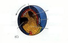
Gastrulation -The invagination formed by cells moving inward is the primitive gut, or archenteron. |
|
|
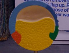
Gastrulation -The archenteron expands. |
|
|
Gastrulation -The blastocoel (white) is pushed aside by the expansion of the archenteron and eventually disappears. Notice that the archenteron is lined with endoderm. |
|
|
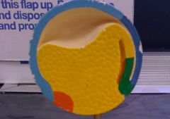
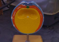
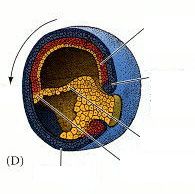
Complete gastrulation -Not the blastopore plug formed at the beginning of the archenteron. All three primitive germ layers are now established, and the green mesoderm is differentiating into the notochord. |
|
|
Early neurulation -Note the green notochord, formed from the mesoderm. The ectoderm above the notochord is beginning to differentiate into the neural tube, due to the induction from the notochord. |
|
|
Neurulation (longitudinal section) -The head region is on the left, the tail is on the right. Not the fully closed neural tube. The gut is ready to open into the mount and anus (formerly the blastopore). |
|
|
Completed neurulation/Neural crest migration -Notice the open gut with the anus. The red mesoderm layer will differentiate into muscles, heart and other tissues. The notochord will eventually be replaced by bone, while the nerves near the head will form the brain. |
|
|
cardiac muscle |
|
|

|
|
|

|
|
|
|
|
|

|
|
|

|
|
|

|
|
|
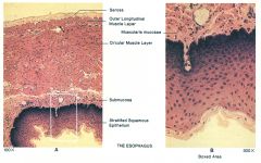
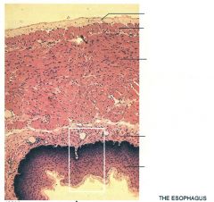
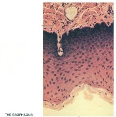
-Top: serosal layer secretes fluid into internal space to lubricate organs -Mucosal layer, stratified, squamous, smooth muscle, circular muscle layer
|
|
|

-Desquamation
|
|
|
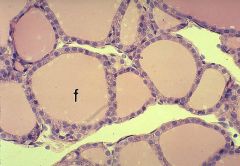
Thyroid follicles -simple cuboidal epithelium (1 layer thick) -store material |
|
|
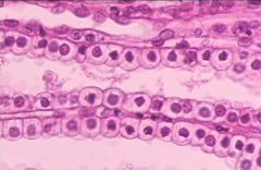
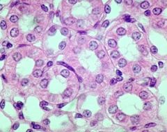
Urinary tubules (longitudinal) -simple cuboidal epithelium |
|
|
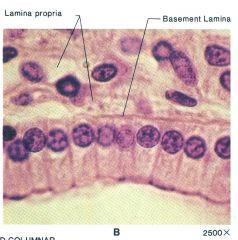
-disorganized, loose connective tissue -nuclei lined up at same distance from basal lamina |
|
|
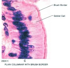
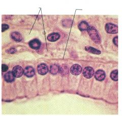
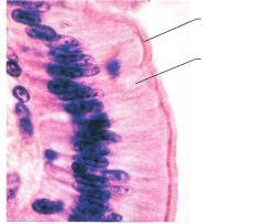
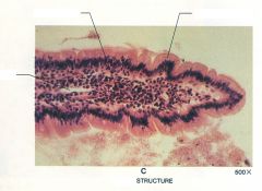
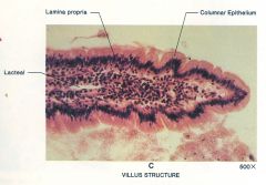
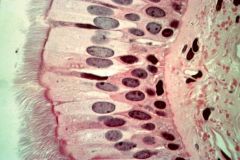
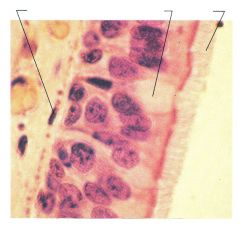
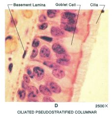
-dense columnar epithelial -goblet cells are clear, secret mucus -layer of microvilli increases surface area
|
|
|
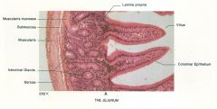
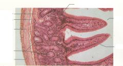
-depressions between villi is the pit |
|
|
-glands at base of folds of villi, have columnar epithelial lining |
|
|
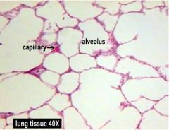
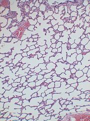
Lung epithelium -thin, simple squamous epithelium -mostly voids -alveoli are the point of O2/CO2 exchange |
|
|
Lung epithelium -thin, simple squamous epithelium -mostly voids -alveoli -blood vessels |
|
|
Hematoxylin-Eosin stain (side view) -loose connective tissue -dark nuclei |
|
|
Hematoxylin-Eosin stain (top view) -loose connective tissue -dark nuclei |
|
|
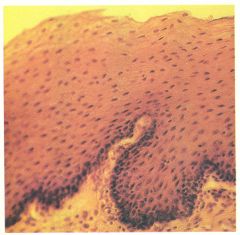
Rectum/mouth -stratified squamous epithelium -top: void -desquamating epithelial -bottom: blood vessels and connective tissue |
|
|
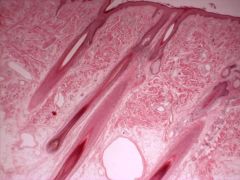
Surrounding the hair follicles -stratified squamous |
|
|
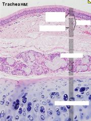
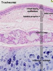
Trachea -ciliated pseudostratified columnar |
|
|
Trachea |
|
|
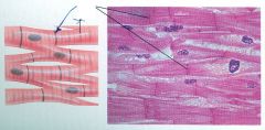
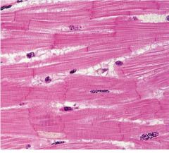
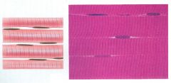
Cardiac -striation visible -somewhat branchy -nuclei spread out -cells connected via intercalated disks
|
|
|
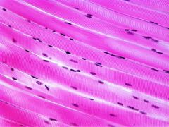
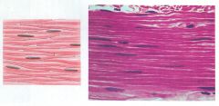
Skeletal: -striation very visible -straight and unbranched -nuclei pushed to the side -cells fused during development |
|
|
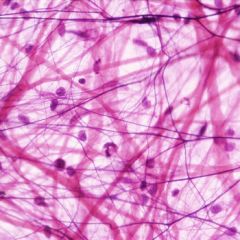
Loose connective tissue -loose is the most common -elastin fibers -collagen fibers -fibroblast cells (generating fibers) -looks disorganized |
|
|
|
|
|
|
|
|
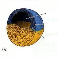
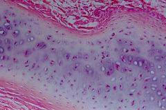
Cartilage in the center (blue/gray) -double nucleus -cells surrounded by matrix -collagen fibers -no blood supply (reason for long healing time) -hyaline found in joints, costocartilages, respiratory tracts |
|
|
|
|
|
|
|
|
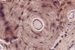
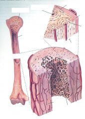
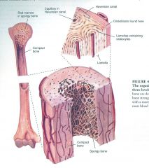
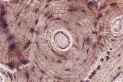
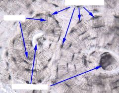
-osteon is center ring with haversian canals that reach to lacunae -lamella are concentric rings
|
|
|
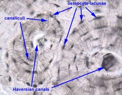
Bone -canalculi canals are how osteocytes communicate |
|
|
Red blood cells -smaller -most numerous -biconcave -no nucleus
|
|
|
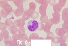
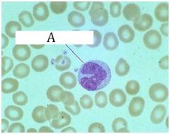
Monocyte -type of white blood cell -large -large nucleus in horseshoe shape |
|
|
skeletal muscle |
|
|
smooth muscle |
|
|
Monocyte |
|
|
Kidney tubules -cuboidal epithelium |
|
|
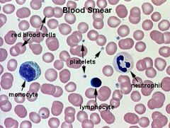
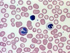
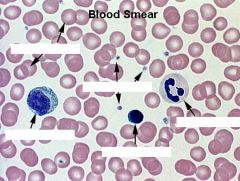
Other Types of white blood cells: -lymphocyte - same size as RBC, mostly nucleus -neutrophil - multi-lobed nucleus -platelets - dark spots |
|
|
|
|
|
|
|
|
Five types of epithelial cells that we are responsible for?
|
simple squamous, simple cuboidal, simple columnar, pseudostratified ciliated, stratified squamous
|
|
|
What are the two types of stratified squamous cells?
|
Non-keratinized and keratinized
|
|
|
What are the three types of muscle?
|
Skeletal, cardiac, smooth.
|
|
|
What are the four types of loose connective tissue that we are responsible for?
|
Loose connective tissue, cartilage, bone, blood
|
|
|
What is the sub-division of simple columnar cell that we need to know?
|
Simple columnar non-ciliated
|
|
|
Where are simple squamous cells found (3 places)?
|
alveoli, blood, and lymph vessels
|
|
|
Where are simple cuboidal cells found (2 places)?
|
kidney tubules, ducts of small glands
|
|
|
Where are simple columnar, non-ciliated cells found? (2 places)
|
digestive tract lining, gall bladder
|
|
|
Where are pseudostratified ciliated cells found?(2 places)
|
trachea, upper respiratory tract
|
|
|
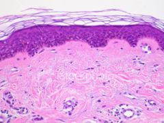
Where are keratinized stratified squamous cells found?(1places)
|
epidermis
|
|
|
Where are non-keratinized stratified squamous cell found (3 places)?
|
esophagus, mouth, vagina
|
|
|
Characterize the skeletal muscle cells?
|
Voluntary & striated.
|
|
|
Characterize the cardiac muscle cells?
|
Involuntary & striated.
|
|
|
Characterize the smooth muscle cells?
|
Involuntary & non-striated
|
|
|
Where are skeletal muscles found?
|
Attached to bone or skin.
|
|
|
Where are cardiac muscle cells found?
|
Heart walls.
|
|
|
Where are smooth muscle cells found?
|
Walls of hollow organs. Not associated with bone.
|
|
|
What is the type of cartilage cell that we’re responsible for knowing?
|
Hyaline
|
|
|
Where is hyaline found (3 places)?
|
Joints, costal cartilages, respiratory tract
|
|
|
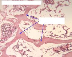
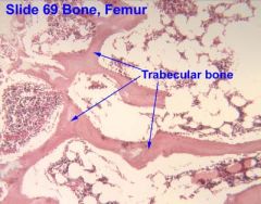
What role do bones play?
|
Structural support throughout the body
|
|
|
What is the other name used for blood?
|
Reticular tissue.
|
|
|
Where is blood found (3 places)?
|
Venous and arterial systems, lymph fluid, throughout tissues
|
|
|
What are the five types of cells found in the blood that we need to know?
|
monocyte, neutrophil, lymphocyte, red blood cells, platelets.
|
|
|
What role do monocytes play?
|
Immune surveillance.
|
|
|
How do monocytes compare in size to red blood cells?
|
They are much bigger than red blood cells. They have a big nucleus in horseshoe shape.
|
|
|
What role do neutrophils play?
|
Immune defense
|
|
|
What is an example of dead neutrophils in the body?
|
Pus is made up of dead neutrophils.
|
|
|
What is distinctive about the appearance of neutrophils?
|
They have a single nucleus, but appear to be divided into lobes.
|
|
|
What role do the lymphocytes play?
|
Immune response.
|
|
|
Characterize the size of lymphocytes and the amount of cytoplasm?
|
They are similar sized to red blood cells and have very little cytoplasm.
|
|
|
What role do red blood cells play?
|
C02 and 02 transport.
|
|
|
What role do platelets play?
|
They are involved with clotting.
|
|
|
How do platelets appear under microsocope?
|
Small, dark staining, looks like debris. Appears to be breaking off bone (but isn’t really).
|
|
|
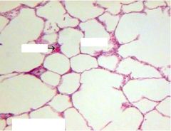
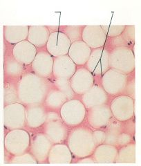
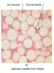
What is the type of loose connective tissue that we are responsible for knowing?
|
Adipose.
|
|
|
Where are adipose cells found (4 places)?
|
Within alveolar connective tissue, subcutaneous tissues, bone marrow, mesenteries.
|
|
|
What is another name for red blood cells?
|
Erythrocytes.
|
|
|
Where is nervous tissue found (3 places)?
|
Brain, spinal cords, peripheral nerves.
|
|
|
What do neurons do?
|
They carry electrical impulses with great speed down a thread-like dendrite through nerve cell body to long non-branching or many branching axons.
|
|
|
Where do electrical signals trigger exocytosis of a chemical message within the nervous system?
|
At tge tips of axons.
|
|
|
What is the synapse?
|
The place between neural cells where the chemical signal is released to the next cell in line. N.B. There is an actual space here.
|
|
|
What are nerves?
|
Bundles of individual neurons.
|
|
|
What are myotubes?
|
Within muscular striated tissue, they are very long tubes that are multinucleate because they arose by the fusion of many individual cells (myocytes.)
|
|
|
What are the cells involved in the formation of myotubes?
|
Myocytes that have fused together.
|
|
|
What are the striations that we see in striated muscle?
|
Overlapping fibrils from the myotubes.
|
|
|
What kind of junctions are present in between myotubes?
|
With the exception of the heart, no gap (communicating) junctions are found between myotubes; only desmosomes are present.
|
|
|
What kind of muscle tissue is polynucleated?
|
Striated muscle.
|
|
|
What is connective tissue?
|
Connective tissue is any group of cells that provides support or connects tissues, or is involved in bodily defense, or in accumulation and transport of materials.
|
|
|
What work do cartilage, fat or adipose, tendon, bone, and loose connective tissue serve?
|
These cells secrete a large amount of extracellular matrix which provides support and adhesion, and thus, the cells themselves tend not to show any specialized junctions.
|
|
|
What does reticular tissue consist of?
|
Special type of connective tissue that consists of the arteries, veins, capillaries and all circulating cells within these vessels in a liquid matrix.
|
|
|
What is reticular tissue responsible for?
|
It is responsible for bodily defense or for the transport or accumulation of materials.
|
|
|
Where are lymphocytes (white blood cells) manufactured?
|
They are manufactured in the lymph nodes, bone marrow, and spleen.
|
|
|
In bone, where do osteocytes sit?
|
They sit in spaces called lacunae.
|
|
|
What function do osteocytes play?
|
They secrete collagen that becomes infused with the precipitates of calcium and phosphate to make the hard structure of bone.
|
|
|
How are the lacunae connected?
|
They are connected by a network of tiny caniculi and large longitudinally running Haversian canals.
|
|
|
What are lamellae?
|
The large amount of mineralized matrix deposited around Haversian canals in concentric rings.
|
|
|
Where are the villi in intestinal cells?
|
They are observed in epithelial intestinal cells surrounding the lumen.
|
|
|
What is the intestinal lumen?
|
The lumen is the cavity of the gut.
|
|
|
What are the intestinal villi composed of?
|
Columnar cells
|
|
|
What are below the villi in the intestine?
|
Connective tissue
|
|
|
What is below the connective tissue layer in the intestine?
|
Circularly arranged circular smooth muscle cells surrounded by a thin layer of longitudinal smooth muscle cells.
|
|
|
What is below the circularly arranged smooth muscle and longitudinal smooth muscle layer in the intestine?
|
Squamous epithelial cells.
|
|
|
Where are chondrocytes and what do they do?
|
They are found in cartilage cells, and they produce and maintain the cartilaginous matrix, which consists mainly of collagen and proteglycans.
|
|
|
What are the spaces for blood vessels in bones called?
|
Haversian canals
|
|
|
What are the concentric rings that surround the Haversian canals called?
|
Lamellae.
|
|
|
What are the black dots on lamellae and what do they signify?
|
The black dots are lacunae and they indicate where the osteocytes were located.
|
|
|
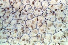
What kinds of organelles are highly preponderant in pancreatic cells?
|
Golgi apparati.
|
|
|
What are the three components of embryonic development?
|
Growth, Differentiation, Morphogenesis.
|
|
|
What is morphogenesis?
|
Establishment of characteristic forms from individual tissue types.
|
|
|
What are the five stages of embryogenesis?
|
Blastulation, gastrulation, neuralation, neural crest formation, organogenesis.
|
|
|
What happens in blastulation?
|
Blastulation is the final result of cleavage, and usually consists of a ball of hundreds of cells surrounding a fluid-filled cavity.
|
|
|
What is gastrulation?
|
Gastrulation is the movement of some of the cells into the cavity to set up three layers of cells (the germ layers), which later gave rise to the specific tissue types.
|
|
|
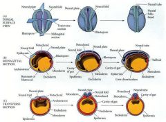
What is neuralation?
|
After the germ layers are established, the ectoderm (on top) rolls into the neural tube. This is essential to forming the characteristic chordate dorsal, hollow nerve cord.
|
|
|
What happens in neural crest formation and migration stage?
|
The neural crest forms along the neural tube, and blocks of cells migrate from the crest. These somites signal the nearby cells to begin developing into other parts of the body.
|
|
|
What happens in organogenesis?
|
Organs of the body become established, after being directed by the action of the somites and other surrounding cells. |
|
|
What stage of embryogenesis is an example of primary induction?
|
Neuralation.
|
|
|
What stage of embryogenesis is an example of secondary induction?
|
Neural crest formation and migration.
|
|
|
What are the three germ layers?
|
Endoderm, mesoderm, ectoderm.
|
|
|
What does endoderm give rise to?
|
Endoderm gives rise to the gut, gut-related organs (such as liver and pancreas)
|
|
|
What does the mesoderm give rise to?
|
It gives rise to muscle, notochord, skeleton.
|
|
|
What does the ectoderm give rise to?
|
It gives rise to skin and neural tube (and therefore brain, spinal cord and nerves)
|
|
|
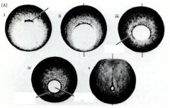
What does the establishment of three germ layers give rise to?
|
A visible primitive streak in the blastoderm. |
|
|
In the amphibian model, what is light blue?
|
Ectoderm
|
|
|
In the amphibian model, what is dark blue?
|
Ectoderm (nervous tissue)
|
|
|
In the amphibian model, what is red?
|
Mesoderm.
|
|
|
In the amphibian model, what is green?
|
Mesoderm (notochord)
|
|
|
In the amphibian model, what is yellow?
|
Endoderm. |