![]()
![]()
![]()
Use LEFT and RIGHT arrow keys to navigate between flashcards;
Use UP and DOWN arrow keys to flip the card;
H to show hint;
A reads text to speech;
126 Cards in this Set
- Front
- Back
- 3rd side (hint)
|
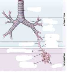
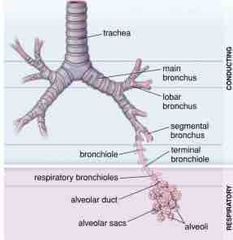
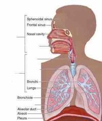
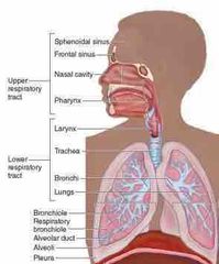
The conducting portion |
Consist of all the components that condition air and bring it into lungs. Consists of nasal cavity, pharynx, larynx, trachea, bronchi, bronchioles and terminal bronchioles |
|
|
|
The respiratory portion |
Is responsible for gas exchange. It consist of : 1.respiratory bronchioles 2. alveolar ducts/sacs 3.alveoli |
|
|
|
The respiratory portion consist of |
1. Respiratory bronchioles 2. Alveolar ducts, sacs 3. Alveoli |
|
|
|
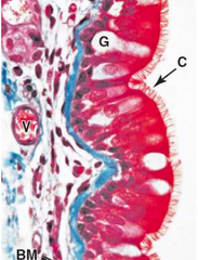
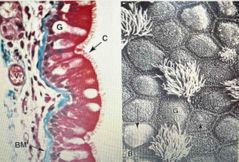
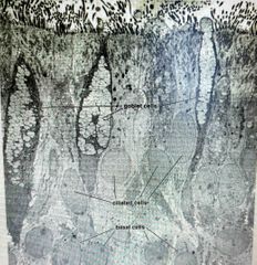
Respiratory epithelium is made up of what tissue type? |
Pseudostratified ciliated columnar epithelium |
|
|
|
Pseudostratified ciliated columnar epithelium |
Varies in different regions of the respiratory tract and rests on a very thick BM |
|
|
|
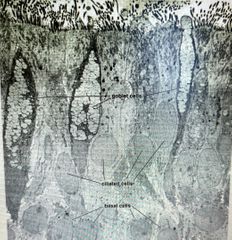
The 3 main cell types in respiratory epithelium are |
1. ciliated epithelial cells extending to surface and have cilia-Most abundant cell type 2. Goblet cells with a mucinogen granules 3. Basal cells confined to the basal portion of the epithelium layer near the CT |
|
|
Front (Term) |
Respiratory epithelium |
|
|
|
Most of the small round cells at the basement membrane are____ which make up 30% of epithelium |
Stem cells |
|
|
|
Among goblet cells, there are intraepithelial |
Lymphocytes and dendritic cells |
|
|
|
The lamina propria of respiratory epithelium is |
lamina propria is the highly vascular, loose connective tissue matrix between the muscularis mucosa and epithelium |
|
|
Front (Term) |
-Note long cilia on their bulging apical ends (ciliated columnar cells) -Round small cells at BM are Stem cells that make up 30% of epi -Mucus secreting goblet cells, INTRAepi lymphocytes and dendritic cells are also present. -Lamina propria is well vascularized |
|
|
|
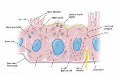
_____have SMALL apical surfaces with SHORT blunt microvilli |
Brush cells |
|
|
|
What is the function of mucus In respiratory epithelium? |
Film of mucus traps most airborne dust particles and micro organisms |
|
|
Front (Term) |
Back (Definition) |
|
|
Front (Term) |
Back (Definition) |
|
|
Front (Term) |
Name |
|
|
Front (Term) |
Name |
|
|
Front (Term) |
Respiratory epithelium: Note thick BM, goblet cells |

|
|
|
Other columnar cells, in RE, are called ____ that have small apical surfaces and short blunt microvilli |
brush cells |
|
|
|
Ciliary movement, in RE, is ___ propelling the sheet of mucus toward the ____ for elimination |
continuously, Pharynx |
|
|
Front (Term) |
Back (Definition) |
|
|
|
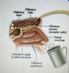
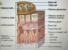
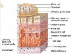
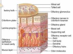
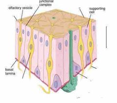
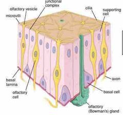
The olfactory epithelium contains |
Olfactory chemoreceptors that sense smell |
|
|
|
Olfactory epithelium covers |
Superior conchae bilaterally |
|
|
|
Olfactory epithelium send axons to the brain via small openings in |
The cribriform plate of the Ethmoid bone |
|
|
|
Olfactory epithelium is what type of tissue? |
Pseudostratified epithelium |
|
|
|
Olfactory epithelium contains the following cells |
1. Basal stem cells 2. columnar supporting cells 3.bipolar olfactory neurons (dendrites of these neuron have cilia with many membrane receptors for odor) |
|
|
|
Olfactory epithelium:_____ of these neurons have cilia specialized with many membrane receptors for odor molecules |
Dendrites |
|
|
Front (Term) |
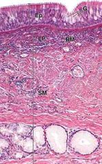
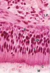
Note: Olfactory nerves (ON) in middle of pseudostratified olfactory epi. with supporthing cells (S) on top. Note a THIN BM that separates the basal cells (B) and underlying lamina propria (LR) |
|
|
|
Binding of odor molecules causes ______which passes along ____axons to the ________of the brain |
Depolarize, basal, olfactory bulb |
|
|
|
Nuclei of the ______olfactory neurons lie in the middle of a pseudo stratified epithelium with a zone of _____below it (their nucleus anyway) |
Bipolar, supporting cells |
|
|
|
At the apical end of each bipolar olfactory neuron is |
Non motile cilia or olfactory hairs and a layer of mucus. No cilia on supporting cells |
|
|
Front (Term) |
Back (Definition) |
|
|
|
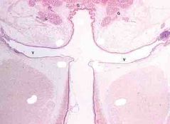
a short air passage b/w pharynx and trachea is ___ Its wall contains skeletal muscles and pieces of ____for sound production or phonation |
larynx cartilage |
|
|
|
Laryngeal vestibule is surrounded by |
Seromucous glands |
|
|
|
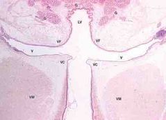
Lateral walls of the laryngeal vestible region bulge as a pair of |
Testicular folds VF |
|
|
|
Vestibular folds contain seromucous glands and ________ |
Areolar CT with lymphoid nodules |
|
|
|
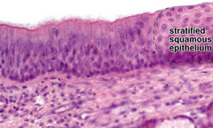
VF are largely covered by____ with regions near the epiglottis having___ |
RE, stratified squamous epithelium |
|
|
|
Larynx: Below ventricle is another pair of lateral folds called |
Vocal folds or cords (VC) |
|
|
|
VF: Note stratified squamous epithelium near epiglottis |
|
|
|
Below each vestibular fold (VF) is a narrow space called |
Ventricle |
|
|
|
Below each ventricle is another pair of lateral folds called _____ or _____ |
Vocal folds or vocal cords |
|
|
|
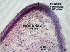
Vocal cords are covered by |
Stratified squamous epithelium |
|
|

Front (Term) |
Vocal cord covered in stratified squamous epi |
|
|
|
Vocal cords contain a large stratified ____and near the surface a small ligament |
Vocalis muscle (VM) |
|
|
|
Muscles cause variable _____of these ligaments that produces different sounds as air is _____across the vocal cords |
tension, expelled |
|
|
Front (Term) |
Back (Definition) |
|
|
Front (Term) |
Back (Definition) |
|
|
|
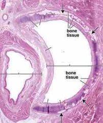
Trachea lined by |
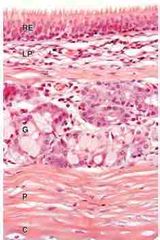
RE |
|
|
|
Trachea: below the RE lies the |
Lamina propria CT (loose areolar CT) |
|
|
|
Trachea: within the lamina propria and submucosa you can find |
Seromucous glands |
|
|
|
Trachea: the submucosa also contains |
C shaped rings of hyaline Cartlidge (C) covered by perichondrium (P) |
|
|
Front (Term) |
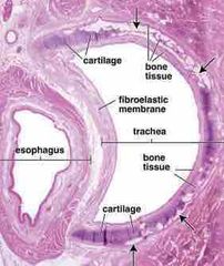
Trachea |
|
|
Front (Term) |
Back (Definition) |
|
|
Front (Term) |
Trachea. RE at top--> Lamina propia (LP)-->Seromucous glands (G)-->Perichondrium (P)->Hyaline cartilage (C) |
|
|
|
Bronchial tree: the trachea bifurcates as right and left primary bronchi. Primary bronchi enter the hilum on the posterior side of each lung along with the _____ |
pulmonary vessels lymphatics and nerves |
|
|
|
Bronchial tree: Within each lung, bronchi divide further to form the bronchial tree which is the ___component of ___ |
last component of the conducting system |
|
|
|
The ____ is a ____air passage between pharynx and trachea |
Larynx, short |
|
|
|
Trachea--> carina-->RL Primarily bronchus--> Secondary/lobar bronchi--> tertiary (segmental bronchus)--> smaller bronchi--> |
bronchioles-->terminal bronchiole-->respiratory bronchioles-->alveolar ducts-->alveolar sacs-->alveoli |
|
|

|
Tertiary (segmental) bronchus |
|
|
|
The lining of RE and the mucosa are ____ due to contraction of its ___ |
folded, smooth muscle (M) |
|
|
|
The wall of RE is surrounded by many pieces of |
Hyaline cartilage |
|
|
|
RE contains many seromucous glands (G) in the _____. These glands drain into the ____ |
submucosa, lumen |
|
|
|
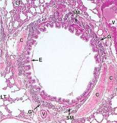
Surrounding the CT of the bronchi is |
arteries and vein (V). These vessels branch smaller and smaller as the approach respiratory bronchioles |

|
|
|
All bronchii are surrounded by distinct |
lung tissue (LT) which is characterized by many empty spaces of pulmonary alveoli |
|
|
|
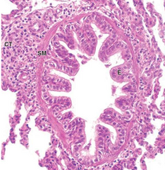
The epithelial lining (E) of bronchi is |
Pseudostratified ciliated columnar cells WITH a FEW GOBLET cells |
|
|

|
epithelial lining of bronchii |
|
|
|
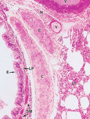
In SMALLER bronchi, the epi is mostly _____ and the ____ has both ____ and _____ near cartilage |
Columnar cells with cilia and FEWER goblet cells. Lamina propria has smooth m. (SM and small serous glands (G) near cartilage (C) |
|
|
|
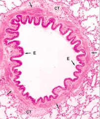
a LARGE bronchiole has |
folded RE (E) Prominent smooth m. supported ONLY by fibrous CT (CT) |
|
|

|
A large bronchiole |
|
|
|
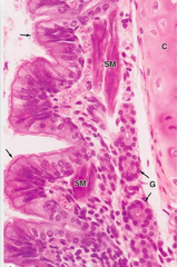
The epithelium of a SMALLER BRONCHIOLES is |
simple ciliated columnar, its smooth m. has elastic fibers with high elastic content (arrow) |

|
|
|
Small Bronchiole. |
|
|
|
small bronchiole has CT that includes |
lymphocytes (L) and nodules |

|
|
|
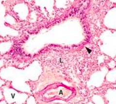
Small bronchioles: ______ are present in the tunica media of large arteriole (A) and to a lesser extent the accompanying venule (V) |
Elastic fibers |
|
|
|
Small bronchioles: ______ are also present in the ______ of a large arteriole (A) and its accompanying veniole (V) |
elastic fibers in the tunica media |

|
|
|
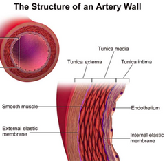
Structure of artery wall. In bronchiolles, elastic fibers invade tunica media in LARGE arteriole |
|
|
|
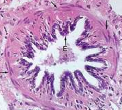
The epi of VERY SMALL bronchioles is composed of |
simple ciliated cuboidal with several layers of smooth muscle |

|
|
|
Very small bronchiole with simple ciliated cuboidal epi big layer of smooth m. |
|
|
|
Terminal bronchiole are the ___ system BEFORE gas exchange occurs |
last part of the air conditioning |

|
|
|
last part of conducting sys- terminal bronchiole |
made of ciliated simple cuboidal and many low simple non-ciliated columnar |
|
|
Terminal bronchiole have only 1-2 layers of ___ surrounded by ___ |
smooth m. surrounded by CT |
|
|
|
Terminal bronchiole epi is made up of |
ciliated simple cuboidal and many low simple non-ciliated columnar |
|
|
|
_____ are seen in the surround LT of terminal bronchiole |
Alveoli |
|
|

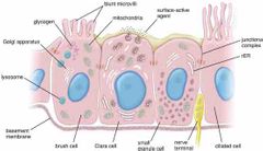
Front (Term) |
Clara cells |
|
|
|
Clara cells are |
non ciliated and have bulging domes of apical cytoplasm containing granules |
|
|
|
Clara functions |
1. Secrete components of surfactant which reduces surface tension and helps prevent collapse of bronchioles 2. Secrete P450 enzyme of their smooth ER detoxifies |
|
|
|
Clara cells also function to |
secrete secretory components for: 1. the transfer of IgA into bronchiolar lumen 2. lysozyme and other enzymes active against bac and viruses 3. Several cytokines that regulate local inflammatory responses |
|
|
|
Clara cell function (Broad) |
Secrete: 1. Surfactant 2. P450 enzyme 3. IgA 4. Lysozyme and other other enzymes, 5.cytokines |
|
|
|
____ cells give rise to all the cells within the bronchiolar epi are also included among Clara cells |
stem cells |
|
|
|
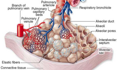
Terminal bronchioles branch into ____ which further branch into |
respiratory bronchioles,
alveolar ducts and individual alveoli |
|
|
|
Respiratory bronchioles are similar to terminal bronchioles except for the presence of |
scattered alveoli along their length |
|
|
|
__ travel with the bronchioles |
Pulmonary blood vessels |
|
|
|
A dense layer of branching __and __ surround each alveolus |
capillaries and elastic fibers |
|
|
|
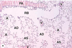
Respiratory Bronchioles have LT that is spongy like. It is abundant wit air passages and packets called |
Alveoli |
|
|
|
|
respiratory bronchioles |
|
|
Respiratory bronchioles (RB) Shows branching continuity with |
alveolar ducts (AD) and sacs (AS) |
|
|
|
Respiratory bronchioles have a layer of ___ and some regions of ____ |
smooth muscle and cuboidal epithelium |
|
|
|
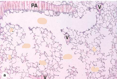
RB runs along a thin-walled branch of the |
pulmonary artery (PA) |
|
|
|
RB branches of the pulmonary vein (V) course elsewhere in the |
parenchyma |
|
|
|
Alveolar ducts consist of |
1. a linear series of alveoli, each with smooth muscle fibers around the opening 2.End in two or more clusters of alveoli called alveolar sacs |
|
|
|
Individual alveoli (A) open to |
alveolar sacs or ducts |
|
|
|
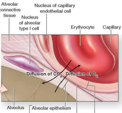
Gas exchange bw air and blood occurs at a membranous barrier bw each__and ___ |
alveolus and capillaries surrounding it |
|
|

|
Air-blood barrier consists of 1. Alveolar type I cell 2. Capillary endothelial cell 3. fused BMs Oxygen and CO2 pass in opposite directions |
|
|
|
The inner lining of alveoli is covered by a layer of ___which ____flud surface tension and ___ |
surfactant , lowers, prevents collapse of alveoli |
|
|
|
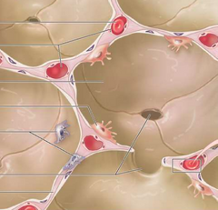
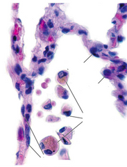
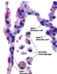
The septa between alveoli (A) contain several cell types : |
|
|
|
|
Capillaries include |
erythrocytes and leukocytes |
|
|
|
The alveoli are lined mainly by |
squamous type I alveolar cells (I) |
|
|
|
___ cells line almost the entire alveolus surface and gas exchange occurs across these cell |
Type I |
|
|
|
|
|
|
|
Type II alveolar cells |
1. Line a bit of each alveolus and are large rounded cells 2. Often bulge into the alveolus (II) 3. Have many functions of Clara cells, including production of surfactant |
|
|
|
Alveolar macrophages (M) also called |
dust cells and present in the alveoli or in the interalveolar septa |
|
|
|
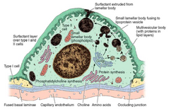
surfactant production by a type 2 cell
|
|
|
|
Surfactant production by a type 2 cell |
1. Secreted continuously by exocytosis and forms an oily film containing phospholipids and surfactant protein 2.contains protein-lipid complexes synthesized initially in the ER and Golgi apparatus |
|
|
|
Surfactant production by a type 2 cell: Surfactant containing protein lipid complexes formed my ER and golgi--> |
form multivesicular bodies-->lamellar bodes (large)-->extruded via exocytosis OR Golgi--> small lamellar body can fuse with lipoprotein vesicle and then get extruded out via extocytosis |
|
|
|
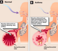
Asthma is |
A chronic inflammatory disease of the airways characterized by episodes of acute bronchoconstriction |
|
|
|
Asthma leads to |
shortness of breath, cough, chest tightness, wheezing, and rapid respiration. |
|
|
|
Airflow obstruction in asthma is due to bronchoconstriction that results from: |
1. contraction of bronchial smooth m., 2 inflammation of the bronchial wall, 3. increased secretion of mucus |
|
|
|
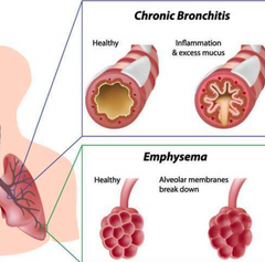
CHRONIC OBSTRUCTIVE PULMONARY DISEASE (COPD) is |
A chronic, irreversible obstruction of airflow that is usually progressive and characterized by persistent symptoms |
|
|
|
Symptoms of COPD include |
1. Cough 2. Excess mucus production 3. chest tightness 4.breathlessness 5. difficulty sleeping 6. fatigue |
|
|
|
____is the greatest risk factor for COPD |
smoking |
|
|
|
What happens to your lungs in COPD? |
There is small airways fibrosis Obstruction, and/or destruction of alveoli and of elastin fibres in the lung parenchyma |
|
|
|
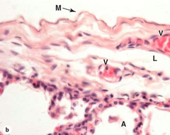
What is Pleura? What types are there? |
Are serous membranes (serosa) associated with each lung and thoracic cavity 1. Parietal pleura 2. Visceral pleura |
|
|
|
lines the inner surface of the thoracic cavity |
Parietal pleura |
|
|
|
covers the outer surface of the lung |
Visceral pleura |
|
|
|
Between parietal and visceral pleura is the narrow space called |
pleural cavity |
|
|
|
histo of pleura |
|
|
|
both layers of pleura consist of a _____ on a thin layer of connective tissue that is rich in ____and ___ CT also contains both __ |
simple squamous mesothelium (M) collagen and elastic fibers blood vessels (V) and lymphatics (L) |
|