![]()
![]()
![]()
Use LEFT and RIGHT arrow keys to navigate between flashcards;
Use UP and DOWN arrow keys to flip the card;
H to show hint;
A reads text to speech;
24 Cards in this Set
- Front
- Back
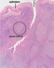
Identify |
Palatine tonsil (Notice deep crypt, stratified squamous epithelium and lymph follicle) |
|
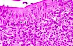
Identify |
Pharyngeal Tonsil (Notice respiratory epithelium (pseudo stratified ciliated columnar) and lack of crypts) |
|
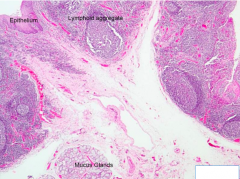
Identify |
Lingual Tonsil (Notice lymphoid aggregates, stratified squamous epithelium, mucus glands, and shallow crypt) |
|
Identify |
Thymus (Notice septae (thin CT capsule) and cortex/medulla) |
|
|
Where are Hassall's Corpuscles found? |
In the medulla of thymus |
|
|
What are Hassall's Corpuscles? |
Degenerated ERCs with eosinophilic stain |
|
|
Epithelioreticular cells |
-Primary support cells of thymus -Form blood-thymus barrier in the thymus cortex |
|
|
What holds ERCs together? |
Desmosomes |
|
|
Blood-thymus barrier components |
Capillary wall -Continuous capillaries with pericytes Perivascular CT -Macrophages EPCs
|
|
|
Lymph Flow |
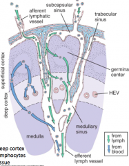
Afferent lymphatic vessel --> capsule --> subcapsular sinus --> trabecular/cortical sinus --> medullary sinus --> hilum --> efferent lymphatic vessel |
|
|
High Endothelial Venules (HEVs) |
-In deep cortex and medulla -Allow for the transition of lymphocytes from blood to lymph tissue |
|
|
Outer cortex of lymph node |
-Mainly B lymphocytes -Some T lymphocytes, reticular cells, APCs and macrophages |
|
|
Paracortex (deep cortex) of lymph node |
-Mainly T lymphocytes -HEVs |
|
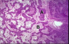
Identify A and B |
A- Medullary cords B- Medullary sinuses |
|
|
Lymph Node Medullary Cords |
Dense lymphoid tissue containing B cells, reticular cells, plasma cells and macrophages |
|
|
Lymph Node Medullary Sinuses |
Spaces with meshwork of reticular cells and macrophages |
|
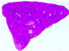
Identify |
Spleen |
|
|
Spleen capsule |
Simple squamous dense CT capsule |
|
|
Spleen Periarterial Lymphoid Sheath (PALS) |
-Ensheaths central artery in T lymphocytes -PALS itself is ensheathed in B lymphocytes |
|
|
Spleen Marginal Zone |
-Separates White Pulp from Red Pulp -Consists of APCS and macrophages to trap antigens |
|
|
Cords of Billroth |
Splenic cords |
|
|
Splenic Cords |
Made from reticular fibers |
|
|
Littoral Cells |
-Elongated endothelial cells that line splenic sinuses -Arranged like planks of barrel -Filters RBCs |
|
|
Where is blood-thymus barrier located? |
Cortex of thymus |