Reading...
![]()
Play button
![]()
Play button
![]()
Use LEFT and RIGHT arrow keys to navigate between flashcards;
Use UP and DOWN arrow keys to flip the card;
H to show hint;
A reads text to speech;
52 Cards in this Set
- Front
- Back
|
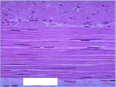
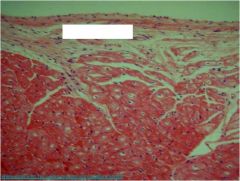
smooth muscle
|
|
What is this?
|
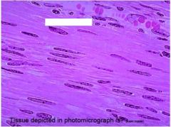
cardiac muscle
|
|
What is this?
|
smooth muscle
|
|
What type of tissue?
|
cardiac muscle
|
|
What type of stuff?
|
cardiac muscle
|
|
|
What is this?
|
cardiac
|
|
|
what is this?
|
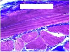
skeletal muscle
|
|
What do we see?
|
smooth my friend
|
|
What type of muscle?
|
skeletal
|
|
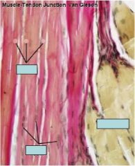
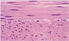
What are we looking at right and left?
|
Left is collagen fiber and right is skeletal muscle
|
|
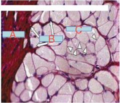
What is A, B, C and what is overall picture of?
|
A. Epimysium
b. Perimysium c. Endomysium Skeletal muscle |
|
What do arrows point to and what is overall structure?
|
skeletal muscle and arrows point to
1. 2. |
|
|
What are the three layers surrounding muscles and what tissues are each composed of?
|
1. Epimysium- is a layer of connective tissue which ensheaths the entire muscle. It is composed of dense irregular connective tissue.
2. Perimysium- is a sheath of connective tissue that groups individual muscle fibers (anywhere between 10 to 100 or more) into bundles or fascicles 3. Endomysium, meaning within the muscle, is a layer of connective tissue that ensheaths a muscle fiber and is composed mostly from reticular fibers. It also contains capillaries, nerves, and lymphatics. It overlies the muscle fiber's cell membrane: the Sarcolemma. |
|
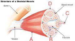
What are a-d?
|
a.epimysium
b. endomysium c. muscle fiber d. perimysium |
|
|
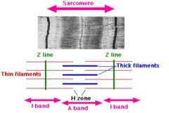
What is role of the following in the Sarcromere organization?
a. z-line b. a-actin c. Nebulin d. M- line "band" |
a. anchors thin filaments and connects adjacent sarcomeres
b. holds actin in to Z-disc c. controls actin polymerization to maintain length d. holds thick filaments (myosin) |
|
|
What is role of the following in the Sarcromere organization?
a. desmin |
a. desmin- anchors adjacent sarcromeres
|
|
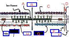
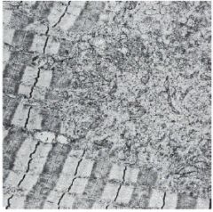
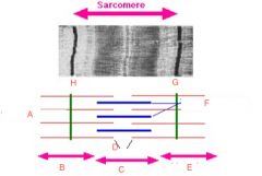
Label A-H with the following terms...
1. troponin, tropomyosin 2. desmin 3. m-band 4. thick filament/myosin 5. thin filament/actin 6. nebulin 7. a-aktinin 8. z-band |
A. thick filament/myosin
B.M-band c. troponin/tropomyosin d. z-band e. desmin f. a-aktinin g. actin thin filament h. nebulin |
|
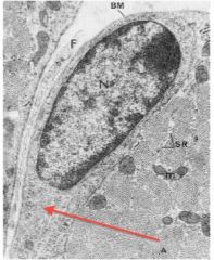
What is the red outline of the muscle cell? What condition occurs from problems related to this protein?
|
problems in dystrophin lead to muscular dystrophy
Protein called Dystrophy on periphery |
|
|
What is sarcoplasmic reticulum made of and function?
|
Structure:
SER striated muscled located inside muscle fibers and located at junction between A & I bands Function: store and release Ca ion. |
|
|
Briefly describe the characteristics of the thin filament...
a. filamentous-actin b. Tropomyosin c. Troponin |
a. filamentous actin
i. double helix of 2 strands of G-actin protein ii. Polar with myosin biding sites b. Tropomyosin i. double helical long, thin polypeptide ii. in grooves of actin double helix iii. covers-myosin binding site on actin c. Torponin i. three globular protein complex (T-tropomyosin, C-calcium, I-inhibits actin:myosin interaction) ii. exposis myosin-binding site on actin |
|
|
Briefly describe the characteristics of the thick filament...
|
1. Myosin- made of 6 polypeptides
i. 2 myosin heavy chain (MHC) on tail that have enzyme and contractile proteins ii. 2 pairs of myosin light chains (MLC) - 1 MHC and 2 MLC makes head that actin bind |
|
|
What general structural name is given to the thick and thin filaments?
|
myofilaments
|
|
|
What are triads and roles?
What separates them? |
- 2 terminal cisternae shaped like a butterfly that expand Sarcoplasmic reticulum (also between A and I junctions)
- Seperated by 1 T (transvers) tubule |
|
What is circled? and what is role?
|
triads- expand sarcoplasmic reticulum and are separated by T-tubule
|
|
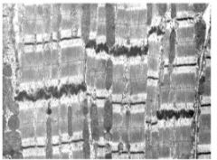
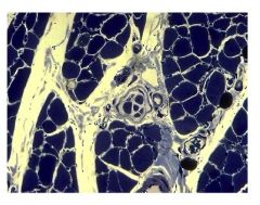
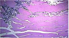
Which one is cardiac and skeletal?
How do you know the difference? Located major organelle that allows you to know the difference... |
skeletal muscle will have more mitochondria... one on Right is cardiac
|
|
What is the arrows pointing to that are dark sense? What type of muscle is this? What determines when more glycogen?
|
Arrow points to glycogen in skeletal muscle.
Fed has more glycogen than starved state Also different fiber types have more or less glycogen (more to come)... "thats what she said" |
|
|
What are the two types of structures provide proprioception for muscles?
|
1. muscle spindle
2. golgi tendon organ |
|
|
What is the function and location of the following?
1. muscle spindles 2. golgi tendon reflex |
1. anchored to permysium and endomysium of extrafusal fibers
functions to detect changes in extrafusal fiber length and rate of change (stretch receptor) sends signals to brain when muscle stretches 2. located at musculo-tendon junction detects when muscle contracts too strenuously |
|
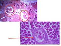
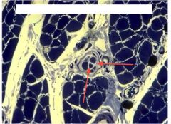
what is circled? and pointed to?
|
Muscle spindles
|
|
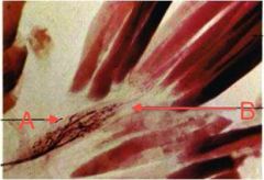
What is A and B and what is main aspect of picture?
|
A. Tension reception organ
B tendon golgi tendon organ |
|
What is the skeleton like structure and how many capsules can you see?
|
muscle spindle and 2 capsule
|
|
|
Are skeletal muscles able to reproduce to heal? If not how do they get replaced?
|
No- muscle fibers are incapable of mitosis, but within the basal lamina there are Satellite cells- which activate myoblasts which develop new muscle
|
|
|
In area arrow points what allows for muscle regeneration or growth and replacement?
|
satellite cells
|
|
What does arrow point to and significance in muscle healing or regeneration?
|
satellite cells- capable of mitosis made of inactive myoblasts
|
|
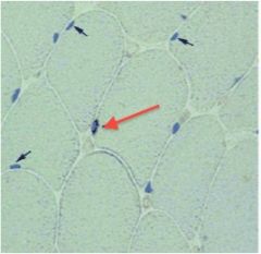
what is red arrow pointing to? Smaller arrows?
|
smaller arrows point to myocyte nuclei
satellite cells are from red arrow |
|
|
What can humans do to increase the number of satellite cells?
How does this action affect muscle fibers? |
resistance training increases number of satellite cells, creatine enhances effect
muscle fibers get thicker with resistance training but nuclei # does not increase |
|
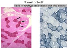
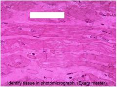
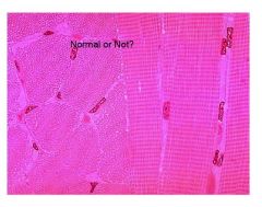
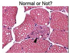
Normal or not normal skeletal muscle?
|
normal
|
|
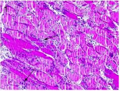
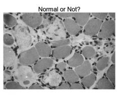
Normal or not? why?
|
not its clumped not same size
|
|
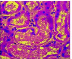
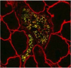
What does this slide of showing?
|
kidney tubules plugged with myosin possibly due to steroid use
|
|
|
What happens to your muscles when they are pushed beyond normal level of excercise?
|
1. Damage
2. Slides show leaked myoglobin which plugs muscle particularly in the liver and kidneys (with steroid users) |
|
What is this picture showing? What do arrows point to and whatcondition is this prevalent in?
|
Shows damaged muscle
- arrows point to myoglobin leaked in This happens in Rhabdomyolysis - kidney damage |
|
What is wrong with this slide? What specific case could this result in? How can you tell?
|
irregular muscle due to necrosis (lack of nuclei gave it away)
Specific to steroid use and rhabdomyolysis may result |
|
|
normal
|
|
normal or not?
|
normal
|
|
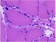
Normal or not?
What does this disorder does this slide represent? What is cause? Symptoms? |
not
Muscular dystrophy Abnormality or absence of the protein dystrophin Symptoms- 1. generalized weakness 2. muscle wasting first affecting the muscles of hips, pelvic area, thighs and shoulders 3. calves are often enlarged |
|
Is this normal? What gives it away? What would you called this?
|
not normal... known as myositis (inflammation of muscle)
- can see large amount of neutraphils (i.e. inflammation and its in muscle) |
|
|
Normal or not?
What gives it away? |
large vacuole (white round things) not normal
|
|
Normal or not?
What gives it away? |
large vacuole (white round things) not normal
|
|
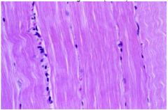
What is this a pic of?
|
dense connective tissue
|
|
Normal or not?
|
not
|
|
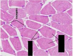
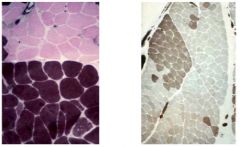
Normal or not?
|
normal look at size of capilaries about the same
|
|
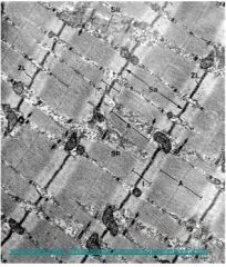
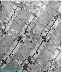
Label A-H...
|
|