Reading...
![]()
Play button
![]()
Play button
![]()
Use LEFT and RIGHT arrow keys to navigate between flashcards;
Use UP and DOWN arrow keys to flip the card;
H to show hint;
A reads text to speech;
174 Cards in this Set
- Front
- Back
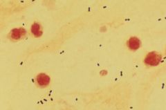
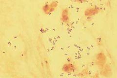
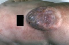
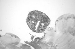
This is a sputum sample from a patient. What is the organism?
|
Streptococcus pneumoniae
Notes: gram-positive diplococci |
|
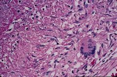
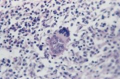
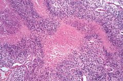
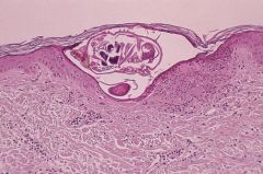
What organism caused this lesion?
|
Mycobacterium tuberculosis
Notes: Langhans' giant cells: multinucleated giant cells in which nuclei collectively form semicircle or circle |
|
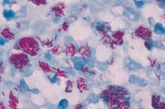
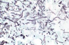
Organism?
|
Mycobacterium tuberculosis
Notes: "red snappers" on acid-fast staining |
|
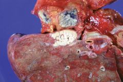
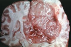
Diagnosis?
|
Miliary tuberculosis
Notes: caseous lesion in upper lobe and miliary lesions in surrounding hilar node |
|
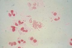
What organism is identified in this sputum sample?
|
Staphylococcus aureus
Notes: Gram positive cocci in clusters |
|
What organism is present in this smear from a urethral discharge?
|
Neisseria gonorrhoeae
Notes: Multiple gram negative diplococci within polymorphonuclear leukocyte |
|
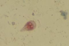
This sample is from the small intestine, what is the organism?
|
Giardia lamblia
Notes: Trophozoite has a classic pear shape with double nuclei giving an "owl's-eye" appearance |
|
Renal biopsy from a neonate, what is the problem?
|
Cytomegalovirus (CMV) infection
Notes: Cowdry type A nuclear inclusions in renal tubular cells resembling owl's eyes |
|
This is a lung biopsy from an individual who recently visited Arizona. What is the diagnosis?
|
Coccidioidomycosis
Notes: Endospores are shown within a spherule in the lung parenchyma. Usually resolves spontaneously except in immunocompromised individuals |
|
Special stain of CSF from an AIDS patient with meningoencephalitis. What is the organism?
|
Cryptococcus neoformans
Notes: India ink preparation shows extracellular polysaccharide capsule |
|
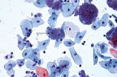
Organism?
|
Candida albicans
Notes: Branched budding and pseudohyphae |
|
Organism?
|
Trichomonas vaginalis
Notes: Trophozoites with flagella |
|
Diagnosis?
|
Herpes genitalis
Notes: Ulcerating vesicles associated with HSV-2 |
|

Diagnosis?
|
Syphilis
Notes: Painless chancre associated with primary syphilis |
|
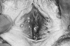
This is a slide from a patient with tertiary syphilis . What is the abnormality?
|
Tabes dorsalis revealing degeneration of dorsal columns and dorsal roots in the thoracic spinal cord.
|
|
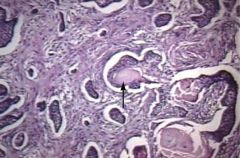
What is the diagnosis and what is the arrow pointing to?
|
Bacterial vaginosis
Clue cell |
|
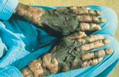
Diagnosis?
|
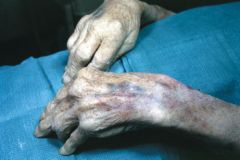
Leprosy, lepromatous type.
Notes: Nodules and thick plaques on dorsa of fingers, wwrists, and forearms with hypopigmentation of overlying skin. |
|
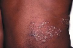
Causal agent?
|
Herpses zoster
Notes: Reactivation spreads across dermatomes. |
|
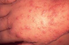
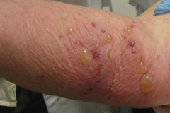
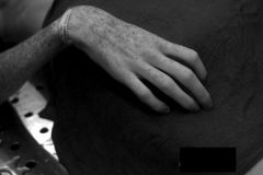
This is the hand of a 3 y/o child. What is the diagnosis?
|
Coxsackie exanthem (hand-foot-mouth disease)
Notes: Diffuse eruptive vesiculopapules |
|
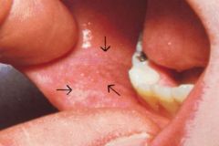
Arrows are pointing to tiny white lesions with erythematous halos. What are these lesions and what are they pathognomonic for?
|
Koplik spots, measles
Notes: Precede generalized rash by 1-2 days |
|
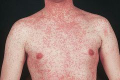
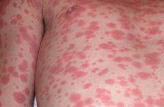
What is the diagnosis?
|
Measles
|
|
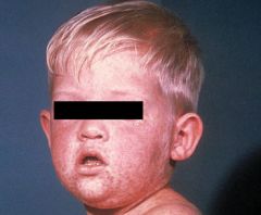
What is the diagnosis?
|
Measles
Notes: Rash often begins in the head or neck area |
|
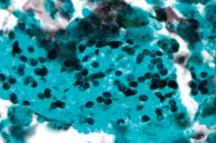
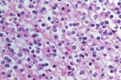
What does this special silver stain of lung epithelium show?
|
Pneumocystis jiroveci (formerly carinii)
Notes: Numerous small, disk-shaped organisms |
|
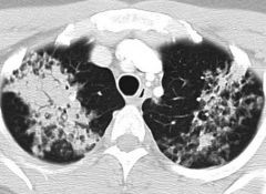
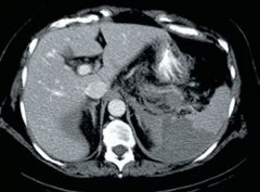
What does this chest CT scan reveal?
|
Pneumocystis pulmonary infection.
Notes: Bilateral confluent air-space opacities in a central distribution |
|
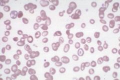
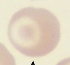
What cells are present hear?
|
Target cells
Notes: Result from increase in SA:volume from iron deficiency anemia (decreased cell volume) or obstructive liver disease (increased cell membrane) |
|
What?
|
Target cell
|
|
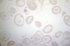
Blood smear of a patient from Cyprus. What is the likely diagnosis?
|
Thalassemia major
Notes: A blood dyscrasia caused by a defect in β-chain synthesis in hemoglobin, target cells present. |
|
What is the disorder? Why?
|
Iron deficiency anemia. Microcytosis and hypochromia.
|
|
Diagnosis?
|
Sickle cell anemia.
Notes: Sickled cells, anisocytosis, poikilocytosis, and nucleated RBCs |
|
This patient has sickle cell anemia, what happened?
|
Splenic infarction
Notes: The spleen is particularly susceptible to infarction due to the lack of collateral blood supply. Autosplenectomy often occurs in sickle cell anemia patients. |
|
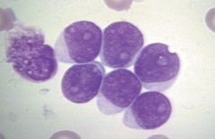
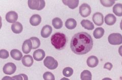
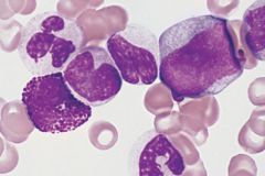
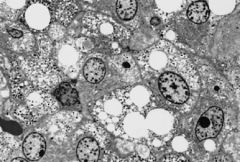
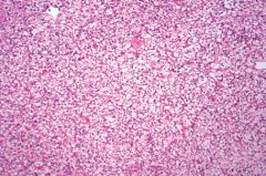
Peripheral blood smear of a 6 y/o child. Diagnosis?
|
Acute lymphocytic leukemia
Notes: Affects children <10 y/o |
|
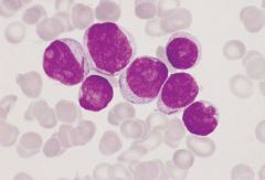
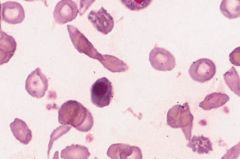
Diagnosis?
|
Acute myeolocytic leukemia
Notes: There are auer rods in two of the cells. Affects adolescents to young adults, but most commonly diagnosis in older adults. |
|
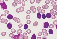
Peripheral blood smear of a 65 y/o patient. Diagnosis?
|
Chronic lymphocytic leukemia
Notes: Lymphocytes are very fragile and form "smudge cells" upon slide preparation. Affects individuals >60 y/o |
|
Blood smear of a 45 y/o patient. Diagnosis?
|
Chronic myeloid leukemia
Notes: Promyelocytes and myelocytes are seen adjacent to a vascular structure. Affects individuals between 30 and 60 y/o |
|
What is another important finding in this disorder?
|
Bence Jones protein in the urine.
Notes: Classic "punched out" lytic lesions seen in multiple myeloma. |
|
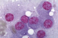
This is a smear from a patient with multiple myeloma. What are these cells?
|
Plasma cells
Notes: RBCs in rouleaux formation commonly seen on peripheral smears |
|
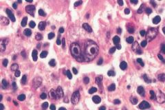
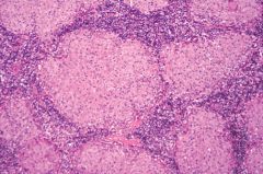
What is the diagnosis?
|
Burkitt's lymphoma
Notes: Classic "starry-sky" appearance. |
|
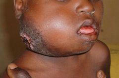
Young nigerian boy with history of jaw swelling unresponsive to antibiotics. Diagnosis?
|
Burkitt's lymphoma
|
|
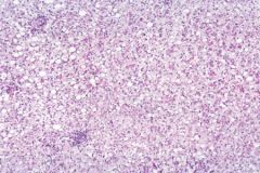
Probable diagnosis?
|
Hodgkin's lymphoma
Notes: Classic Reed-Sternberg cells are necessary but insufficient pathologic finding for the diagnosis of Hodgkin Lymphoma |
|
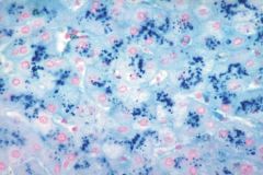
Diagnosis?
|
Hemochromatosis
Notes: Prussian blue iron stain shows hemosiderin in the liver parenchyma. Deposition occurs throughout the body causing organ damage and darkening of skin. |
|
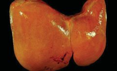
Diagnosis?
|
Macrovesicular steatosis of liver
Notes: Early reversible change associated with alcohol consumption is seen. |
|
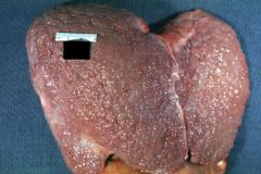
What?
|
Fatty liver.
Notes: Enlarged, yellow appearance |
|
What?
|
Micronodular cirrhosis of the liver
Notes: Fine granular appearance due to alcoholism. Later stages show irregularly shrunken liver with larger nodules giving it a "hobnail" appearance |
|
What?
|
Cirrhosis
Notes: Regenerative lesions are surrounded by fibrotic bands of collagen (bridging fibrosis) froming characteristic nodularity. |
|
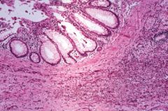
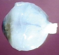
This was excised from the colon, what is it?
|
Tubular adenoma (type of colonic polyp)
Notes: Smaller and rounded in morphology and have less malignant potential than do villous adenomas |
|
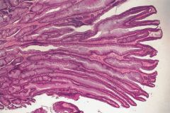
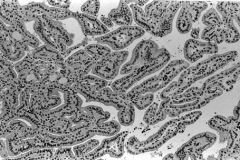
What is this?
|
Villous adenoma
Notes: Composed of long, fingerlike projections |
|
This patient presented with LLQ pain. What is the diagnosis?
|
Diverticulitis
Notes: My cause perforation, peritonitis, abscess formation, or bowel stenosis |
|
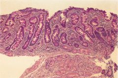
This is biopsy of the small intestines. Diagnosis?
|
Celiac sprue
Notes: Gluten-sensitive enteropathy showing blunting of the villi and crypt hyperplasia |
|
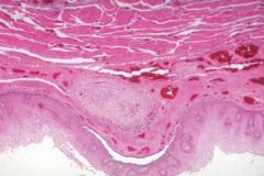
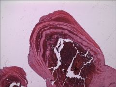
This is a sample from the esophagus. What is shown?
|
A sclerosed esophageal varix.
Notes: Overlying espohageal mucosa is generally normal. |
|
What?
|
Intussusception of infant gut
|
|
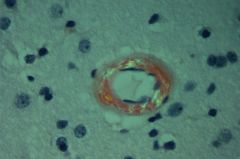
Diagnosis?
|
Pulmonary embolism
Notes: Large pulmonary thromboembolus shown. Most often arises from deep venous thrombosis. |
|
What is seen in this pulmonary thromboembolus?
|
Lines of Zahn
Notes: These interdigitating areas of pale pink and red represent layers of red cells, platelets, and fibrin that are laid down in the vessel as the thrombus forms. |
|
Biopsy of a mass in a pulmonary hilar lymph node. Diagnosis?
|
Small (oat) cell carcinoma
Notes: Almost all of these tumors are related to tobacco smoking. They can arise anywhere in the lung, most often near the hilum, and quickly spread along the bronchi. |
|
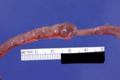
Section of procine myocardium. What?
|
Taenia solium (pig tapeworm)
Notes: When humans ingest this meat, the larvae attach to the wall of the small intestine and mature to adult worms. |
|
Diagnosis?
|
Acute respiratory distress syndrome (ARDS)
Notes: Persistent inflammation leads to poor pulmonary compliance and edema; note both alveolar fluid and hyaline membranes |
|
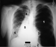
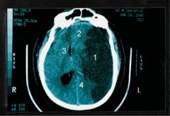
What is the diagnosis?
|
Tension pneumothorax
Notes: 1-Hyperlucent lung field 2-Hyperexpansion lowers diaphragm 3-Collapsed lung 4-Deviation of trachea 5-Mediastinal shift 6-Compression of opposite lung |
|
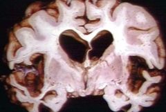
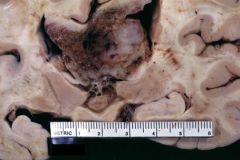
Diagnosis?
|
Alzheimer's disease
Notes: Key histologic features include "senile plaques" (note pictured); a coronal section showing atrophy, especially of the temporal lobes |
|
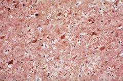
What?
|
Senile plaques
Notes: Focal masses of interwoven neuronal processes around an amyloid core; neurofibrillary tangles are evident |
|
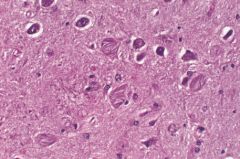
Describe
|
Remnants of neuronal degeneration associated with Alzheimer's disease, the most common cause of dementia in older persons
|
|
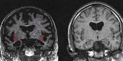
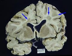
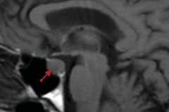
What?
|
Alzheimer's disease
Notes: T1-weighted MRI on left shows bilateral temporal lobe atrophy (arrows) as compared with normal appearance on right. |
|
Prussian blue stain of the lung. What is the diagnosis?
|
Asbestosis
Notes: Ferrunginous bodies in the lung; inhaled asbestos fibers are ingested by macrophages |
|
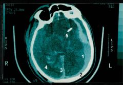
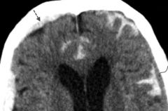
What?
|
Subdural hemorrhage with concamitant subarachnoid hemorrhade
Notes: 1-subdural blood, layering 2-skull 3-falx 4-subarachnoid blood 5-shunt catheter |
|
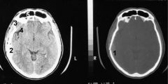
Diagnosis?
|
Epidural hematoma from skull fracture
Notes: 1-skull fracture 2-hematoma in epidural space 3-temporalis muscle 4-Sylvian fissure 5-frontal sinus |
|
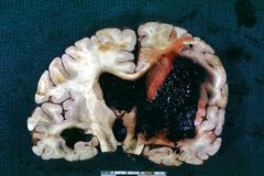
What?
|
Brain with hypertensive hemorrhage in the region of the left basal ganglia
|
|
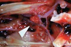
What?
|
Berry aneurysm located on the anterior cerebral artery.
Notes: small, saclike structure can easily rupture during periods of hypertension or stress |
|
What?
|
Berry aneurysm histology showing lack of internal elastic lamina
|
|
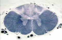
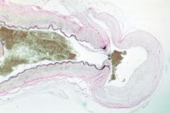
Diagnosis?
|
Multiple sclerosis
Notes: Lumbar spinal cord with mostly random and asymmetric white-matter lesions |
|
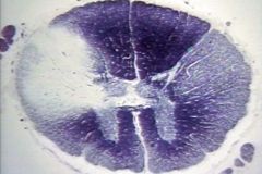
This slide shows periventricular white-matter plaques of demyelination, what is the diagnosis, and what clinical findings are classically associated?
|
Multiple sclerosis. Nystagmus, scanning speech, and intention tremor.
|
|
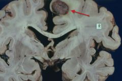
Diagnosis?
|
Glioblastoma multiforme.
Notes: It is extending across the midline of the cerebral cortex |
|
This is a section from a cerebral mass. Diagnosis? Salient histological findings?
|
Glioblastoma multiforme. Necrosis with surrounding pseudopalisading of malignant tumor cells.
|
|
Diagnosis?
|
Oligodendroglioma.
|
|
Section of cerebral mass. Diagnosis?
|
Oligodendroglioma
Notes: Classic "fried egg" appearance with perinuclear halos and "chicken-wire" capillary pattern |
|
Diagnosis?
|
Left middle cerebral artery stroke.
Notes: 1-ischemic brain parenchyma; 2-subtle midline shift to the right; 3-right frontal horn of the lateral ventricle; 4-left lateral ventricle obliterated by edema |
|
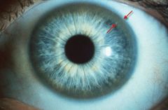
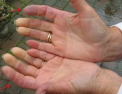
What is between the arrows? Diagnosis?
|
Kayser-Fleischer ring. Wilson's disease.
|
|
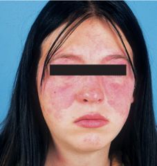
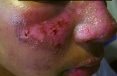
Diagnosis?
|
Systemic Lupus Erythematosus (SLE).
Notes: Malar rash in young female |
|
The arrows are pointing to ulcerations of the fingertips. Diagnosis?
|
Scleroderma.
Notes: Progressive tightening of the skin contracts the fingers. Fibrosis is widespread and may involve esophagus (dysphagia), lun (restrictive disease), and small vessels of the kidney (HTN). |
|
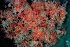
Joint biopsy. Diagnosis?
|
Gout
Notes: Tophi (stones) in joints consist of urate crystals surrounded by an inflammatory reaction consisting of macrophages, lymphocytes, and giant cells. |
|
Diagnosis?
|
Gout
Notes: Typically involves PIP joints growing like tubers from the bones |
|
Diagnosis?
|
Erythema multiforme.
|
|
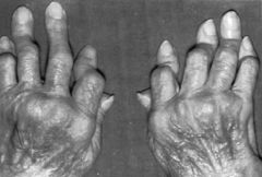
Diagnosis?
|
Rheumatoid arthritis
Notes: Swan-neck deformities of digits and involvement of PIP joints |
|
This is the arm of a 6-month old boy. What could cause these confluent erythematous papules and nodules?
|
Arteriovenous malformation (AVM)
|
|
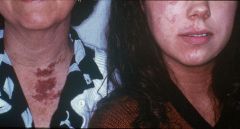
What could cause these patches and plaques on this mother and daughter?
|
Capillary malformation, port-wine type
|
|
Diagnosis?
|
Scabies
|
|
Skin biopsy, diagnosis?
|
Squamous cell carcinoma
Notes: arrow is pointing to a keratin pearl |
|
Diagnosis?
|
Malignant melanoma
Notes: pigmented and non-pigmented cells. Not all melanomas make melanin. |
|
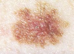
Diagnosis?
|
Malignant melanoma
Notes: irregular border and pigmentary variation |
|
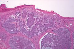
Skin biopsy, diagnosis?
|
Basal cell carcinoma
Notes: Nests of basaloid cells are present within the dermis with peripheral palisading and prominent retraction artifacts. |
|
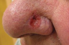
Diagnosis?
|
Basal cell carcinonma
|
|
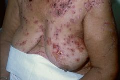
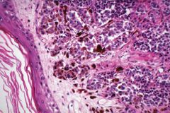
Diagnosis?
|
Pemphigus vulgaris
Notes: numerous crusted, denuded, and weepy erythematous plaques are seen on the chest, breast, abdomen, and arms |
|
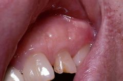
This images is showing vesicles on the gingiva. Diagnosis?
|
Pemphigus vulgaris
|
|
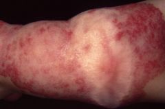
Diagnosis?
|
Bullous pemphigoid
Notes: tense bullae and urticarial plaques |
|
Diagnosis?
|
Psoriasis
|
|
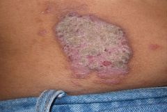
Diagnosis?
|
Acanthosis nigricans
Notes: Extensive hyperpigmented plaques on arms of a patient with lipodystrophy. This is an unusual distribution for acanthosis nigricans |
|
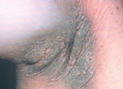
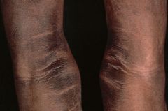
Diagnosis?
|
Acanthosis nigricans, typical underarm distribution
|
|
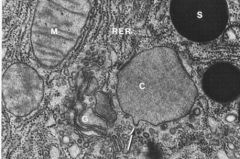
Pancreatic cell on EM. Notes
|
A condensing vacuole (C) is receiving secretory product (arrow) from the golgi complex (G). M-mitochondrion; RER-rough endoplasmic reticulum; S-mature condensed secretory zymogen granules
|
|
This patient had primary hyperaldosteronism. What is the diagnosis?
|
Adrenocortical adenoma
|
|
EM of an adrenal tumor cell. Diagnosis?
|
Pheochromocytoma
Notes: neurosecretory granules are present |
|
Diagnosis?
|
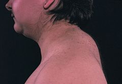
Cushing's disease
Notes: Moon facies and buffalo hump |
|
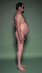
Diagnosis?
|
Cushing's disease
Notes: Truncal obesity and abdominal striae |
|
This patient had abdominal striae, truncal obesity, and hyperpigmented skin, what is the diagnosis?
|
ACTH-producing pituitary adenoma
|
|
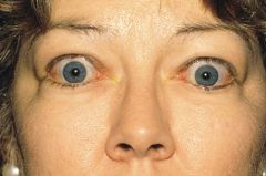
Diagnosis?
|
Grave's disease
Notes: Exophthalmos, proptosis, and periorbital edema |
|
Diagnosis?
|
Graves' disease
Notes: CT shows extraocular muscle enlargement at the orbital apex |
|
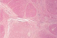
Thyroid biopsy, diagnosis?
|
Graves' disease
Notes: Stimulation of follicular cells by autoantibodies that stimulate TSH receptors causes the normal uniform architecture to be replaced by hyperplastic papillary, involuted borders, and decreased colloid. Treat with propylthiouracil to inhibit production of thyroid hormone as well as peripheral conversion of T4 to T3 |
|
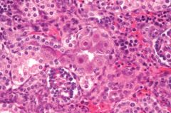
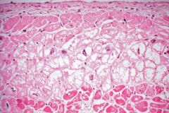
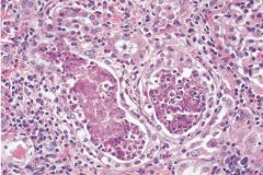
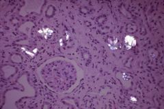
Renal biopsy, what is the problem?
|
Arteriolar sclerosis
Notes: Efferent and afferent arterioles are plugged with masses of hyaline material. This is from a diabetic patient |
|
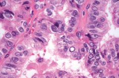
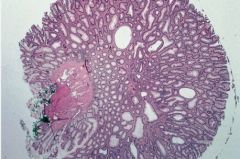
Thyroid biopsy, diagnosis?
|
Papillary carcinoma
Notes: Orphan annie eye nuclei and often psammoma bodies are present. Associated with neck irradiation. |
|
Notes:
|
Papillary carcinoma. Metastatic solitary lesion seen on gross specimen in coronal plane
|
|
This mass was found in the uterus of a pregnant women. Diagnosis?
|
Hyaditidiform mole.
Notes: "bunch of grapes", most common precursors of choriocarcinoma. Complete moles: 46, XX with all chromosomes from sperm. Partial moles: triploid or tetraploid with some fetal parts. |
|
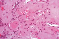
Biopsy of the heart. Describe what has happened.
|
Myocytolysis and coagulative necrosis beneath the endocardium
|
|
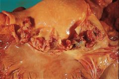
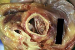
Diagnosis?
|
Acute bacterial endocarditis.
Notes: virulent organisms (e.g., S. aureus) infect previously normal valves, causing vegetations (here, in the aortic valve) and potentially giving rise to septic emboli |
|
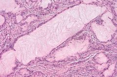
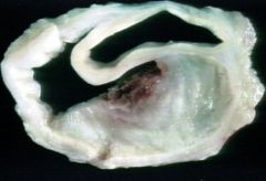
Diagnosis?
|
Calcified bicuspid aortic valve
Notes: The abnormal shape of the valve make its leaflets susceptible to otherwise ordinary hemodynamic stresses, which ultimately leads to valvular thickening, dystrophic calcification, increased rigidity, and stenosis |
|
This is a section of the aorta. Describe the problem.
|
A blood clot compressed the aortic lumen. A tear in the intima allowed blood to surge through the muscular layer to the adventitia, forming an aortic dissection.
|
|

What is the lesion?
|
Intracranial berry aneurysm .
|
|
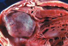
Diagnosis?
|
Left atrial myxoma
Notes: Most common primary cardiac tumor; known to produce VEGF |
|
|
Diagnosis?
|
Left atrial myxoma
|
|
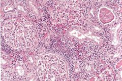
Kidney biopsy, diagnosis?
|
Acute pyelonephritis
Notes: Neutrophilic infiltration and abscess formation within the renal interstitium. |
|
Renal biopsy, diagnosis?
|
Chronic pyelonephritis
Notes: Lymphocytic invasion and fibrosis |
|
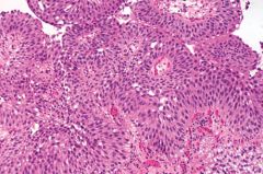
Diagnosis?
|
Transitional cell carcinoma
Notes: Image shows a papillary growth lined by transitional epithelium with mild nuclear atypia and pleomorphism. |
|
Diagnosis?
|
Lupus erythematosus
NotesA: Enlarged, very pale kidneys with "flea bitten appearance" |
|
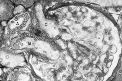
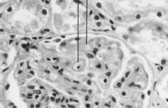
Notes
|
Normal glomerulus
A-macula densa B-afferent arteriole C-efferent arteriole |
|
Diagnosis?
|
Minimal change disease (Lipoid nephrosis).
Notes: Effacement of foot processes. Treat with corticosteroids |
|
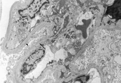
Kidney from a patient with SLE. What has occurred?
|
Wire-loop thickening as a result of immune complex deposition within the glomerulus. It is associated with subendothelial deposits.
|
|
Diagnosis?
|
SLE, malar rash
|
|
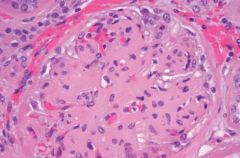
Diagnosis?
|
Diabetic glomerulosclerosis
Notes: Kimmelstiel-Wilson syndrome characterized by acellular ovoid nodules in the periphery of the glomerulus |
|
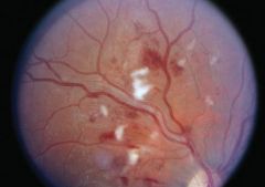
Diagnosis?
|
Diabetic retinopathy
Notes: Cotton wool spots and macular edema |
|
Kidney biopsy with partially crossed light polarizers. Diagnosis?
|
Calcium oxalate crystals
Notes: Tubular failure in oxalate nephropathy can result from vitamin C or antifreeze abuse |
|
Diagnosis?
|
Staghorn calculus
Notes: Although any stone may form a staghorn calculus, it is most commonly triple phosphate (struvite) and associated with recurrent Proteus infections |
|
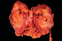
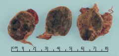
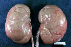
Diagnosis?
|
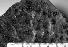
Renal cell carcinoma
Notes: Nodular, golden-yellow tumor with areas of hemorrhage and necrosis |
|
Biopsy of a renal mass. Diagnosis?
|
Renal cell carcinoma
Notes: Polygonal cells with small nuclei and abundant clear cytoplasm with a rich, delicate branching vasculature |
|
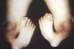
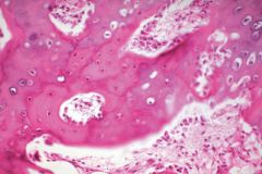
Diagnosis?
|
Osteogenesis imperfecta
Notes: Blue sclera caused by translucency of connective tissue over the choroid. |
|
Diagnosis?
|
Osteogenesis imperfecta
Notes: Abnormal collagen synthesis results from a variety of gene mutations and causes brittle bones and connective tissue malformations |
|
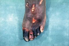
Diagnosis?
|
Foot gangrene.
|
|
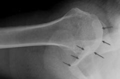
What is taking place?
|
New bone formation with osteoblasts following a fracture
|
|
Diagnosis?
|
Meningiomyelocele
Notes: A neural tube defect in which the meninges and spinal cord herniate through the spinal canal. This is a gross image of an infant's lower back |
|
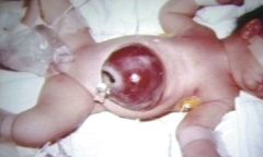
Diagnosis?
|
Omphalocele in a newborn
Notes: Defect is midline and is covered by peritoneum as opposed to gastroschisis which is not covered by peritoneum and often not midline. |
|
Diagnosis of this lymph node biopsy?
|
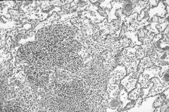
Sarcoidosis
Notes: numerous tightly formed granulomas |
|
Diagnosis?
|
Amyloidosis
Notes: Congo red stain demonstrates amyloid deposits in artery wall that show apple-green birefringence under polarized light |
|
|
Diagnosis?
|
Raynaud's disease
|
|
|
Diagnosis?
|
Raynaud's disease
|
|
Diagnosis
|
Raynaud's disease
|
|
Diagnosis?
|
Colon cancer
Notes: Circumferential tumor with heaped-up edges and central ulceration |
|
|
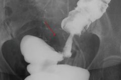
Diagnosis?
|
Colon cancer
Notes: Classic apple core lesion of the sigmoid colon seen on barium enema |
|
Diagnosis?
|
Colon cancer
Notes: Classic apple core lesion seen in sigmoid colon on barium enema |
|
What disorder does this patient likely have?
|
Marfan's syndrome
|
|
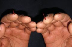
What disorder does this patient likely have?
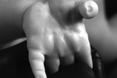
|
Down syndrome
Notes: The simian crease is a characteristic feature of patients with Down syndrome |
|
What is the diagnosis?
|
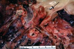
Hydatid cyst
Notes: Echinococcus eggs develop into larvae in the intestine, penetrate the intestinal wall, and disseminate throughout the body. The larvae commonly form hydatid cysts in the liver |
|
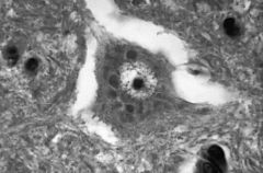
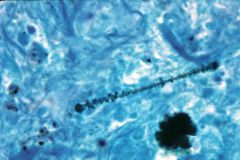
This is a neuron, what is the diagnosis?
|
Rabies infection.
Notes: Negri bodies in neuronal cytoplasm are pathognomonic for rabies virus infection |
|
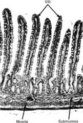
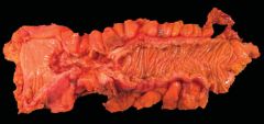
What section of the GI tract is this?
|
Small intestine
|
|
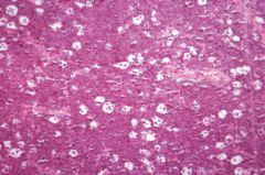
There is a clear distinction between the left and right halves of this liver biopsy slide. Describe what is going on.
|
Coagulative necrosis of hepatocytes on the right side. Cells are disorganized, the nuclei are pyknotic or absent, and the cytoplasm is homogeneous
|
|
Diagnosis?
|
Multinodular goiter
Notes: Image shows follicles distended with colloid and lined by a flattened epithelium with areas of fibrosis and hemorrhage |
|

What is the diagnosis of this bone mass?
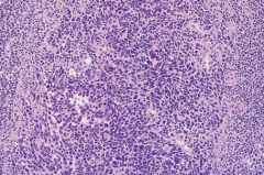
|
Ewing's sarcoma
Notes: This malignant tumor of bone occurs in children and is characterized by a t(11;22) that results in the fusion gene, EWS-FLI1. The tumor is composed of sheets of uniform small, round cells. |
|
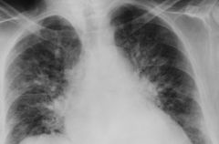
What does this gross picture of the lung reveal?
|
Pneumonia.
Notes: There is a large area of consolidation at the base plus multiple small areas of consolidation involving bronchioles and surrounding alveolar sacs throughout the lungs. |
|
What is the diagnosis of this lung biopsy?
|
Pneumonia
Notes: Neutrophils in alveolar spaces |
|
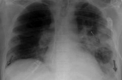
Diagnosis?
|
Cavitary pneumonia
Notes: CXR showing left-sided pneumonia with cavitation and bilateral pleural effusions |
|
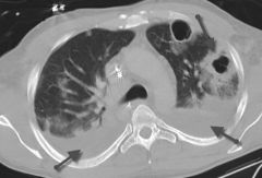
Diagnosis?
|
Cavitary pneumonia
Notes: Chest CT scan showing cavitary lesions with bilateral pleural effusions |
|
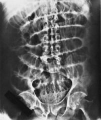
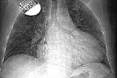
Diagnosis?
|
Small bowel obstruction
Notes: Dilated loops of small bowel in a ladder-like pattern. Air-fluid levels may be seen on upright x-ray |
|
Diagnosis?
|
Small bowel obstruction
Notes: Abdominal CT scan showing multiple dilated loops of small bowel with air-fluid levels. |
|
Lung biopsy, diagnosis?
|
Emphysema
Notes: Abnormal, permanent enlargement of airspaces distal to the terminal bronchiole. Enlarged alveoli are seen separated by thin septa, some of which appear to float within the alveolar spaces. |
|
Diagnosis?
|
Emphysema.
Notes: Multiple cavities lined by heavy black carbon deposits, typical of smoking |
|
CXR finding?
|
Pulmonary edema
Notes: Acute pulmonary edema due to left ventricular failure. Note the bat's-wing density, cardiac enlargement, increased size of upper lobe vessels, and pulmonary venous congestion. |
|
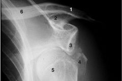
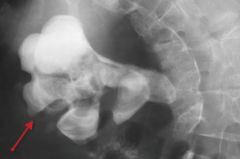
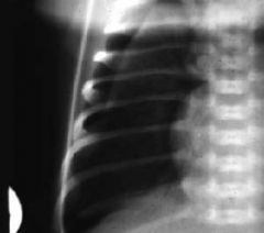
What are the radiological findings?
|
Anterior shoulder dislocation.
Notes: The humeral head (5) is inferior and medial to the glenoid fossa (3) and fracture fragments of the greater tuberosity (4) are evident. 1-acromion, 2-coracoid, 6-clavicle |
|
What are the radiological findings?
|
Anterior shoulder dislocation.
Notes: Axillary view showing humeral head dislocated anteriorly with respect to the glenoid fossa. |
|
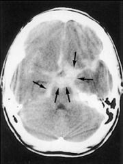
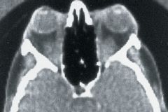
Diagnosis?
|
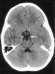
Subarachnoid hemorrhage
Notes: blood in the subarachnoid space, compare to this normal image |
|
What are the radiological findings?
|
Subdural hemorrhage (straight arrow), subarachnoid hemorrhage (dotted arrow), and chronic isodense subdural hemorrhage (curved arrow)
|
|
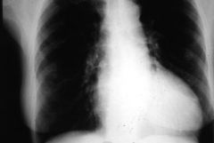
List one major radiological finding.
|
Left ventricular hypertrophy
Notes: Mediastinum is wider than 50% of the width of the chest |
|
What drug could cause the findings in this CXR?
|
Amiodarone toxicity
Notes: Pulmonary fibrosis is evident by the diffuse interstitial bilateral pulmonary markings in a reticular nodular pattern most prominent in the lung bases and posteriorly |
|
What is the radiological finding?
|
Pneumothorax.
Notes: The right lung has collapsed and the arrow indicates the edge of the collapsed lung |
|
|
What is the diagnosis?
|
Pneumothorax
Notes: CT scan shows collapsed lung with ipsilateral increased density of the lung parenchyma |
|
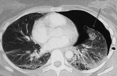
What is the diagnosis?
|
Pneumothorax
Notes: CT scan shows collapsed left lung with ipsilateral increased density of the lung parenchyma |
|
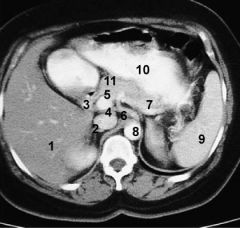
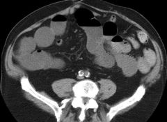
Label structures
|
1-liver; 2-IVC; 3-Portal vein; 4-hepatic artery; 5-gastroduodenal artery; 6-celiac trunk; 7-splenic vein; 8-aorta; 9-spleen; 10- stomach; 11-pancreas
|
|
|
Label structures
|
1-large left adrenal mass; 2-kidney; 3-verterbal body; 4-aorta; 5-IVC; 6-pancreas; 7-spleen; 8-liver; 9-stomach; 10-colon, splenic flexure
|
|
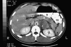
Label structures
|
1-large left adrenal mass; 2-kidney; 3-verterbal body; 4-aorta; 5-IVC; 6-pancreas; 7-spleen; 8-liver; 9-stomach; 10-colon, splenic flexure
|
|
Diagnosis?
|
Chronic pancreatitis
Notes: Punctate calcifications are seen in the head, body, and tail of the pancreas. |
|
Diagnosis?
|
Osteoarthritis
Notes: Increased fibrosis of the joint and a decreased amount of cartilage are seen. |
|
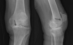
Diagnosis?
|
Osteoarthritis
Notes: Frontal and lateral x-rays of the knee showing medial and patello-femoral joint space narrowing and sclerosis |