![]()
![]()
![]()
Use LEFT and RIGHT arrow keys to navigate between flashcards;
Use UP and DOWN arrow keys to flip the card;
H to show hint;
A reads text to speech;
33 Cards in this Set
- Front
- Back
|
Germinal Center in Secondary follicle of LN * Contains B cells and macrophages |
|
|
Medullary Cord/Sinus of LN * Contains B cells and Plasma cells |
|
|
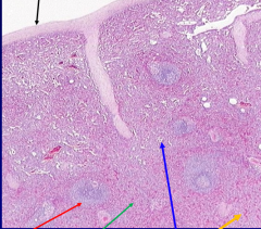
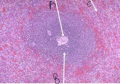
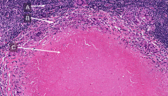
Lymph node A = subcapsular sinus B = germinal center w/ B cells C = mantle zone w/ naive B cells |
|
|
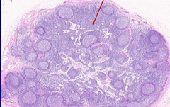
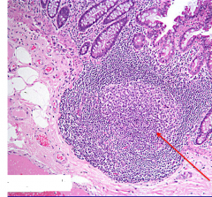
Lymph Node Black arrow = subcapsular sinus White arrows = germ centers of secondary follicle Red circle = primary follicle |
|
|
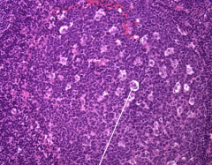
Tingible-body Macrophage in germinal center of LN *remove apoptosed B cells that fail selection |
|
|
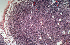
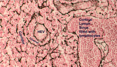
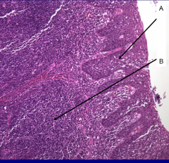
Paracortex of LN *Contains T cells |
|
What are the black fibers? |
Reticular fibers (type 3 collagen) made by reticular cells in LN |
|
|
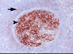
Arrowhead = Follicular Dendritic cell in LN Arrow = Mantle |
|
|
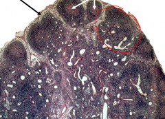
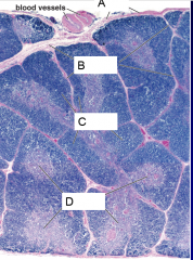
Infant Thymus (ie no fat) A - capsule B - Trabeculae (separate lobes) C - Cortex (darker) D - Medulla (lighter center) |
|
|
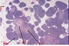
Adult Thymus (fatty) A - Fat B - cortex C - Hassall's Corpuscle D - Medulla |
|
|
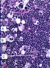
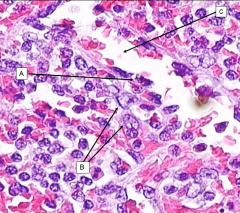
Thymus Cortex A- Thymocytes (immature CD4- and CD8-) B - Macrophage C - epithelial reticular cell |
|
|
Thymus Medulla Upper - Mature (CD4+ or CD8+) T cell Mid - Epithelial Reticular cell Lower - Hassall's Corpuscle |
|

|
Hassall's Corpuscle - concentric arrangement of epi. reticular cells and keratin. Found in thymus medulla |
|
|
Spleen Black - Capsule Red - White Pulp (lymphoid aggregate) Green - Red Pulp (cords) Blue and Orange - Sinuse Orange |
|
|
Spleen A - Central artery B - Periarterial Lymphatic Sheath (PALS) - contain T-cell C -Sinus |
|
|
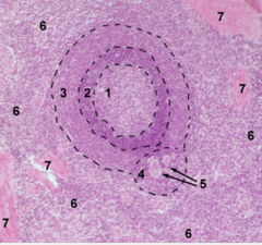
Spleen - white pulp 1 - Germ. center 2 - mantle 3 - Marginal zone 4 - PALS 5 - aterioles 6 - Red pulp 7 - trabeculae |
|
|
Spleen - red pulp A - Endothelial cell B -Macrophages C - SInus |
|
|
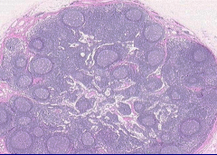
Peyer's Patch *GALT in ileum Arrow = germinal center |
|
|
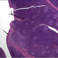
Tonsil (oral MALT) Upper arrow = crypt Lower arrow = epthelium |
|
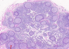
Sample taken from tonsils |
Benign Tonsilitis from an Acute Infection
A - Inflammatory Cells in epithelium B - Hyperplasia of lymphoid tissue |
|
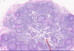
Sample taken from lymph node |
Chronic Lymphadenitis w/ Follicular Hyperplasia
*Increase in # of follicles and variety in size *Seen in RA and early HIV
|
|
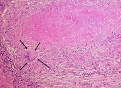
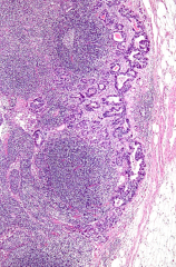
Taken from a mass in a lymph node |
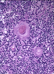
Necrotizing Caseous Granuloma
A - Lymphocytes B - Histeocytes (macrophages and epitheloid cells) C - Caseous (necrotic) material |
|
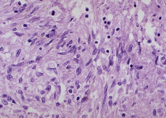
Taken from a mass in a lymph node |
Langhans-type giant cell in a Necrotizing caseous granuloma |
|
Taken from a mass in a lymph node |
Epitheloid Cells (activated macrophages) in a caseous granuloma *Have elongated nuclei and lots of pink cytoplasm |
|
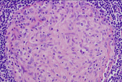
Taken from a mass in a lymph node |
Non-necrotizing caseous granuloma
*unlike necrotizing type, these granulomas have cells in center (not amorphous substance) |
|
Taken from a mass in a lymph node |
Metastatic carcinoma in subcapsular sinus *abnormal glandular tissue in capusle |
|
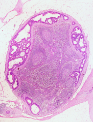
Taken from a mass in a lymph node |
Metastatic carcinoma in subcapsular sinus *abnormal glandular tissue in capusle |
|
Taken from mass in anterior mediastinum |
Thymoma
*Tumor of epithelial cells of thymus, usually benign |
|
Taken from mass in anterior mediastinum |
Thymoma
*Tumor of epithelial cells of thymus, usually benign |
|
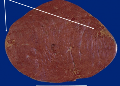
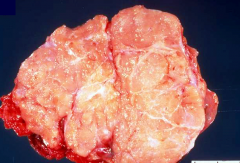
Cross section of a spleen. Spleen was measured to be 1,500 grams. |
Splenic Infarct and Splenomegaly (normal is 150g) |
|
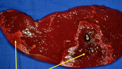
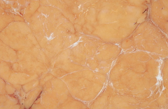
Cross section of a spleen |
Splenic infarct w/ coagulative necrosis |
|
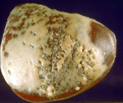
Spleen |
Perisplenitis - fibrous capsule that occurs after peritonitis or ascites |
|
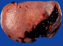
Spleen |
Lacerated spleen |