![]()
![]()
![]()
Use LEFT and RIGHT arrow keys to navigate between flashcards;
Use UP and DOWN arrow keys to flip the card;
H to show hint;
A reads text to speech;
41 Cards in this Set
- Front
- Back
|
Left Coronary artery |
anterior interventricular branch circumflex branch Supply: ventricles and left atrium |
|
|
Right coronary artery |
sinoartrial nodal branch atrioventricular nodal branch Right marginal branch -> right ventricle Posterior interventricular branch |
|
|
Caridac Veins |
Coronary sinus Great cardiac vein (from apex) into sinus Middle cardiac vein int posterior interventricular goove. Small cardiac vein (between right atrium and ventricle). Posterior vein of left ventricle. Left marginal vein |
|
|
Fetal Circulation |
Placent as connection between mom and baby. Umbilical Vein in the umbilical cord through the umbilicus. Left branch of the portal vein -> ductus venosus -> inferior vena cava. Inferior cava -> right atrium through foramen ovale -> left atrium -> left ventricle. Left ventricle throught aortic arch to head and arms -> superior vena cava -> right atrium. Right atrium -> inferio cava -> left ventricle. |
|
|
Fetal circulation |
Pulmonary trunk -> pulmo arteries -> pulmo veins -> left ventricle. Through ductus arteriosum -> aortic arch -> umbilical artieries back to placenta. |
|
|
Innervation of heart |
Autonomic Fibers Cardiac plexus -> symapa and para F: Electrical conducting system Atrial and ventricular myocardium Cornary vasculature |
|
|
Parasympathetic fibers |
Vagus N CN X F: Decrease in heart rate Decrease of cardiac contraction |
|
|
Sympathetic fibers |
T1 - T5 Left sympathetic trunk, Right sympathetic trunk F: Incresing heart rate Increasing force of contraction |
|
|
Function of Cardiac Skeleton |
Fibrous structural support for the heart chambers. F: Anchors heart valve cusps to interior walls. Prevents them from malfunctioning. F: atrioventricular valves and semilunar valves opened. F: Connects heart muscles and separates the artia from ventricles. F: Electrical insulator |
|
|
Anatomy of cardiac skeleton |
Fibers from collagen and elastic fibers. Made of four rings, two trigones and one ligament. |
|
|
Atrioventricular bundel |
Collection of heart muscle cells specialized for elevtical conduction. F: Transmits electrical impulses from AV node to the the apex via the bundle branches. |
|
|
Bundle of His |
Left and right bundle branches that run along the interventricular septum. Left Branch: left ant and left post fascicles. It gives of fibers call Purkinje fibers that give impulses to ventricular muscle. |
|
|
bundle of his |
|
|
Conducting system of Heart |
5 Elements Sinoatrial node Atrioventricular node Bundle of his, with left and right branches Terminal strands |
|
|
Sinoatrial Node |
Pacemakeer of the heart Location: Superior to sulcus terminalis of right atrium next to opening of vena cava. F: Governs sinus rhythm F: If it fails the atrioventricular node can become pacemaker. Inn: atria of the heart |
|
|
Atrioventricular node |
Continues the action potential L: right atrium, next to septal cusp of tricuspid valve. Inn: atria of the heart |
|
|
Bundle of His |
superior edge of interventricular septum to septal muscle portion of apex Divides into left and right bundles |
|
|
Right Bundle Path way |
Emerges in the right ventricular endocardium Near the base of the anterior paillary muscle |
|
|
Right Bundle Path way |
Emerges in the right ventricular endocardium Near the base of the anterior paillary msucle |
|
|
Left bundle of his |
Anterior & posterior fascicles Medial fascicle activates septal myocardium |
|

|
. |
|
|
Outer layer pericardiumq |
Fibrous layer, consists of dense connective tissue. Attached to central tendon of the diaphram via the pericardiacophrenic ligament. Attach to the ligaments of sternum. F: Stops the heart from overfilling.
|
|
|
Outer layer pericardium |
Fibrous layer, consists of dense connective tissue. Attached to central tendon of the diaphram via the pericardiacophrenic ligament. Attach to the ligaments of sternum. F: Stops the heart from overfilling. |
|
|
Serous layer |
Inner layer of pericardium. Direct contact with the pericardial fluid. Mesothelial layer (simple epethilium tissue) runs directly over the external surface of the heart known as visceral pericardium. |
|
|
Pericardial cavity |
Pericardial fluid in here. Allowing for the two surfaces to be lubricated and rub against another. |
|
|
Blood supply |
Pericardiocophrenic arteris Internal thoracic arteries
|
|
|
Blood suply |
Pericardiocophrenic arteris Internal thoracic arteries |
|
|
Inn of pericardium |
Phrenic N |
|
|
Mediastinum |
Area in midline of thorax that is surrounded by left and right pleural sacs. Divided into Superior and inferior mediastinum Inferior media: anterior middle and posterior |
|
|
Superior mediastinum |
First rib unti horizontal plane of T4 Esophagus, trachea and Thymus. Vagus N, Left recurrent layrngeal |
|
|
Anterior infreior mediastinum |
T4 until diaphragm at level T9 It extends posteriorly from sternum to fibrous pericardium. |
|
|
Middle inferior mediastinum |
T4 -T9/T10 Phrenic Nerve The heart and pericardium ascending aorta, pulmonary trunk |
|
|
Posterior inferior mediastinum |
T4 to T12 All of the above structure exept heart and pericardium. Azygos venous system, esophageal plexus and so on. |
|
|
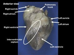
Anterior view of Heart (Structures) |
Aortic Arch Pulmonary trunk left pulmonary arteriy, and veins anterior interventricular sulcus coronary sulcus right atrial appandage cardiac apex left ventricle, right ventricle |
|
|
Posterior view of Heart (Structures) |
right pulmonary arteriy inferior vena cava coronary sinus right and left ventricle posterior interventricular sulcus Left and right pulmonary veins left and right atrium |
|
|
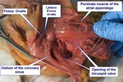
Right ventricle |
Supraventricular crest septomarginal trabecula Trabeculae carneae tricuspid valve chordae tendineae pailary muscles conus arteriosus pulmonary valve
|
|
|
Right ventricle |
Supraventricular crest septomarginal trabecula Trabeculae carneae tricuspid valve chordae tendineae pailary muscles conus arteriosus pulmonary valve |
|
|
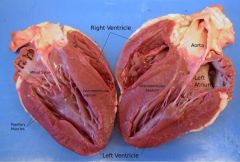
Left ventricle Aorta with left and right coronary artery Left coronary artery -> anterior inter and circumflex Thick walls Mitral valve |
|
|
|
Left ventricle Aorta with left and right coronary artery Left coronary artery -> anterior inter and circumflex Thick walls Mitral valve |
|
|
|
Left atrium Posterior Side 4 Pulmonary veins |
|
|
|
Right Venticle Much thinner walls Tricuspid valve |

|