![]()
![]()
![]()
Use LEFT and RIGHT arrow keys to navigate between flashcards;
Use UP and DOWN arrow keys to flip the card;
H to show hint;
A reads text to speech;
103 Cards in this Set
- Front
- Back
|
Name the fascia that surrounds entire neck and envelops the trapezius, SCM and parotid glands |
Investing fascia |
|
|
Cervical fascia consists of: (4 fascial planes) |
Investing Carotid Pretrachial Preverterbral |
|
|
Name the fascia: anterior aspect of neck; encloses thyroid, trachea, esophagus and infrahyoid (strap) muscles |
Pretracheal Fascia |
|
|
Name the fascia: surrounds the vertebral column and surrounding muscles |
Prevertebral fascia |
|
|
Name the facia: contains common carotid and internal carotid arteries, internal jugular vein and vagus nerve |
Carotid sheath |
|
|
In what fascia would you find the parotid gland? |
Investing fascia |
|
|
What fascia encloses the thyroid gland? |
Pretracheal fascia |
|
|
Why are the cervical fascia important? |
They are a route of infection.
eg. if inflammation occurs in the parotid gland, the investing fascia would stretch and this can become painful |
|
|
In what fascia would you find the muscles surrounding the vertebral column? |
Prevertebral fascia |
|
|
Which fascia encloses the vagus nerve? |
Carotid sheath |
|
|
What fascia encloses the internal jugular vein? |
Carotid sheath |
|
|
Function of the hyoid bone? |
1. attachment for anterior neck muscles
2. prop to keep airways patent |
|
|
Name the bones and cartilages of the neck |
Cervical vertebrae Hyoid bone Thyroid cartilage Cricoid cartilage Tracheal rings |
|
|
These parts are characteristic of which cervical vertebrae?
posterior tubercle, superior articular facet, facet for dens, transverse ligament |
Atlas (C1)
* recall Atlas has no body |
|
|
These parts are characteristic of which cervical vertebrae?
Spinous process, transverse process, transverse foramen, body |
Typical cervical vertebrae C3-C6 |
|
|
These parts are characteristic of which cervical vertebrae?
Spinous process, Superior articular facet, body of axis, facet for atlas, dens |
Axis (C2) |
|
|
Function of dens on Axis? |
attaches to body of axis |
|
|
Facet of dens on Atlas facilitates what motion? |
shaking head "no" motion |
|
|
Superior articular facet facilitates what motion? |
shaking head "yes" motion |
|
|
What "special joint" facilitates nodding "yes"? |
atlanto-occipital joint |
|
|
What "special joint" facilitates nodding "no"? |
atlanto-axial joint |
|
|
What structures would you find in the upper portion of the carotid sheath? |
internal carotid internal jugular vagus sandwiched between the above (external carotid has branched off, not here) |
|
|
What structures would you find in the lower portion of the carotid sheath? |
common carotid internal jugular vagus sandwiched between the above |
|
|
Which sheath would you find the vessels in your neck? |
Carotid sheath |
|
|
What is a possible clinical implication of the fascial planes of the neck? |
keeps things within fascial plane, but nothing stopping it vertically… connection between neck and mediastinum |
|
|
What happens in mediastinitis? |
Massive dental infections can track into retropharyngeal and alar spaces down to mediastinum... |
|
|
Mediastinitis can occur between what spaces? |
Retropharyngeal and alar "danger" spaces
- once an infection gets to these spaces, there is nothing stopping it from going to the heart |
|
|
What are the large branches of the carotid? |
Common carotid Carotid sinus Internal carotid External carotid |
|
|
What are the branches of the external carotid? |
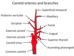
1. posterior auricular 2. occipital 3. superficial temporal 4. maxillary 5. facial 6. lingual 7. superior thyroid 8. ascending pharyngeal |
|
|
Superior thyroid meets with what artery from the subclavian subdivision? |
inferior thyroid artery from the thyrocervical trunk
|
|
|
What part of the carotid arteries has the following properties?
- mechanoreceptors for blood pressure |
Carotid body of the carotid sinus bulge |
|
|
Name the subclavian arteries and branches |
Subclavian has 1. a branch to common carotid from brachiocephalic 2. vertebral artery 3. thyrocervical trunk with branch to.... 4. inferior thyroid |
|
|
Name the veins of the neck |
Subclavian External jugular Internal jugular brachiocepalic
|
|
|
Thyrocervical trunk gives a branch to which artery? |
inferior thyroid |
|
|
The subclavian artery has which branches? |
thrycervical trunk with branch to inferior thryoid vertebral artery
|
|
|
The SCM is sandwiched between which two veins? |
internal jugular (deep) and external jugular (superficial)
... both of these go to brachiocephalic and then to the superior vena cava |
|
|
The neck is a transitional region, serving as conduit from the head to the rest of the body. What is a disadvantage of this setup? |
vessels are vulnerable to injury |
|
|
Which artery would most likely be damaged in a cut to the mid neck? |
Common carotid |
|
|
What does the thoracic duct drain? |
it collects most of the lymph in the body, other than the right thorax, arm, head |
|
|
The thoracic duct is the largest lymphatic vessel in the body, and is also known as the left lymphatic duct. Where does it drain? |
the thoracic duct drains to the left subclavian vein |
|
|
What does the right lymphatic duct drain? |
right thorax, arm, head |
|
|
The cervical plexus consists of? |
- ventral rami of C1-C4 - cutaneous nerves to neck, posterior scalp, upper part of thoracic wall - motor nerves to infrahyoid (strap) muscles - phrenic nerve to diaphragm - is deep to SCM and branches emerge from posterior triangle |
|
|
The cutaneous nerves in the neck are all sensory. Name the 4 branches of cutaneous nerves. |
Lesser occipital Greater auricular Transverse cervical Supraclavicular |
|
|
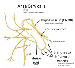
Name the 3 muscular branches of the cervical plexus. |
Ansa cervicalis (C1-4) Phrenic nerve (C3-5, mainly C4) Segmental branches (C1-4)
|
|
|
What does the ansa cervicalis (C1-4) innervate? |
- for C1 only - geniohyoid and thyrohyoid C1-3 - sternothyroid, sternohyoid, omohyoid
superior root = C1 inferior root = C2 + C3 |
|
|
What does the phrenic nerve (C3-5) innervate? |
- motor only to diaphragm - also supplies sensory and sympathetic branches along the way down - ventral rami of C3-5 |
|
|
What do the segmental nerves (C1-4) innervate? |
Anterior and middle scalenes |
|
|
What makes up the LOOP of the ansa cervicalis? |
|
|
|
What does the phrenic nerve travel along? |
anterior scalene muscle |
|
|
What 3 cranial nerves are found in the neck? |
Hypoglossal (CN XII) (motor to tongue)
Spinal accessory (CN XI) (SCM and trapezius muscles)
Vagus (CN X) (in carotid sheath sandwiched between internal carotid and internal jugular - goes to larynx, pharynx, heart, gut) |
|
|
What 3 sympathetic ganglions will you find in the neck? |
Superior cervical ganglion (anything that goes to the head and neck will synapse here!)
Middle cervical ganglion
Inferior cervical ganglion |
|
|
What does the hypoglossal nerve (CN XII) innervate? |
Hypoglossal (CN XII) (motor to tongue)
|
|
|
The spinal accessory nerve (CN XI) innervates what muscles? |
Spinal accessory (CN XI) (SCM and trapezius muscles)
|
|
|
The vagus nerve (CN X) in the neck goes on to innervate what? |
Vagus (CN X) (in carotid sheath sandwiched between internal carotid and internal jugular - goes to larynx, pharynx, heart, gut) |
|
|
Pupillary constriction, ptosis (drooping of eyelid), sunken eye, vasodilation and lack of sweating on face and neck are characteristic of what syndrome? |
Horner's syndrome.... lack of sympathetic supply to head and neck |
|
|
Which of the following nerves are not found in the neck? |
a. auriculotemporal <— innervates TMJ, symp to parotid |
|
|
What are the 2 landmark muscles of the neck and what nerve innervates them? |
SCM (sternocleidomastoid) Trapezius
Innervated by spinal accessory nerve (CN XI) |
|
|
Name the infrahyoid (strap) muscles |
sternohyoid sternothyroid thyrohyoid superior belly of omohyoid inferior belly of omohyoid |
|
|
Which two infrahyoid muscles have attachments on the oblique line of the thyroid cartilage? |
sternothyroid and thyrohyoid |
|
|
Why are omohyoid muscles given this name? |
They insert on the scapula of the shoulder (omo = shoulder) |
|
|
Is the sternohyoid deep or superficial? |
superficial and medially located |
|
|
Are the thyrohyoid and sternothyroid deep or superficial? |
deep and medially located |
|
|
Name the triangles of the neck. |
anterior posterior submental submandibular muscular carotid |
|
|
What are the borders of the anterior triangle? |
inferior border of the mandible anterior border of SCM midline of neck |
|
|
What are the roof and floor of the anterior triangle? |
Roof: subcutaneous fascia (contains platysma)
Floor: pharynx, larynx and thyroid gland |
|
|
What are the borders of the posterior triangle? |
posterior border of SCM anterior border of trapezius clavicle |
|
|
What are the roof and floor of the posterior triangle? |
Roof: investing fascia
Floor: - prevertebral muscles (lower, from cervical vertebrae): levator scapulae, scalenus medius, scalenus anterior
-splenius capitis and semispinalis capitis - intrinsic muscles of the back (upper)
- fascia (deep fascia, distinct from investing fascia) |
|
|
What muscle emerges deep from SCM and crosses the lower part of the posterior triangle en route to the scapula? |
inferior belly of omohyoid |
|
|
What nerves are found in the posterior triangle? |
- Accessory nerve (CN XI)
- branches of Cervical plexus - sensory nerves, as they emerge deep to the SCM (lesser occipital, greater auricular, transverse cervical, supraclavicular)
- phrenic nerve (C3-5) - crosses ant surface of scalenus anterior, deep to SCM (not strictly within triangle)
- brachial plexus - roots (C5-T1) emerge between scalenus anterior and medius - trunks (upper, middle, lower) are in lower part of triangle |
|
|
What vessels are found in the posterior triangle? |
arteries - subclavian and its suprascapular (with nerve supplies arm) and transverse cervical branches
veins - subclavian - receives external jugular before leaving triangle... external jugular receives suprascapular and transverse cervical veins
|
|
|
What nerve travels along the levator scapulae and pierces the trapezius muscle, and what does it innervate? |
spinal accessory (CN XI) - trapezius and SCM |
|
|
What nerve travels along the anterior scalene? |
Phrenic nerve |
|
|
You hit your friend on the neck just posterior to the SCM. He says he can't breathe, why? |
Could have hit the phrenic nerve that controls diaphragm, and cutaneous nerves there too that could produce numbness. |
|
|
Which triangle is not found bilaterally (on the midline)? |
submental triangle |
|
|
What are the borders of the submental triangle? |
anterior belly of the digastric (left and right) hyoid bone
|
|
|
What makes up the floor of the submental triangle? |
mylohyoid muscles (that attach to the inner aspect of the mandible and hyoid bone) |
|
|
What are the main contents of the submental triangle? |
submental lymph nodes |
|
|
What are the smaller divisions of triangles of the anterior triangle? |
submental submandibular muscular triangle carotid triangle |
|
|
What are the borders of the submandibular triangle? |
inferior border of mandible digastric (anterior belly) digastric (posterior belly) |
|
|
What does the submandibular triangle contain? |
stylohyoid muscle - attaches to styloid process (projection on base of skull) and inserts into the hyoid bone
superficial part of submandibular gland
submandibular lymph nodes
facial artery and vein |
|
|
What are the borders of the muscular triangle? |
midline superior belly of omohyoid anterior border of SCM |
|
|
What does the muscular triangle contain? |
infrahyoid (strap) muscles:
superior belly of omohyoid sternohyoid sternothyroid thryohyoid |
|
|
The superior belly of the omohyoid, sternohyoid and sternothyroid are involved in what action/movement? |
depression (lowering) of the pharynx and larynx during swallowing
supply: cervical plexus |
|
|
What action/movement does the thyrohyoid do? |
shortens the distance between the hyoid bone and thyroid cartilage, so it can either depress the hyoid bone or elevate the thyroid cartilage
supply: cervical plexus |
|
|
the sternohyoid and sternothyroid muscles lie over what structures? |
larynx, trachea and thyroid gland |
|
|
The borders of the carotid triangle are? |
posterior belly of digastric anterior border of SCM superior belly of omohyoid |
|
|
What are the contents of the carotid triangle? |
carotid sheath: common and internal carotid arteries, internal jugular vein, vagus nerve (CN X), part of the external carotid artery and some of its branches
Nerves: sympathetic truck is deep to this** - superior cervical ganglion |
|
|
which tri is bounded by ant and post bellies of
what is in that triangle? |
submandibular triangle
submandibular gland, submandibular lymph nodes, stylohyoid muscles, facial artery and facial vein |
|
|
subclavian vein puncture and internal jugular vein punctures are performed on which side of the body? |
right side - because superior vena cava is on right side |
|
|
In a subclavian vein puncture, what else do you worry about potentially puncturing? |
brachioplexus, apex of lung (pleura), subclavian artery... too deep, could get pneumothorax |
|
|
Why would you preferentially puncture the right internal jugular vein instead of the subclavian? |
Internal jugular is large and straight down, use SCM as reference.... can get into right side of heart if needed… use catheter for this after the needle. |
|
|
A cervical rib is a super-numary rib and can occur on the R/L or both sides. What can be squished here? |
subclavian artery is here and could get squished… brachioplexus gets squished
.. for these people, arm can go numb if they put arm up/can't put arm up very high |
|
|
Where is the thyroid gland? |
found between the thyroid cartilage and the 5th tracheal cartilage
|
|
|
This gland in the neck is responsible for cellular metabolism, and has two lobes joined in the anterior midline by an isthmus. What gland is it? |
thyroid |
|
|
What vessels supply the thyroid gland? |
superior thyroid artery (from external carotid) superior thyroid vein (from internal jugular)
inferior thyroid artery (from subclavian artery) inferior thyroid vein (from brachiocephalic vein)
- plexus of veins most at risk! |
|
|
How many parathyroid glands are there and where are they located? |
2 pairs - superior and inferior |
|
|
The parathyroid glands are endocrine glands (calcium and inorganic phosphate metabolism) on the posterior aspect of the thyroid glands... what vessels are they supplied by? |
same vessels as thyroid!
superior thyroid artery (from external carotid) superior thyroid vein (from internal jugular)
inferior thyroid artery (from subclavian artery) inferior thyroid vein (from brachiocephalic vein) |
|
|
What might be compressed by an enlarged thyroid (goiter)? |
surrounding trachea, so could compress airway, and other vessels |
|
|
Describe where a cricothyrotomy is performed... |
between rings 1 and 2
- in particular danger with veins that form plexus if you nick the thyroid gland
….eg. trach done around 7th ring is close to the arch of the aorta — if you get a bleed there… you are worried about carotid and subclavians |
|
|
The esophagus is a continuation of the pharynx... what level does it begin at? |
The cricoid cartilage - rests on prevertebral fascia, posterior to trachea - nerve supply and blood supply similar to the trachea |
|
|
Where does the trachea begin? |
lower border of cricoid cartilage |
|
|
what covers the trachea? |
pretracheal fascia (deep fascia) - attaches to laryngeal cartilages and below it merges with the pericardium |
|
|
What supplies the trachea (vessels and nerves)? |
inferior thyroid arteries inferior thyroid veins - front of trachea - makes a plexus before entering left brachiocephalic vein in thorax
recurrent laryngeal nerve
|