Reading...
![]()
Play button
![]()
Play button
![]()
Use LEFT and RIGHT arrow keys to navigate between flashcards;
Use UP and DOWN arrow keys to flip the card;
H to show hint;
A reads text to speech;
99 Cards in this Set
- Front
- Back
|
How many cervical vertebra?
|
7
|
|
|
How many thoracic vertebra?
|
12
|
|
|
How many lumbar vertebra
|
5
|
|
|
How many vertebra make up the sacram?
|
5
|
|
|
How many vertebra make up the coccyx?
|
4
|
|
|
Spinal Nerve C1 exits between what?
|
skull and C1 vertebra
|
|
|
Spinal nerve C3 exits between what?
|
C2 and C3
|
|
|
Spinal nerve C7 exits between what?
|
C7 and C6
|
|
|
Spinal nerve C8 exits between what?
|
C7 and T1, there is no C8 vertebra
|
|
|
Spinal nerve T1 exits between what?
|
T1 and T2
|
|
|
How many spinal nerves are found in the body?
|
62 (31 pairs)
|
|
|
How many cranial nerves are there?
|
12 pairs
|
|
|
Describe a typical neuron
|
cell body (nucleus and organelles)
axon (process conducting away from the cell body) dendrite (process conducting toward the cell body) |
|
|
In the central nervous system
-a cell body is called: -a neuronal process is called: |
nucleus (cell body)
tract (process) |
|
|
In the PNS
-cell bodies are called: -processes are called |
ganglion (cell bodies)
nerves (cell processes) |
|
|
Describe a unipolar neuron, when are they found?
|
1 process, found during development
|
|
|
Describe a Pseudo-unipolar neuron, where are they prevalent?
|
starts as 1 process, but branches into 2
found in the PNS |
|
|
What kind of neuron has 2 true processes?
|
Bipolar Neuron
|
|
|
Multipolar Neuron is used to describe what? Where are they found?
|
Any neuron with 2 or more processes
Found in both CNS and PNS |
|
|
Neurons that carry impulses away from the CNS
|
efferent neurons
(e.g. from CNS to muscle) |
|
|
Neurons taht carry impulses toward the CNS
|
afferent neurons
(e.g. from skin to brain) |
|
|
What are some somatic (or parietal) structures?
|
skeletal muscle
skin bones joints |
|
|
From what are somatic (or parietal) structures derived?
|
somites
|
|
|
What are the main functions of somatic structures
|
provide framework for body
concerned with protection and voluntary movement |
|
|
What are some visceral (or splenchnic) structures?
|
organs
glands blood vessels |
|
|
What are the main fucntions of visceral (aka splenchnic) structures?
|
concerned with involuntary activities
carry out internal functions (digestion, respiration, excretion) |
|
|
Four Functional Types of Neurons
|
GVE (General Visceral Efferent)
GVA (General Visceral Afferent) GSE (General Somatic Efferent) GSA (General Somatic Afferent) |
|
|
Mixed Nerves
|
have at least 1 efferent and 1 afferent neuron
|
|
|
What type of neuron carries impulses from skin to CNS?
|
GSA
|
|
|
What type of neuron is responsible for proprioception?
|
GSA (awareness of body position)
|
|
|
If your blood pressure drops, what type of neurons send a message to brain to increase heart rate?
|
GVA
|
|
|
What type of neurons are invovled in visceral reflexes?
|
GVA
|
|
|
What type of neurons carry motor impulses to skeletal muscle?
|
GSE
|
|
|
What type of neurons aid digestion, secretion, and cardiac activity?
|
GVE
|
|
|
Where are the cell bodies of GSA neurons found?
|
In Ganglia in the PNS
|
|
|
What is the structure of afferent neurons?
|
Pseudounipolar, with cell body in ganglia in PNS
|
|
|
What is the structure of efferent neurons?
|
most are multipolar
|
|
|
Where are cell bodies of GVE neurons found?
|
Both in CNS and in PNS (autonomic nervous system) Neuron originating in CNS synapses with cell body of neuron in PNS
|
|
|
What type of neuron has no cell bodies outside of the CNS?
|
GSE
Somatinc Efferent Motor Neurons Innervate skeletal muscle |
|
|
Neuroepithelial Cells of the Neural Tube proliferate into what 3 layers?
|
Ventricular (innermost layer of original cells)
Mantle Layer Marginal Layer (outermost) |
|
|
What is derived from the mantle layer of the neural tube?
|
mantle cells become the gray matter of the spinal cord
mantle cells clump into: 2 basal plates= that become the ventral horns 2 alar plats= that become the dorsal horns |
|
|
What is derived from the ventricular layer of the neural tube?
|
ependymal layer that lines the central canal
|
|
|
What is derived from the marginal layer of the neural tube?
|
undergoes myelination to beomce the white matter
|
|
|
Where does the spinal cord begin?
|
the spinomedullary junction (jxn with brainstem)
|
|
|
Where does the spinal cord end in adults? in infants? Why?
|
in adults it ends in the upper lumbar region (L1-L2)
in infants it may extend as far as L3 growth rate of spinal cord and vertebral column are not equal |
|
|
define conus medullaris, where is it found?
|
terminal part of the spinal cord
(upper lumbar region in adults L1-L2) |
|
|
What are the two swellings of the spinal cord?
Where are they found? |
Both are regions with lots of cell bodies that provide innervation to the limbs
Cervical Enlargement (C5-T1) Lumbar Enlargement (L1-S3) |
|
|
Gray Matter is organized into
|
Dorsal and Ventral Horns (sometimes lateral horns)
Contains neuron cell bodies |
|
|
White matter generall contains
|
axons (fiber tracts)
|
|
|
what is the meninges?
|
the 3 CT layers surrounding the spinal cord
dura mater arachnoid mater pia mater |
|
|
what is the epidural (extradural) space?
|
space between the dural mater and the vertebral canal
filled with fat contains the internal vertebral venous plexus |
|
|
where does the epidural space begin and end?
|
begins at the foramen magnum and ends inferiorly at the sacral hiatus
|
|
|
what is an epidural block?
|
local anesthesia can be injected into the epidural space to anesthetize the nerve roots
|
|
|
What forms the dural sac, where is it found?
|
formed by the dura
begins at the foramen magnum is continuous with the dura mater surrounding the brain extends to ~the S2 vertebral level |
|
|
what is the external filum terminale?
|
(coccygeal ligament) the extension of the dural sac below the sacral hiatus, anchored to the coccyx
|
|
|
what forms sleevelike projections around the spinal nerve roots as they exit the vertebral canal?
|
dural sac
|
|
|
Where is the subdural space?
|
*potential space* could contain blood
between dura and arachnoid mater |
|
|
Where is the arachnoid Mater
|
lines the dural sac (but is not attached to the dura)
|
|
|
Where is the subarachnoid space?
Where does it end? What does it contain? |
-located between the arachnoid mater and the pia mater
-extends as far as the dural sac (~S2) -contains CSF |
|
|
Where does one do a spinal tap? What layers does one go through?
|
between L3/L4 or L4/L5
goes through Supraspinous Ligament, Interspinous ligament, and Ligamentum Flavum, then through the dura mater and arachnoid mater to collect CSF from subarachnoid space |
|
|
define the internal filum terminale? where does it begin?
|
thin strand of pia mater that continues inferiorally beginning at the conus medullaris (L1/L2)
joins the external filum at the inferior limit of the dural sac (~S2) |
|
|
what are denticulate ligaments? what are they made of?
|
"Little teeth"
lateral extensions of pia mater fuse the dura and help anchor the spinal cord within the vertebral canal |
|
|
What is a spinal cord segment?
|
portion of spinal cord associated with each somite (each has a pair of associated spinal nerves)
|
|
|
what connects the spinal nerves to the spinal cord
|
rootelets (collectively dorsal root and ventral root)
|
|
|
What kind of neurons are in the dorsal root?
|
sensory neurons "sensory root"
|
|
|
What kind of neurons are in the ventral root
|
motor neurons "motor root"
|
|
|
Spinal Ganglion
|
aka Dorsal Root Ganglion
collection of sensory (GSA and GVA) neuronal cell bodies in the Dorsal Root |
|
|
Dorsal Primary Ramus (DPR)
|
branch of spinal nerve that goes to the back region, innervates the Intrinsic muscles of the back
|
|
|
Ventral Primary Ramus (VPR)
|
branch of the spinal nerve that travels around the body wall and is distributed primarily to the neck, trunk, and limbs
|
|
|
What is the major difference between Rami and Roots?
|
ROOTS: contain either sensory neurons (dorsal root) or motor neurons (ventral root)
RAMI: are mixed nerves, contain both sensory and motor neurons |
|
|
What are the "typical" spinal nerves? Why do they get that distinction?
|
T2-T12
They travel in typical spinal nerve pattern around body wall DO NOT FORM PLEXUSES |
|
|
What are the "Atypical" Spinal Nerves? What do they form?
|
The VPR of Spinal nerves in the Cervical, T1, Lumbar and Sacral regions form NERVE PELXUSES and travel out into the extremities
|
|
|
What is a nerve plexus?
From what is it formed? |
network or mixing of nerves
formed by VPR from the T1, Cervical, Lumbar and Sacral Spinal Nerves |
|
|
cauda equina
|
"horse's tale"
collection of nerve roots (corresponding to Lumbar and Sacral Vertebrae) extending below the conus medullaris travelling to their increasingly distant intervertebral foramina |
|
|
dermatome
|
somite derivative, area of skin that is innervated by the spinal nerve pair
(branches that supply skin are cutaneous nerves) |
|
|
Cutaneous nerves
|
nerves that innervate teh skin
contain SENSORY FIBERS (GSA) that innervate the skin itself and detect pressure, temperature, etc... and MOTOR FIBERS (GVE) that innervate structures found within the dermis of the skin (i.e. sweat glands, arector pili muscles, blood vessels, etc..) |
|
|
What type of neuron innervates skin of the arm? What does the fiber look like and where are its parts located?
|
GSA
Pseudounipolar receptor= dendrite in forearm cell body in dorsal root ganglion ends in dorsal horn |
|
|
afferent signals travel through which spinal nerve root?
|
dorsal root
|
|
|
Describe the path of a signal to a skeletal muscle in the back...
What type of neuron is involved? |
GSE (multipolar nueron)
begins in ventral horn travels through ventral root (efferent) goes through mixed spinal nerve and enters dorsal rami (innervating muscle in back) |
|
|
what type of neurons travel through spinal nerve
|
all 4 functional types
|
|
|
What determines which root a neuron travels through?
|
afferent "toward CNS" (dorsal root)
efferent "away from CNS" (ventral root) |
|
|
What determines which rami a neuron travels through?
|
DPR= true back
VPR= neck, trunk, limbs |
|

Identify The Following:
The L5 vertebral body Which nerve root exits the L4/L5 intervertebral foramen? |
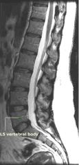
Lumbar Nerve 4
|
|
Identify the L2 spinal process
What typically ends near this vertebrae? |
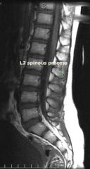
The spinal cord (the conus medullaris)
|
|
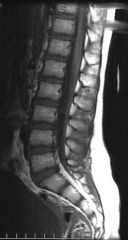
What is this section from?
Identify the dorsal root ganglia. |
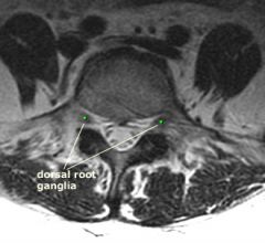
Caudal section of the spine, inferior to spinal cord, cauda equina visible
|
|
|
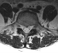
Ventral Root of Spinal Nerve
Dorsal Root of Spinal nerve Spinal Sensory (Dorsal Root) Ganglion White and Gray rami communicantes to and from sympathetic trunk |
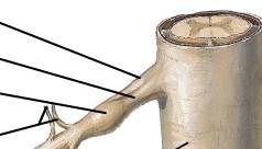
What are the marked structures?
|
|
|
What functional component consists of a two neuron chain?
|
GVE
General Visceral Efferent Neurons |
|
|
Cell bodies of GVA neurons are located where?
|
Dorsal Root Galglion (along with all afferent neuronal cell bodies)
|
|
|
Which of the following describes the correct number of spinal nerves?
A. 8 cervical, 12 thoracic, 5 lumbar, 5 sacral, 1 coccygeal B. 7 cervical, 12 thoracic, 5 lumbar, 5 sacral, 1 coccygeal C. 8 cervical, 12 thoracic, 5 lumbar, 4 sacral, 1 coccygeal D. 7 cervical, 12 thoracic, 4 lumbar, 4 sacral, 1 coccygeal |
A. 8 cervical, 12 thoracic, 5 lumbar, 5 sacral, 1 coccygeal
|
|
|
Which of the following branches of the spinal nerve typically forms nerve plexuses?
A. dorsal root B. ventral root C. dorsal ramus D. ventral ramus |
D. ventral ramus
|
|
|
Which functional component forms the autonomic system?
A. GSA B. GVA C. GSE D. GVE |
D. GVE
autonomic system is a visceral motor system |
|
|
Which of the following activities is NOT mediated by the parasympathetic nervous system?
A. sweating B. excretion C. digestion D. pupillary constriction |
A. Sweating
|
|
|
Where are the cell bodies of preganglionic sympathetic neurons located?
A. sympathetic chain ganglia B. brain C. spinal cord (regions T1-L2/L3) D. prevertebral ganglia |
C. spinal cord (regions T1-L2/L3)
|
|
|
Where are the cell bodies of preganglionic parasympathetic neurons located?
|
in the brain
and the sacral region of the spinal cord |
|
|
Preganglionic sympathetic nerve fibers enter the sympathetic trunk via the:
|
white ramus communicans
|
|
|
Which of the following is TRUE regarding GVA nerve fibers?
A. the GVA component consists of a preganglionic neuron and a postganglionic neuron B. GVA nerve fibers are either sympathetic or parasympathetic C. GVA nerve fibers often travel with autonomic fibers D. GVA nerve fibers synapse in the dorsal root ganglion |
C.
Although GVA fibers often travel with GVE fibers, they are not part of the autonomic system. Therefore, they do not consist of a two neuron chain, and there is no such thing as "sympathetic" or "parasympathetic" GVA neurons. The cell bodies of GVA neurons are found in the dorsal root ganglion, but a synapse does not occur here (since there is only one neuron in the GVA component). |
|
|
Where are the cell bodies of postganglionic parasympathetic neurons located?
|
A. in peripheral ganglia that are near the target organ
|
|
|
Where are the cell bodies of postganglionic sympathetic neurons located?
A. in sympathetic chain (paravertebral) ganglia B. in dorsal root ganglia C. in prevertebral ganglia D. both A and C are correct |
D. both A and C are correct
|
|
|
True or False?
Sympathetic neurons traveling in the gray ramus communicans (GRC) are always postganglionic |
TRUE
|