![]()
![]()
![]()
Use LEFT and RIGHT arrow keys to navigate between flashcards;
Use UP and DOWN arrow keys to flip the card;
H to show hint;
A reads text to speech;
27 Cards in this Set
- Front
- Back
|
Coulter principle - Impedance or direct current |
*counting & sizing cells *based on: *2 compartment container w/ aperture between *electrodes suspended from the top of each compartment connected to an ohmmeter measuring resistance between them *saline soln covers electrodes *cells are added to one side *spigot is opened *increase in resistance detected by ohmmeter as a pulse when a cell passes through aperture displacing electrolyte (saline soln) *# of cells (needle flicks) per mL can be measured *replace ohmmeter w/ a battery and oscilloscope-electrical pulse appears as spike on oscilloscope screen (height of spike is proportional to cell size) |
|
|
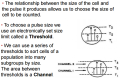
Thresholds & channels in cell counting |
|
|
|
Aperture impedence systen - sweep flow principle |
|
|
|
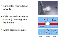
Hydrodynamic focusing principle |
*laminar flow ensures single file cell passage *coincidence effects minimized *diluted blood is injected into center of a "sheath" stream of buffered saline which is forced through a tapered flow chamber *electro-optical flow cytometer provides concurrent electronic & optical measurements |
|
|
Conductivity (radio/other high freq wave) principle |
*measures internal cell structures (nucleus & granules) using radiographic imaging similar to ultrasound *proprietary tech |
|
|
Laser light principle |
*light scatter measures cell surface granularity using broad range of angles, 60+angles of light scatter are analyzed |
|
|
Fluorescent flow cytometry principle |
*unique to Sysmex *fluorescent stain for nucleic acid & cytoplasmic organelles *measures fluorescence & side angle light scatter to differentiate cells *side fluorescence light: RNA/DNA info |
|
|
Histogram principle |
*each spike is 1 cell *spike height is proportional to cell size *grouped together into size categories |
|
|
Scatterplot/Scattergram principle |
Coulter: 3 probes (DC, RF & scatter) interrogate each of the cells simultaneously *every cell treated in the same manner and each is given an X, Y, & Z coordinate * all cell pops are directly measured Sysmex: measures forward scatter (size), side scatter (internal structure) & side fluorescence (RNA/DNA info) *placement is based on size and internal structure |
|
|
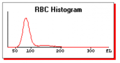
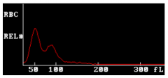
Normal RBC histogram |
|
|
|
Abn RBC histogram - cold agglutinins |
|
|
|
Abn RBC histogram - macrocytic cells, possibly dimorphic RBCs |

|
|
|
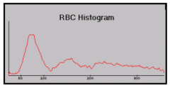
Abn RBC histogram - fragments (schistocytes, microcytes, giant plts, nrbcs) |

|
|
|
Abn RBC histogram - dimorphic RBCs (due to transfusion) |
|
|
|
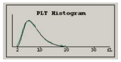
Normal plt histogram |
|
|
|
Abn plt histogram |

Top & middle: giant plts can show up in WBC histogram Bottom: small plts (shifted curve) |
|
|
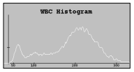
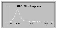
Normal WBC histogram |

|
|
|
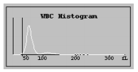
Abn WBC histogram - immNE1 & immNE2 |
|
|
|
Abn WBC histogram - lymphocytosis |
|
|
|
Abn WBC histogram - variant lymph |

|
|
|
Abn WBC histogram - immNE2 |

|
|
|
Abn WBC histogram - eosinophilia |

|
|
|
Abn WBC histogram - blasts |
|
|
|
Normal Dataplot (Coulter) |
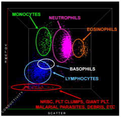
|
|
|
Abn Dataplot (Coulter) |

|
|
|
Normal Scattergram (Sysmex) |
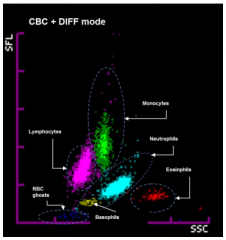
vs. Coulter *Neus & eos are lower *Baso more separate from lymphs |
|
|
Abn scattergram (Sysmex) |

|