Reading...
![]()
Play button
![]()
Play button
![]()
Use LEFT and RIGHT arrow keys to navigate between flashcards;
Use UP and DOWN arrow keys to flip the card;
H to show hint;
A reads text to speech;
89 Cards in this Set
- Front
- Back
|
What are the three types of white matter?
|
1. projection
2. association 3. commissural |
|
|
What is the centrum seminovale?
|
Areas of the white matter that contain all three white matter fiber types. (projection, association and commissural)
|
|
|
What are projection fibers?
|
Fibers that convey impulses either from the cortex to distant loci (efferent fibers) or from distant loci to the cortex (afferent fibers).
|
|
|
What are the corona radiate?
|
Projection fibers that are located near the cortex.
|
|
|
What is the internal capsule?
|
White matter projection fibers in the diencephalic area that funnel together.
|
|
|
What does the internal capsule consist of?
|
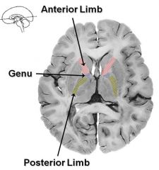
The anterior limb, posterior limb and genu
|
|
|
What major projection fiber pathways contain both efferents and afferents?
|
1. corona radiate
2. internal capsule |
|
|
What does the posterior limb of the internal capsule contain?
|
"Thalamocortical Radiations" and "Corticospinal Tract"
Ascending sensory fibers traveling from the extremities to the cortex via the thalamus, as well as the descending motor fibers going from the motor cortex to the spinal cord |
|
|
What does the genu contain?
|
"Corticobulbar Tract"
Descending motor fibers from the motor cortex to the brain stem. |
|
|
What are the optic radiations?
|
Convey visual information from the thalamus (lateral geniculate nucleus) to the striate cortex.
|
|
|
Where do the optic radiations convey information from? to?
|
They convey information from the lateral geniculate nucleus of the thalamus to the striate cortex.
|
|
|
What are association fibers?
|
Interconnect cortical regions of the same hemisphere either w/in the same lobe or from one lobe to another.
|
|
|
What does the arcuate fasciculus do?
|
Connects the receptive language area (Wernicke's area) to the expressive language area (Broca's area).
|
|
|
What are commissural fibers?
|
Interconnect cortical regions of the left and right hemisphere.
|
|
|
What kind of structure is the corpus callosum?
|
Commissural fibers of white matter.
|
|
|
What does the corpus callosum consist of?
|
Rostrum, genu, body and splenium.
|
|
|
What does the genu interconnect?
|
The rostral portions of the frontal lobes.
|
|
|
What does the body of the corpus callosum interconnect?
|
The rest of the frontal lobes and parietal lobes.
|
|
|
What does the splenium of the corpus callosum interconnect?
|
The temporal and occipital lobes.
|
|
|
What does the corpus callosum form the floor of?
|
The interhemispheric fissure and the roof of much of the lateral ventricles.
|
|
|
What does the anterior commissure interconnect?
|
The hippocampal formations.
|
|
|
What border does the posterior commissure of the corpus callosum mark?
|
The distal border b/e the midbrain and the diencephalon.
|
|
|
What is the pathway b/e the midbrain and the diencephalon important for?
|
Pupillary constriction to light.
|
|
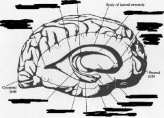
Who are we?
|
Internal capsule w/ anterior limb (blue), genu (red) and posterior limb (yellow)
|
|
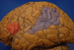
Who are we?
|
Broca's area/expressive language area (red) and Wernicke's area/receptive language area (grey)
|
|
|
What structure is important for the pupillary constriction to light reflex?
|
Posterior commisure
|
|
|
What are the basal ganglia?
|
Subcortical structures that are integrally involved w/ the motor system. They allow the cortex to select wanted patterns of movement and suppress unwanted patterns of movement.
|
|
|
Where in the brain is defective in Parkinson's disease?
|
Parkinson's disease is a movement disorder where unwanted movement is not suppressed so it is a disease of the basal ganglia.
|
|
|
Where is the basal ganglia derived from embryologically?
|
The telencephalon.
|
|
|
What telencephalic structures compose the basal ganglia?
|
The caudate nucleus, putamen and globus pallidus.
|
|
|
What structures compose the lentiform nucleus?
|
The putamen and globus pallidus.
|
|
|
What structures compose the striatum?
|
The caudate nucleus and the putamen
|
|
|
What does the striatum do?
|
Receives information from the cortex
|
|
|
What does the globus pallidus do?
|
It is the major source of output from the basal ganglia back to the cortex.
|
|
|
Where is the putamen?
|
The most lateral portion of the basal ganglia, lying b/e the globus pallidus (medially) and the external capsule (laterally)
|
|
|
What is the putamen connected to rostrally?
|
The caudate nucleus (striatum)
|
|
|
What does the caudate nucleus consist of?
|
A head, body and tail
|
|
|
Where is the head of the caudate nucleus located?
|
In the anterior horn of the lateral ventricle
|
|
|
Where are the body and tail of the caudate nucleus located?
|
In the inferior horn of the lateral ventricle.
|
|
|
Where does the caudate nucleus terminate?
|
In the amygdaloid nuclear complex.
|
|
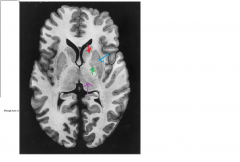
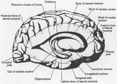
Who are we?
|
Red: Caudate nucleus
Blue: Putamen Green: Globus Pallidus Purple: Thalamus |
|
|
What is the globus pallidus?
|
The medial portion of the lenticular nucleus
|
|
|
What forms the lateral border of the globus pallidus?
|
The Putamen
|
|
|
What forms the medial border of the globus pallidus?
|
The internal capsule
|
|
Who are we?
|
|
|
|
Where is the substantia nigra located? What is the embryological origin of the substantia nigra?
|
In the midbrain and is of mesencephalic origin
|
|
|
What is the embryologic origin of the subthalamic nucleus?
|
Diencephalon
|
|
|
What disease is caused by degeneration of the substantia nigra?
|
Parkinson's disease
|
|
|
What does the limbic system consist of?
|
Consists of cortical and subcortical prosencephalic structures on the medial aspects of the hemispheres.
|
|
|
What does the limbic system do?
|
Controls emotional responsiveness and affective behavior resulting in an individualized interpretive response to external and internal stimuli.
It also plays an important role in incorporating new memory. |
|
|
What is the "limbic lobe"?
|
The hippocampus, parahippocampal gyrus and the cingulate gyrus
|
|
|
What does the Papez circuit do?
|
Necessary for incorporating new memory and is an important part of the limbic system.
|
|
|
What is the Papez circuit?
|
hippocampus => fornix => mammillary bodies => cingulate gyrus => ant. nucleus of the thalamus
|
|
|
Where is the major output of the limbic system?
|
Via the hypothalamus
|
|
|
What does the hypothalamus do?
|
The control center of the autonomic nervous system and neuroendocrine secretions.
|
|
|
What does the olfactory system consist of?
|
The olfactory bulb and tract, olfactory striae and the uncus (primary olfactory cortex)
|
|
|
Where is the primary olfactory cortex located?
|
In the uncus
|
|
|
What is the only primary sensory system that is able to bypass the thalamus and project directly to the cortex?
|
The olfactory system
|
|
|
What is the diencephalon?
|
A paired structure that lies on each side of the third ventricle.
|
|
|
What forms the lateral border of the diecephalon?
|
The internal capsule
|
|
|
Where is the diencephalon in relation to the midbrain?
|
Just rostral
|
|
|
What separates the midbrain from the diencephalon?
|
The posterior commissure.
|
|
|
What are the parts of the diencephalon?
|
1. epithalamus
2. thalamus 3. hypothalamus 4. subthalamus |
|
|
What does the epithalamus consist of?
|
Forms the dorsal part of the diencephalon and consists mainly of the pineal gland and the habenular nuclei.
|
|
|
What subdivision of the diencephalon has significant limbic connections?
|
The epithalamus
|
|
|
What part of the diencephalon is an important marker on CT scans b/c it calcifies?
|
The pineal gland.
|
|
|
What does the pineal gland do?
|
Secretes melatonin (a skin-lightening pigment)
|
|
|
What would a patient with a tumor of the pineal gland present with? what does the tumor compress?
|
The tumor compresses the pretectum and cerebral aqueduct which results in paralysis of upgaze and obstructive hydrocephalus.
*This is parinaud's syndrome |
|
|
Where is the thalamus located?
|
B/e the third ventricle and the posterior limb of the internal capsule.
|
|
|
Where do afferent impulses to the cortex first have to synapse before entering the cortex?
|
In the thalamus w/ olfaction being the exception.
|
|
|
What is the function of the thalamus?
|
Filters out irrelevant information from the body and only "talks" to the cortex when necessary.
It is the "executive secretary" of the brain. |
|
|
Where is the hypothalamus located?
|
Ventral to the thalamus
|
|
|
What forms the inferior and lateral walls of the third ventricle?
|
Hypothalamus
|
|
|
Where does the hypothalamus extend from and to?
|
Extends from the optic chiasm to the mammillary bodies
|
|
|
What is the major output center of the limbic system?
|
The hypothalamus by virtue of its two major functions (autonomics and neuroendocrines).
|
|
|
What is the neuroendocrine function of the hypothalamus?
|
Controls the release of anterior pituitary hormones into the blood.
|
|
|
What region of the brain controls various visceral activities such as feeding, drinking, sleeping, reproduction and thermoregulation?
|
The hypothalamus
|
|
|
What are the boundaries of the subthalamus?
|
Bound by the thalamus dorsally, the hypothalamus medially and the internal capsule laterally
|
|
|
What is the function of the subthalamus?
|
Forms an important functional part of the basal ganglia circuitry.
|
|
|
What does the subthalamus communicate with mainly?
|
The globus pallidus
|
|
|
What would be caused by a lesion to one of the subthalamic nuclei?
|
Would result in an involuntary movement disorder in the contralateral limbs known as hemiballismus.
|
|
|
What must all of the information "traffic" going to and from the cerebrum pass through?
|
The brain stem
|
|
|
Where do cranial nerves 3-12 originate?
|
The brain stem
|
|
|
What is the reticular formation? what is its function?
|
Forms the core of the entire brain stem is responsible for maintaining consciousness (it "wakes up" the cortex); maintaining general muscle tone and posture; processing noxious (painful) stimuli and regulating major visceral functions (BP, resp, GI function and cardiac function)
|
|
|
What is the midbrain? where is it located?
|
The smallest part of the brain stem. It lies b/e the pons and the diencephalon.
|
|
|
What marks the anterior surface of the midbrain?
|
The cerebral peduncles.
|
|
|
What pathway is the continuation of the motor fibers in the posterior limb and genu of the internal capsule?
|
The cerebral peduncles.
|
|
|
Where does the oculomotor nerve emerge? (CNIII)
|
B/e the cerebral peduncles in a space called the interpeduncular space.
|
|
|
Where does the trochlear nerve emerge? (CNIV)
|
The only cranial nerve which exits on the dorsal surface of the brainstem and is found lateral to the cerebral peduncles.
|