![]()
![]()
![]()
Use LEFT and RIGHT arrow keys to navigate between flashcards;
Use UP and DOWN arrow keys to flip the card;
H to show hint;
A reads text to speech;
44 Cards in this Set
- Front
- Back
|
This condition is characterized by an elevation of intraocular pressure (IOP) that results in progressive loss of retinal ganglion cells and their axons leading cause of blindness in the middle-aged dog. |
Glaucoma |
|
|
The pressure of the fluid inside the front or anterior chamber of the eye is known as the _________. |
intra-ocular pressure (IOP) |
|
|
Normal intraocular pressure for dogs & cats |
10-25 mmHg – normal (dogs & cats) |
|
|
Values of intraocular pressure for patients with glaucoma |
30 mmHg and higher |
|
|
Values of intraocular pressure that rapidly cause blindness, and are painful and cause the eye to stretch and enlarge |
above 50 mmHg |
|
|
It is the fluid that fills the anterior chamber of the eye. |
Aqueous Humor |
|
|
It is produced by the epithelial cells of the ciliary body, flows through the pupil, and circulates in the anterior chamber. |
Aqueous humor |
|
|
Optically clear, but contains various compounds such as carbohydrates, amino acids, glucose, lactate, small proteins, ions, and urea, which provide the nutritional support to the avascular lens and cornea. |
Aqueous humor |
|
|
Drainage of AH occurs through the _________ which is a complex structure formed by the junction of the peripheral iris and cornea. |
iridocorneal angle (ICA) |
|
|
This determines the intraocular pressure (IOP). |
Balance between AH formation and drainage |
|
|
It is caused by inadequate drainage of aqueous fluid when this angle, or drain, is obstructed for any reason, aqueous humor builds up inside the eye and results in increased IOP. |
Glaucoma |
|
|
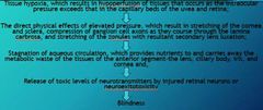
Pathophysiology of glaucoma |
|
|
|
Classification of glaucoma |
▫Primary glaucoma ▫Secondary glaucoma |
|
|
It is due to an inherited malformation in the ICA that, over time, leads to a reduced capacity for AH drainage without any other underlying cause. Usually begins in one eye, but in most patients it eventually involves both eyes, leading to complete blindness. |
Primary glaucoma |
|
|
This results in increased intra-ocular pressure due to disease or injury to the eye. |
Secondary glaucoma |
|
|
Types of primary glaucoma |
◾Primary open-angle glaucoma ◾Angle closure glaucoma ◾Goniodysgenesis |
|
|
The reduced outflow of aqueous humor in POAG is caused by reduction in the function of the trabecular meshwork, raising IOP |
Primary open-angle glaucoma |
|
|
Genes affected by mutation in primary open-angle glaucoma |
✔ADAMTS10 (Beagles and Norwegian Elkhounds) ✔ADAMTS17 (Petit Basset Griffon Vendéen and Basset Hound) |
|
|
It is painless and gradual development of blind spots or loss of vision over a long period of time. |
Primary open-angle glaucoma |
|
|
The aqueous does not reach the trabecular meshwork because access to the anterior chamber angle is obstructed by the iris or sometimes other tissues (some secondary glaucomas), leading to elevated IOP levels-associated with pectinate ligament dysplasia (PLD), an abnormality affecting the iridocorneal angle that has been shown to be highly heritable. |
Angle closure glaucoma |
|
|
The onset of this type of primary glaucoma is characterised by a very rapid (acute) increase in intraocular pressure, often literally overnight, that is extremely painful and frequently leads to sudden and irreversible blindness. |
closed angle glaucoma |
|
|
This is also known as Pectinate Ligament Dysplasia (PLD) |
Goniodysgenesis |
|
|
It is a congenital Iridocorneal angle abnormality in which there is a lack of fenestration of the iridocorneal angle (ICA) during development of the eye. |
Goniodysgenesis |
|
|
Breeds genetically predisposed with Open angle glaucoma |
🐶Beagle 🐶Great Dane 🐶Keeshound 🐶Norwegian Elkhound 🐶Poodle (Toy and Miniature) 🐶Samoyed 🐶Siberian Husky |
|
|
Breeds genetically predisposed with closed angle glaucoma |
🐶Akita 🐶American Cocker Spaniel 🐶Basset Hound 🐶English Cocker Spaniel 🐶English Springer Spaniel 🐶Flat Coated Retriever 🐶Golden Retriever 🐶Poodle (Toy and Miniature) 🐶Samoyed 🐶Shar Pei 🐶Welsh Springer Spaniel |
|
|
Breeds genetically predisposed with goniodysgenesis |
🐶Basset Hounds 🐶Chow Chows |
|
|
This is the most common cause of secondary glaucoma: |
✔anterior lens luxation (movement of the entire lens into the front of the eye)✔uveitis (inflammation inside the eye)✔advanced cataracts✔intraocular masses ✔trauma to the eye✔Tumors✔Anterior dislocation of lens ✔Intra-ocular bleeding✔Pigmentary glaucoma |
|
|
TRUE OR FALSE. Glaucoma in cats is usually secondary to chronic uveitis |
True |
|
|
Although primary glaucoma is less commonly seen in cats, breeds at risk include: |
🐱Siamese 🐱Burmese 🐱Persian 🐱Domestic shorthairs |
|
|
Duration of glaucoma |
▫acute ▫chronic |
|
|
It is defined as an elevation in IOP of < 24-48 hours duration. If patients are treated during this phase, vision may be salvageable. |
Acute glaucoma |
|
|
This develops after the IOP elevation is sustained for days or longer. Medical therapy may be effective at reducing the IOP, however vision cannot be regained. |
Chronic glaucoma |
|
|
Early clinical signs of glaucoma |
-sluggish to slightly dilated pupils -mild bulbar conjunctival venous congestion -early enlargement of the eye (buphthalmia) |
|
|
Classic signs of glaucoma |
Hazy / cloudy cornea Dilated pupil Episcleral injection Buphthalmos Optic nerve cupping |
|
|
Signs of glaucoma |
✔Corneal edema ✔Dilated pupil ✔Episcleral and conjunctival blood vessel congestion ✔pain ✔blindness ✔enlargement of the globe corneal ✔insensitivity ✔cupping of the optic disc ✔thinning of the retina causing hyperreflectivity especially around the optic disc. ✔Corneal striae (Haab's striae) |
|
|
The cloudy appearance is caused by interference with the pumping mechanisms of the corneal endothelial cells. |
Corneal edema |
|
|
TRUE OR FALSE. The high pressure causes the pupil to dilate and be less responsive to light, and dilation is thought to be due to neurologic or vascular damage to the ciliary body and iris as well as impaired retinal and optic nerve function. |
True |
|
|
In this condition, the pressure elevation causes blockage of normal venous drainage and engorgement of anterior ciliary veins. |
Episcleral and conjunctival blood vessel congestion |
|
|
These are cracks in Descemet's membrane from stretching of the globe. Their presence indicates previous glaucoma regardless of the current IOP. |
Corneal striae (Haab's striae) |
|
|
TRUE OR FALSE. By the time eye enlargement, cupping of the disc, or hyperreflectivity of the retina are obvious, there is essentially no chance for return of sight. |
True |
|
|
This tool is useful in the treatment and monitoring of intraocular pressure elevations associated with the disease process. |
Tonometer |
|
|
This procedure helps determine how predisposed the remaining visual eye is to develop glaucoma when primary glaucoma is present in the other eye. |
Gonioscopy |
|
|
A special contact lens used in gonioscopy of the eye, which allows examination of the drainage angle. |
“goniolens” |
|
|
Tx for glaucoma |
✔Osmotic Diuretic: dehydrate to vitreous—used in emergency therapy only; crucial that the patient is know to be capable of producing urine prior to using osmotic diuretics.✔Miotics: increase outflow of aqueous typically by opening the drainage angle✔Carbonic Anhydrase Inhibitors: decrease production of aqueous✔Autonomic Agents: Sympathomimetic--(increases aqueous outflow; reduces aqueous humor production). ✔Beta-blockers reduce the formation of AH via their effects on beta receptors present in the ciliary body. Undesirable cardiac and respiratory effects can be seen with topical beta-blockers, including bradycardia, and bronchoconstriction. Thus, these medications should be avoided in patients with cardiovascular disease and asthma. ✔Chemical Ciliary Body Ablation / Intravitreal Gentocin Injection: indicated for blind, painful eyes. The most commonly described procedure involves an intravitreal injection of gentocin, which is aimed at pharmacologic ablation of the ciliary body. This in turn causes decreased production of aqueous. |