Reading...
![]()
Play button
![]()
Play button
![]()
Use LEFT and RIGHT arrow keys to navigate between flashcards;
Use UP and DOWN arrow keys to flip the card;
H to show hint;
A reads text to speech;
15 Cards in this Set
- Front
- Back
|
Describe the teeth in the oral cavity and the muscles of chewing?
|
The incisors cut food and the molars crush food which mixes with saliva to form a bolus that can be swallowed. Muscles of mastication include the masseter innervated by the trigeminal nerve (cranial nerve V).
|
|
|
Describe the structure of the oropharnx and oesophagus and outline their respective functions.
|
The oropharynx extends from the uvula to the level of the hyoid bone. It transmits food as well as air to the trachea and oesophagus. When swallowing is occurring, the epiglottis closes over the glottis, sealing of the larynx, to prevent aspiration into the lungs. The oesophagus is a 25cm tube ( mouth to cardia = 40cm – needs to be know when inserting nasopharyngeal tubes).
They transmit food to the stomach. |
|
|
Describe the contents and functions of the saliva.
|
Saliva is not essential to eat but is essential for protection of the oral cavity. 1.5l of salvia are produced each day. The roles include:
1. Release amylases which begin digestion of carbohydrates 2. Aids to protect the oral environment by keeping it moist and relatively alkaline 3. Aids swallowing by helping to form a bolus Also: 4. Neutralises acid secretions from bacteria 5. Lubricates and wets food 6. Keeps non keratinsed stratified squamous epithelium of mucosa moist 7. Washes teeth 8. Maintains high calcium concentration – limits extent to which teeth dissolve into saliva Without salvia (zerostomia), moist food can still be eaten but dental caries may occur (teeth decay) and mucosa will decay. Saliva is an innate physical mechanism of defence in the GI. It contains lysozyme, lactoperoxidase, complement, IgA and polymorphs and washes toxins including bacteria down into the stomach. |
|
|
Define zerostomia.
|
Zerostomia is where no saliva is produced. It may be caused by nerve damage eg glossopharyngeal nerve or otic ganglion. The individual can still eat moist food but dental hygiene will be decreased leading to dental caries ( breakdown of bone) and infections within oral cavity. It will be more difficult to chew and swallow as food bolus not as well formed and it may be difficult to speak. ( sijorens syndrome).
|
|
|
Name 3 situations when saliva production would be decreased.
|
- Dehydration
- Muscarinic antagonists eg mebeverine used to treat irritable bowel syndrome - Disease such as sjorgren syndrome – autoimmune disease, inflammation in lacrimal glands and saliva glands causing dry mouth – xerostomia |
|
|
List the constituents of saliva.
|
Water, electrolytes, HCO3, bacteriostats, mucus, enzymes.
Saliva is a hypotonic solution comprised mainly of water with Na and Cl usually at lower levels than plasma and K, Ca2+, I are at higher levels than plasma. It is an alkaline solution and [HCO3] is greater than in the plasma. It comprises of amylases for digestion of carbohydrates, lysosomes and lactoperoxidase, for bacterial digestion, mucus for lubrication and bacteriostats to limit growth of bacteria. As well as complement, IgA and polymorphs. Saliva washes toxins including bacteria down into the stomach |
|
|
List the components of saliva secreted by each pair of salivary glands
|
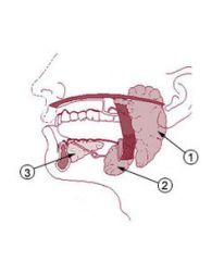
The ducted exocrine glands have central acini which lead to a system of ducts. The acinar and duct cells have different functions.
Submaxillary glands (under cheeks): mixed serious and mucous saliva – serous demilunes present. 70% of secretions Parotid glands (neck): largest glands found overlying mandibular ramus anterior and inferior to the external acoustic meatus. it releases serous saliva– watery secretions with lots of enzymes but little mucus. 25% of secretions although is the largest gland Sublingual (under tongue): mucus saliva- no enzymes but lots of mucus. 5% of secretions. |
|
|
Explain the mechanisms of secretion of serous saliva.
|
Saliva is ALWAYS hypotonic but must be made from more concentrated extracellular fluid. There is no mechanism that secretes water. A concentrated solution is secreted and ions are removed to make it dilute.
1. The acinar cells determine the volume of salvia. They actively secrete ions and water follows producing an isotonic solution to the plasma: same [Na],[K],[HCO3], slightly more[I] and slightly less [Cl]. 2. The composition of saliva is determined by ductal modification. The duct cells remove Na in exchange for K and HCO3. Water can’t follow due to tight junctions between ductal cells. The duct cells secrete some K into the salvia. The [HCO3] falls when resting but duct cells secreted more when stimulated due to food (sight, smell). Duct cells have a maximum rate of modification and the faster saliva is produced, the less modified it is as it is not exposed to cells for long. Therefore it is very salty when produced quickly ie like sweat in hot environment as sodium ions have not been removed . |
|
|
Compare resting saliva and stimulated saliva.
|
Resting saliva: it has a smaller volume( acini not stimulated) which has been highly modified by ductal cells and is thus very hypotonic. It is neutral or slightly acidic
- Na: 7-10 - K: 20 (leaks into saliva due to Na K atpase) - A few enzymes Stimulated saliva: larger volume which has been less modified by ductal cells and is thus less hypotonic. It is very alkaline (50-60mmol/l) - Na : up to 80mmol.l-1 - K: 8-10mmol.l1 - Lots of enzymes |
|
|
Describe the control of salivary secretion.
|
Volume is controlled by activity of acinar cells and composition is controlled by ductal cells.
It is controlled by the ANS. Sympathetic fibres via the superior cervical ganglion travel via the middle meningeal artery ( maxillary artery) and passes through the otic ganglion in the infratemporal fossa to hijack onto auriculotemporal nerve ( V3) to supply parotid gland and causes reduced blood flow to acinar cells so that they don’t have enough energy to actively pump out ions into the ducts. Viscous mucus (less water) is produced. This causes a dry mouth Parasympathetic activity: - Medulla oblongata receives afferents from sensory structures – mouth, tongue, nose ( cephalic phase- prepares GI for ingestion) and from conditional reflexes eg pavlars dogs - Releases efferent impulse from inferior salvatory nucleus via tympanic branch of glossopharyngeal nerve(9th cranial) and to lesser petrosal nerve which synapses in otic ganglion and post-synaptic PSNS fibres hijack onto the auriculotemporal nerve to the parotid gland to the acinar glands. Here it releases acetylcholine which acts on M3 receptors, increasing volume of saliva. It also acts on duct cells triggering release of more HCO3. Co transmitters also stimulate increased blood flow. the submandibular and sublingual gland are innervated by differnet routes - postganglionic sympathetic fibres from superior cervical ganglion enter head via the facial artery and pass through the submandibular ganglion to supply the glands - preganglionic parasymapthetic fibres from superior salvatory nucleus attach to the chorda tympani nerve ( facial nerve) and then travel via the lingual nerve ( V3) to synapse in the submandibular ganglion to form post ganglionic PS fibres that innervate the glands: SUBMANDIBULAR, UBLINGUAL AND MUCOSAL GLANDS OF PALATE |
|
|
Describe the processes of swallowing
|
Voluntary phase: mastication produces a bolus which is pushed into the pharynx and triggers the swallowing reflex.
Pharyngeal phase: pressure receptors in palate and anterior pharynx relay signals to the swallowing centre in the medulla oblongata to stop breathing to avoid aspiration of food. This causes the larynx to raise, the glottis to close and the upper oesophageal sphincter to open. Oesophageal phase: the upper 2/3 is skeletal muscle and the lower 1/3 is smooth muscle. The smooth muscle performs peristalsis to transport the bolus towards the stomach which only takes 9 seconds. It is co-ordinated by the vagus nerve which synapses with neurones in the myenteric plexus (enteric NS). The lower oesophageal sphincter then opens to allow entry into cardia of stomach. |
|
|
Describe the anatomical relationships of the oesophagus and how disordered swallowing may occur as a consequence of primary oesophageal disorder or a condition in a closely related structure.
|
The oesophagus is 25cm long extending from the cricopharyngeal sphincter to the cardiac orifice. The upper 1/3 comprises of striated skeletal muscle and the lower 2/3 comprises of smooth muscle which performs peristalsis to force food down into the cardia of the stomach in 9 seconds. The trachea lies anterior to the oesophagus.
A primary oesophageal disorder: - Strictures within the oesophagus caused by fibrous rings or from gastric acid erosions - Achalasia – failure of lower oesophageal sphincter to relax - Oesophageal squamous cell carcinoma Condition in closely related structure - Gastric adenocarcinoma in cardia of stomach - Oropharyngeal squamous cell carcinoma - Bronchial carcinoma If dysphagia is due to malignancy the patient might also complain about cachexia – weight loss, haematemessis , malaise, malaena . |
|
|
Differentiate between oropharyngeal dysphagia and oesophageal dysphagia.
|
Dysphagia is the difficulty in swallowing which may refer to food or fluids.
Difficulty in swallowing liquids is called oropharyngeal dysphagia and may be caused by - bulbar palsy – damage to the swallowing centre in the medulla oblongata eg guillian barre syndrome - pseudobulbar palsy – damage to nerves in the cortex leading to or away from the swallowing centre eg stroke, parkinsons Therefore there is no stimulation that fluid is present and so breathing is not stopped and aspiration can occur. Food would be easier to swallow as it would stimulate peristalysis in oesophagus, stimulating the swallowing centre. The swallowing reflex is usually triggered by fluid and food hitting receptors in the palate and anterior pharynx. This signals the medulla oblongata which stimulates breathing to stop so that food and fluid do not go down the trachea, raises larynx, closes glottis, opens upper oesophageal sphincter. The food bolus moves down the oesophagus by peristalsis. The inner circular smooth muscle contracts to form a ram behind the bolus and the longitudinal smooth muscle contracts the squeeze the bolus down the tube. The transit time to the stomach is about 9secs. Oesophageal dysphagia is difficulty in swallowing solids. There must be a physical blockage or narrowing of the oesophagus which obstructs the passage of the bolus. - Oesophageal adenocarcinoma/squamous cell carcinoma - Carcinoma of cardiac part of stomach preventing the opening of the lower oesophageal sphincter - Cancers outside of the oesophagus obstructing the oesophagus - bronchial carcinoma - Cancer of the oropharynx - Strictures of the oesophagus caused by chronic acid reflux |
|
|
What investigations may be useful to reveal the cause of oesophageal and oropharyngeal dysphagia.
|
Oesophageal dysphagia: upper GI endoscope or Barium swallow – will show dilated proximal oesophagus
Oropharngeal dysphagia: Video fluoroscopy because it allows all the phases of swallowing to be evaluated which is particularly useful when assessing neurological disease. |
|
|
Define odynophagia and name possible causes of it.
|
Odyngophagia is painful swallowing and may be caused by oesophagitis due to oesophageal candidiasis, peptic ulceration or cancers within oropharynx or oesophagus.
|