![]()
![]()
![]()
Use LEFT and RIGHT arrow keys to navigate between flashcards;
Use UP and DOWN arrow keys to flip the card;
H to show hint;
A reads text to speech;
23 Cards in this Set
- Front
- Back
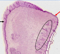
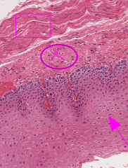
Lip tissue |
Black = Keratinized Stratified squamous epithelium of external lip * Notice the Orbicularis oris MM at bottom left
Red = NON-keratinized stratified wet squamous epithelium of oral mucosa *circle = salivary glands in submucosa
|
|
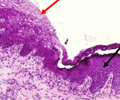
Taken from lip |
Vermillion Zone
Transition from external keratinized epithelium of skin (red) to internal non-keratinized epithelium of oral mucosa (Black)
*Vermillion zone lacks salivary glands and thus needs to be continuously moistened |
|
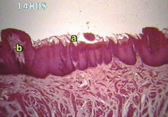
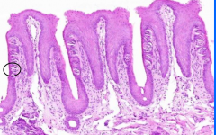
Dorsal Tongue |
A = Filliform Papillae = Rough keratinized papillae w/ no taste buds
B = Fungiform Paillae = rounded/elevated non-keratinized papillae w/ taste buds on upper surface |
|
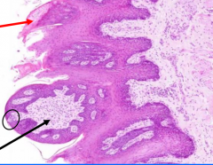
Dorsal Tongue |
Black = Fungiform Papillae with taste bud at surface (circle)
Red = Filliform Papillae (lack taste buds) |
|
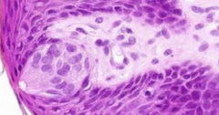
Dorsal Tongue from papillae upper surface |
Taste bud at the upper surface of a fungiform papillae |
|
Dorsal Tongue |
Circumvallate Papillae - taste buds on lateral wall
*Located in a V-shaped line at posterior 1/3 of tongue |
|
Tongue |
Foliate Papillae
*Vertical Ridges on sides of tongue *Taste buds on dorsal surface (circle) *Occur in rows (vs fungiform that occur singly) |
|
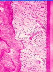
Taken from tooth |
Peridontal Ligament
*made of Sharpey's fibers of collagen that connect alveolar bone to to cementum *shock absorbers |
|
esophagus |
Mucosa Layer of Esophagus, comprised of...
1) non-keratinized stratified squamous epipthelium (arrow) 2) Lamina propria (circle) of loose CT and immune cells 3) Muscularis mucosa (box) |
|
|
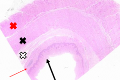
Esophagus Layers
*Black arrow = Stratified squalmous epithelium of mucosa *Red arrow = lamina propria (indicated by lymphoid nodule) *white X = submucosa CT *Black X = Inner circular smooth mm of muscularis externa *Red X = Outer longitudinal smooth mm of muscularis externa |
|
|
Myenteric (aka Auerbach) Nerve Plexus
*located b/w inner circular and outer longitudinal layers of the muscularis externa *regulates peristalsis |
|
|
Where are the Meissner's nerve plexi located and what are their function? |
Submucosa of Esophagus
*fxn to regulate glandular secretions |
|
|
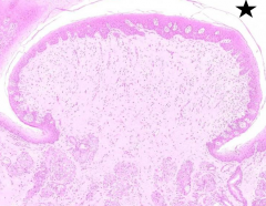
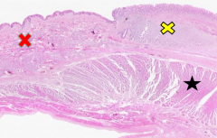
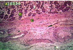
Esophageal-Gastric Junction
*Esophagus (red X) shows 3 layers of mucosa, submucosa, and Muscularis externa *Cardiac Stomach (yellow x) is pale staining glandular tissue *LES = black star |
|
|
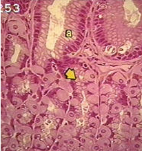
Stomach Fundus
A = mucous surface cells (line the most superficial part of gastric pit.
Arrow = Mucous neck cells (w/ interspersed parietal cells) |
|
|
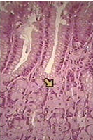
Parietal Cell
*Located in neck of gastric gland - spherical, often binucleate *Secrete H+ via intracellular canalicculi as well as intrinsic factor to find cobalamin in SI |
|
|
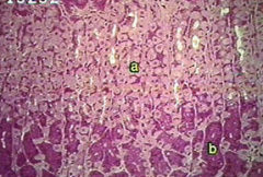
A = parietal cell
B = Cheif Cell - located at base of pit, basophilic cytoplasm due to large amount of RER to make pepsinogen |
|
|
Enteric Endocrine Cell
*scattered along gastric gland to release endocrine and paracrine substances *Can be closed or open type |
|
|
A = pareital cell B = cheif cell C = Muscularis mucosa |
|
|
What stomach layer contains Lymph vessels, aa, VV?
Gastric pits and stem cells? |
1) Submucosa
2) Mucosa |
|
|
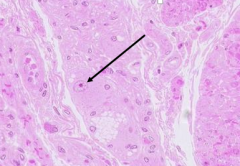
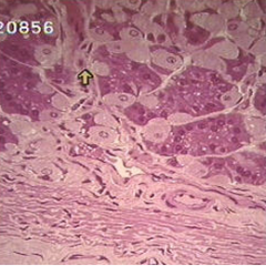
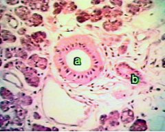
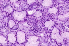
Parotid Gland - completely serous gland
A = striated gland - process initial secretions *serous acini to left of A B = intercalated gland
Path of Drainage = acini -> striated -> intercalated |
|
|
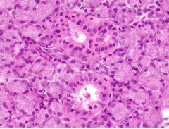
Parotid
*all serous acini |
|
|
Submandibular Gland
Mostly Mixed Acini (mucinous w/ serous demilunes at tips)b w/ some scattered serous |
|
|
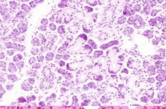
Sublingual
*Mostly mucinous glands w/ few scattered mixed acini |