![]()
![]()
![]()
Use LEFT and RIGHT arrow keys to navigate between flashcards;
Use UP and DOWN arrow keys to flip the card;
H to show hint;
A reads text to speech;
52 Cards in this Set
- Front
- Back
|
What is anatomy? |
It is the study of structures that can be seen without a microscope, microscope is for histology. It can be seen with the naked eye. It is the basis for medicine |
|
|
The two primary techniques students should use when studying anatomy! |
Observation and visualization |
|
|
Shortcut for anatomy definition |
Anatome= Up(ana) + Cutting (tome) |
|
|
Two Major types of anatomy and examples on them |
Regional: parts of the body like head, neck, abdomen, lower and upper limb.old
Systemic: systems in the body like respiratory, cardiovascular and urinary systems. New |
|
|
Other types of anatomy other than the 2 major types |
1) development anatomy (general embryology and systemic embryology) 2) Applied anatomy (clinica, where they feel pain) 3) radiological anatomy (CT, x-ray, MRI) 4) surface anatomy (without cutting the body) e.x measuring the heart rate |
|
|
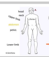
Body region names |
Head and neck Abdomen and pelvis Lower limb Upper limb Thorax Brain and spinal cord |
|
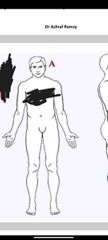
Name the body regions on this body |
|
|
|
What are the anatomical position of the human body |
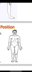
1) body is erect (person is standing) 2) face directed forward 3) limbs at the side of the body 4) legs and feet close together 5) palms directed forward
|
|
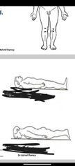
Difference between supine and prone |
Prone is laying on the stomach supine is laying on the back |
|
|
Median plane or midsagittal plane is |
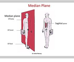
Vertical in Medline, which means it divides the into right and left equal parts. |
|
|
Coronal plane |
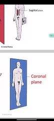
Vertical plane perpendicular to the median, divides the body to anterior and posterior parts, unequal. |
|
|
Horizontal (transverse) |
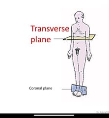
Perpendicular to the median and coronal plane, divides the body into upper and lower parts. |
|
|
Parasagittal plane |
Vertical, parallel to the median. Whichever side this plane is on that plane is smaller than the other side |
|
|
The four anatomical plan |
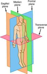
Frontal is coronal |
|
|
Difference between medial and lateral |
Medial is closer to the medial plane (middle line). Lateral further away from the medial line. |
|
|
Difference between anterior and posterior |
Anterior ( ventral): towards the front of the body. Posterior (dorsal): towards the back of the body. |
|
|
anterior and posterior for the hand and foot |
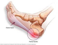
Anterior: palm of the hand Posterior: knuckle side of the hand Dorasal of the foot is the upper surface of the foot. Plantar is the lower side of the foot. |
|
|
Superior and inferior |

Superior (cranial and cephalic) towards the head even if the body is unside down. Inferior (caudal) towards the feet. |
|
|
Proximal and distal |
Proximal is closer to the trunk or point of origin, Distal is away from it. |
|
|
Superficial and deep |
Superficial closer to the surface and deep is further away from the body. |
|
|
External (outer) and Internal (inner): |
External (outer): means towards the surface and applies to the hollow-out structure. * Internal (inner): means towards the cavity of a hollow-out structure. |
|
|
Central and Peripheral: |
Central: means towards the center of the body. * Peripheral: means away from the center of the body. |
|
|
Ipsilateral and Contralateral : |
Ipsilateral: means of the same side of the body. * Contralateral : means of the opposite side of the body. |
|
|
Abduction and adduction |
Abduction Æ moving a part away from midline. Adduction Æ moving a part towards the midline. |
|
|
Abduction and adductionin the fingers |
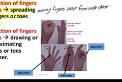
Abduction of fingers & toes Æ spreading of fingers or toes apart. Adduction of fingers & toes Æ drawing or approximating fingers or toes together. And opposition putting the thumb in the opposite way of the finger |
|
|
Circumduction |
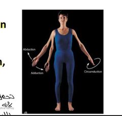
the combination in sequence of movements of flexion, abduction, extension & adduction. |
|
|
Medial rotation and Lateral rotation |

Medial rotation Æ brings anterior surface to face medially Lateral rotation Æ brings anterior surface to face laterally |
|
|
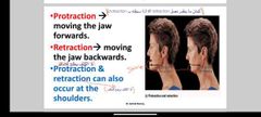
Protraction and retraction |
|
|
|
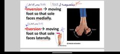
Inversion and eversion |
|
|
|
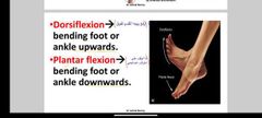
Dorsiflexion and plantar flexion |
|
|
|
The five body cavities |
1) cranial cavity; inside the skull, contains the brain. 2) vertebral cavity; in the vertebral, contains the spinal cord. 3) thoracic cavity; contains the heart inside the pericardial cavity and the lungs inside the pleural cavity. 4) abdominal cavity, contains organs such as the kidneys and gastrointestinal tract organs. 5) pelvic cavity; contains urinary bladder, rectum, (uterus and ovaries), in females. |
|
|
What's the skeleton made of |
@ It comprises cartilages, bones, ligaments & joints. @ The bones are rigid and heavier than cartilages. @ Cartilages are more flexible and lighter. @ The younger the age, the greater is the contribution of cartilage to the skeleton. |
|
|
Two divisions of skeleton |
1) Endo skeleton; a) axiapl skeleton B) the appendicular skeleton 2) exo skeleton nails and enamel of teeth |
|
|
What are bones made of |
Hard type of connective tissue( hardness due to calcium) |
|
|
What are bones composed of |
1) living osteocytes cells. 2) intercellular collagenous matrix( organic compounds). 3) Cement substance and mineral salts (inorganic component). |
|
|
Regional classification of bones |
1) axial skeleton; the skull, vertebral column, ribs and sternum. 2) appendicular skeleton (upper & lower limbs) & their girdles (shoulder & pelvic). |
|
|
What's the basic structure of the skull |
1) cranium, which contains the brain and meninges and the mandible |
|
|
Basic structure of the ribs, sternum and vertebral column |
12 ribs and and 12 sternum cartilages. The vertebral column made up of 33; 7 cervical (neck) 12 thoracic (thorax) 5 lumbar abdomen 5 sacral 4 coccygeal @ The vertebral column: 1. Forms the axial skeleton of the body. 2. Supports the weight of the body. 3. Protects & surrounds the spinal cord
|
|
|
Types of curves of the vertebral column |
1) primary curve The vertebral column is concave anteriorly at birth. 2) the secondary curve; (a) The cervical curve: becomes convex anteriorly when the child extends his head at the 3rd - 4th month. (b) The lumbar curve: becomes convex anteriorly when the child begins to walk between 12-18 months due to strengthening of the muscles of the back. |
|
|
Appendicular skeleton |
The skeleton of the upper limb resembles that of the lower limb; each consists of 3 segments: 1. Proximal segment: formed of one bone. Arm and thigh 2. Intermediate segment: formed of 2 bones. Forearm and leg 3. Distal segment: formed of 3 regions. Hand and foot |
|
|
Names of the Upper limb and lower limb |
@ Bones of Upper limb: 1. Shoulder (Pectoral) girdle: Clavicle & Scapula. 2. Upper arm: Humerus. 3. Forearm: Ulna (medially) & Radius (laterally). 4. Hand: Formed of 3 regions (from proximal to distal); Carpus , Metacarpus & phalanges. @ Bones of Lower limb: 1. Pelvic girdle: Hip bone. 2. Thigh: Femur. 3. Leg: Tibia medially & Fibula laterally. 4. Foot: Formed of 3 regions (from proximal to distal); Tarsus, Metatarsus & Phalanges. |
|
|
Functions of bones: |
1. Supporting framework of the body. 2. Attachment of muscles. 3. Act as Biomechanical levers for movements. 4. Protection of underlying organs. 5. Storehouses for Ca, P & Mg (mineral reservoir). 6. Synthesis of RBCs within the red bone marrow (in ends of long bones, sternum, ribs & vertebrae). 7. Transmission of body weight. |
|
|
Morphological (Anatomical) classification according to shape of bone: |
1. Long bones: have 2 ends & a shaft as bones of proximal & intermediate segments of the limbs (humerus, radius, ulna, femur, tibia & fibula). 2. Short bones: as carpal & tarsal bones. These bones are strong & help in limited movements. 3. Flat bones: as scapula, sternum & skull cap. These have wide surface for muscle attachment or protection. 4. Irregular bones: as vertebrae & facial bones. 5. Pneumatic bones: contain air-filled spaces lined with mucous membrane (paranasal sinuses) in skull bones (maxilla & frontal bones) to reduce the weight of skull, help in resonance of voice & warm air. 6. Sesamoid bone: are small nodules of bone found in the tendons of certain muscles to reduce friction over bony surfaces. e.g. patella & pisiform bones |
|
|
Parts of a growing long bone: |
1. 2 ends called epiphysis. 2. A shaft called diaphysis. 3. Epiphyseal plate of cartilage between the diaphysis & epiphysis. This is the most important factor for the growth of bone in length. 4. The part of the shaft close to the plate is called metaphysis. |
|
|
@ The Periosteum: |
***This is a fibrous strong membrane which covers the shaft of the bone. ** It consists of an outer fibrous layer & inner (deeper) cellular layer (osteogenic). ***It has the following functions: 1. Protection of the bone. 2. Provides muscular attachment. 3. Carry blood supply & sensory nerves to the bone. 4. Has an osteogenic power (bone forming ability) which plays an important role in the growth of bone in width & the healing of bones after fractures. ** Note that the bone grows in length by epiphyseal plate of cartilage & in thickness (breadth) by the periosteum. |
|
|
Histological classification according to structure of bone: |
1.Compact (Ivory) bone: Outer hard layer in outer surface of shaft of long bones. 2. Spongy (Cancellous) bone: Has pores & consists of irregular trabeculae that form a spongy network such as in sternum. |
|
|
Developmental classification according to ossification of bone: |
This means the transformation of the mesodermal tissue into bone. It has 2 types:1. Intracartilagenous (endochondral) ossification (Cartilagenous bone):@ In most of long bones, vertebral column & thoracic cage.@ Early in the intrauterine life (6-8 weeks), a 1ry center of ossification appears in the shaft, where calcification spreads (cartilage model) to help the ossification (bone formation). @ After birth, 2ry centers of ossification appear at the 2 ends of the bone. The shaft & the 2 ends became completely ossified but still separated by a plate of cartilage (epiphyseal plate) to help the growth of bone in length.@ Finally the bone become ossified at certain age (15-22 years) (in females it is earlier by 1-2 years). This is called synostosis. 2. Intramembranous or mesenchymal ossification (membranous bone): @ In flat bones (as skull cap or vault of skull & scapula), facial bones & clavicle (except its ends). * N.B.: Clavicle, although, it is a long bone; a. It is ossified from a membrane. b. It has no medullary cavity. c. It is first bone to begin ossification (5-6th week of I.U.L.). @ Centre of ossification develops at the mesenchymal tissue then transformed into bone without cartilage formation. |
|
|
Bone marrow |
It occupies the medullary (marrow) cavity of long bones & the cancellous bones in flat, short & irregular bones. @ 2 types: Red type (blood forming or hematopoietic) & Yellow type. @ With aging, the red marrow decreases & the yellow marrow replaces it. @ In adults, the red marrow is restricted to the bones of the skull, vertebral column, thoracic cage, girdle bones & the head of the humerus & the femur. |
|
|
Arterial blood supply of bone: |
Arterial blood supply of bone: 1. Periosteal arteries. 2. Metaphyseal & epiphyseal arteries. 3. Nutrient artery: A large blood vessel which enters the shaft of the bone near its middle through a nutrient foramen. It enters the bone perpendicular to the surface. As the bone elongates unequally from both ends, the artery becomes deflected away from the growing end. *** The final direction of the nutrient artery of a limb bone is summarized by the following rule (to the elbow I go, from the knee I flee). *** The growing end of the bone is opposite the direction of the nutrient artery. |
|
|
Cartilages |
Avascular dense flexible connective tissue. @ It is formed of chondrocytes (cartilage cells) & gel- like matrix which is responsible for its firmness & resilience). @ Perichondrium is a fibrous membrane that covers the cartilage. @ According to nature of the matrix, there are 3 types of cartilages. |
|
|
3 types of cartilages |
1. Hyaline cartilage Has a high content of homogenous & transparent matrix. @ Sites: 1. Foetal bones. 2. Articular cartilages of synovial joints. 3. Costal cartilages of ribs. 4. Epiphyseal plates of long bones. 5. Cartilages of respiratory passages eg. Trachea. 2)2. White fibrocartilage @ The matrix is little & rich in collagenous bundles which add strength & durability to this cartilage. @ Sites: 1. Symphysis pubis & Intervertebral discs. 2. Labrum of some synovial joints (Hip & Shoulder). 3. Discs within joints (temporo-mandibular, sterno- clavicular & knee joints). 3-Yellow elastic cartilage@ The matrix is rich in elastic fibers which provide flexibility to this form of cartilage. @ Sites: Auricle of ear, Tip of nose & epiglottis. |
|
|
Functions of cartilage |
1. It helps in maintaining patency of the respiratory passages. 2. It shares in the formation of the skeleton of the body. 3. It forms smooth firm articular surface of joints. |