![]()
![]()
![]()
Use LEFT and RIGHT arrow keys to navigate between flashcards;
Use UP and DOWN arrow keys to flip the card;
H to show hint;
A reads text to speech;
69 Cards in this Set
- Front
- Back
|
Tissue type in upper GI tract |
striated skeletal muscle |
|
|
End of the GI tract |
anus |
|
|
Esophagus tissue type |
Striated to smooth muscle |
|
|
Abdominal GI tube tissue type |
Smooth muscle |
|
|
Anus tissue type |
Skeletal muscle |
|
|
Name of the GI tract's nerve network |
Enteric Nervous System |
|
|
Roles of the ENS |
Peristalsis and secretion control |
|
|
Enteric nervous system is (parasympathetic/sympathetic) |
BOTH! trick question |
|
|
Sympathetic portions of the ENS will have synapses here |
celiac, superior mesenteric and inferior mesenteric ganglia (sympathetic splanchnic ganglia) |
|
|
Parasympathetic portions of the ENS will derive from these two places |
Vagus nerve (cranial nerve X) and pelvic nerve (S2-4) |
|
|
GI is a tube that is formed next to the ________. |
Aorta |
|
|
Foregut's blood supply |
Celiac trunk |
|
|
Midgut's blood supply |
Superior Mesenteric Artery |
|
|
Hindgut parasympathetic nerve supply |
S2-4 (pelvic splanchnics) |
|
|
Hindgut blood supply |
Inferior mesenteric artery |
|
|
Foregut parasympathetic nerve supply |
Vagus nerve |
|
|
Sympathetic nerve supply of the foregut region |
Greater splanchnic nerve |
|
|
Sympathetic supply of the hindgut (nerves) |
Least splanchnic (thoracic) and lumbar splanchnic nerves |
|
|
What are the borders of the hindgut? |
Last 1/3 of transverse colon to rectum |
|
|
All elements of the GI tract were (intra/retro) peritoneal at one time |
intraperitoneal |
|
|
Peritoneum linking one organ to another |
ligament |
|
|
Connecting stomach to the spleen |
Gastrosplenic ligament |
|
|
Connects stomach to the liver |
Lesser omentum |
|
|
Connects liver to the wall of the peritoneal cavity |
Falciform ligament |
|
|
Blood vessels (are/are not) covered with peritoneum |
ARE |
|
|
Parietal peritoneal pain is described as.... |
sharply localized
Parietal periotoneum is innervated like the body wall. Pain fibers ate components of somatic sensory (i.e. intercostal) nerves. |
|
|
Visceral peritoneum's pain fibers are components of these |
Vagus or splanchnic nerves |
|
|
Mild stretching (is/is not) detected in the visceral peritoneum |
IS NOT (i.e. the growth of a tumor goes undetected until the tumor is large. This contributes to high mortality of pancreatic and colon cancers). |
|
|
True or false: Pain in the visceral peritoneum is never intense |
FALSE. The pain can be dull to intense but is not well localized (i.e. stomach ache) |
|
|
Ancillary organs |
Kidneys, Spleen, Pancreas, Duodenum |
|
|
Transpyloric plane demarcates |
Goes through the kidneys, start of the duodenum and the body of the pancreas, and bottom of the spleen
Right kidney lower than the left kidney |
|
|
Umbilicus demarcates |
Upper quadrant v Lower quadrant |
|
|
Spleen's dimensions and location in the abdominal cavity |
1x3x5 inches Weighs 7oz Ribs 9-10 Usually not able to be palpated unless enlarged |
|
|
Spleen is (intra/retro) peritoneal |
intraperitoneal |
|
|
If a person has a longstanding infection, you will see an enlarged _______. |
Spleen |
|
|
Blood supply of the spleen |
Branch of the celiac trunk named the splenic artery |
|
|
Splenic artery will continue past spleen to do this. |
Will travel in the gastrosplenic ligament, branch to supply part of the stomach |
|
|
Splenic vein will eventually go into the _____________. |
Portal vein |
|
|
Blood goes into the spleen and back out to the ____________. |
Liver |
|
|
If there is too much damage to the spleen (i.e. after cracking ribs in a car wreck), you can do the following things: |
Ligate the blood vessels and remove the spleen
Person has had some immune response compromise so he/she must now be careful about infections. |
|
|
Uncinate process- what is it and where is it located? |
Part of pancreas like a fish hook-
Between superior mesenteric vessels/aorta/IVC |
|
|
Pancreas functions |
Digestive enzyme secretion Insulin secretion by the Islets of Langerhans
|
|
|
The uncinate process loops (dorsally/ventrally) in development |
ventrally |
|
|
Pancreas is developing as part of these two embryonic regions |
Foregut and Midgut
Thus, the blood supply reflects this. Gets blood from celiac trunk and superior mesenteric artery. |
|
|
Parts of Pancreas |
Head portion (cradled in curve of duodenum) Body (extends across the superior mesenteric vessels) Tail (extends across to the spleen) Uncinate process (between aorta/IVC and superior mesenteric vessels) |
|
|
Main tube of pancreas |
Pancreatic duct |
|
|
Pancreas blood flow- Head and Uncinate process |
Head and uncinate processes: 2 pancreaticoduodenal arches (from the branches of the gastroduodenal artery (celiac trunk) and the superior mesenteric artery. |
|
|
Liver is (intra/retro) peritoneal |
Intraperitoneal
|
|
|
Place where an infection can develop after abdominal surgery |
Subphrenic space |
|
|
Surfaces of the liver |
Diaphragmatic (upper) Visceral (contact with all other viscera) |
|
|
Lobes of the Liver-- and how to differentiate |
Right Left Quadrate (bound to gallbaldder to the right and ligamentum teres in fissure) Caudate (close to tail, next to IVC) |
|
|
Where all vessels will enter in liver |
Hepata Portis |
|
|
Liver is in direct contact with the diaphragm here |
Bare Area |
|
|
Where leftover umbilical vein will end up in the liver |
Ligamentum teres in the fissure |
|
|
Right triangular ligament is much more (anterior/posterior) than the left triangular ligament |
Posterior |
|
|
Which triangular ligament is more easily seen? |
Left |
|
|
Apical surface of cells will be in contact with (blood/bile). |
Bile |
|
|
Basolateral surface of hepatocytes is in contact with the (blood/bile). |
Blood |
|
|
Blood flow in the liver |
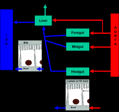
Aorta-->celiac trunk--> Proper hepatic artery--> Left or right hepatic artery
|
|
|
Veins flowing into (through) the liver |
Splenic vein, Superior mesenteric vein, Inferior mesenteric vein
AFTER LIVER: goes to hepatic veins then back to IVC |
|
|
To take out gallbladder you need to ligate this |
Cystic artery |
|
|
Portal triad |
Hepatic artery, Portal vein and Bile duct |
|
|
Branching of the portal triad |
Right lobe: will be supplied by right branches of portal triad Left, quadrate lobe: will have left branches of portal triad Caudate: could have either one |
|
|
Will quadrate lobe be supplied by left or right branches of the portal triad? |
Left |
|
|
Will caudate lobe be supplied by left of right branches of the portal triad? |
Could be either one |
|
|
Blood pressure elevated in the vessels of the liver due to alcoholism/other damage to liver |
Portal hypertension |
|
|
Pancreas blood flow- body and tail |
Pancreatic branches of the splenic artery from the celiac trunk |
|
|
Venous return of pancreas |
Collateral veins to the portal system |
|
|
Lymphatic drainage of the pancreas |
Celiac and superior mesenteric nodes |