![]()
![]()
![]()
Use LEFT and RIGHT arrow keys to navigate between flashcards;
Use UP and DOWN arrow keys to flip the card;
H to show hint;
A reads text to speech;
85 Cards in this Set
- Front
- Back
|
to support continued ATP production, cells have to obtain ________and expel excess ________ continuously. |
1. oxygen 2. Carbon dioxide |
|
|
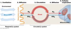
gas exchange involves 5 steps:
|
1. ventilation 2. Diffusion at the respiratory surface 3. Circulation 4. Diffusion at the tissues 5. Cellular respiration |
|
|
Ventilation:
|
the movement of air or water through a specialized gas exchange organ, such as a gill or lungs - air or H2o moves through gas-exchange organs |
|
|
Gas exchange/ diffusion at the respiratory surfaces |
where O2 moves from the air to water into the blood and CO2 moves from the blood into the air or water - CO2 & O2 diffuse between air or H2O & blood at the respiratory surface |
|
|
Circulation
|
the transport of dissolved O2 &CO2 throughout the body - along with nutrients, wastes, and other types of molecules- via the circulatory system - Dissolved O2 & CO2 are transported throughout the body |
|
|
Diffusion at the tissues
|
where O2 moves from blood into the tissues and CO2 moves from the tissues into the blood - O2 moves from blood tissues, CO2 moves from tissues into blood |
|
|
cellular respiration |
the cell's use of O2 and production of CO2. in tissues, where cellular respiration has led to low O2 levels and high CO2 levels, gas exchange occurs between blood and cells. - cells use O2 & produce CO2 - Low O2/ high CO2 cells = gas exchange between blood and cells |
|
|
5 steps of gas exchange
|
|
|
|
Ventilation and Diffusion at the respiratory surface are accomplished by the ____________. |
respiratory system, the collection of cells, tissues and organs responsible for gas exchange between the animal and its enviro. In essence, respiratory system consist of structures for conducting air or water to a surface where gas exchange takes place. |
|
|
understand the diff types of gas exchange surfaces in animals and what types of animals exhibit each type.
|
1. skin: amphibians, flatworms 2. Lungs: tetrapods, lungfishes 3. Tracheae: insects 4. gills; mollusks, arthropods, crustaceans, fishes and amphibians - in some animals the gas exchange is located in the skin , but in most species it is located in gills, tracheae, and lungs. |
|
|
understand why O2 tends to move into tissues and CO2 tends to diffuse out
|
-gas exchange betw the enviro and cells is based on diffusion. normally, O2 is high in the enviro and low in tissues, while CO2 levels are relatively high in tissues and low in the enviro. -so... O2 tends to move from enviro into tissues and CO2 tends to move from tissues into the enviro. |
|
|
difference of air in high altitudes vs. low altitudes |
the % of O2 in the atmosphere does not vary with elevation. the key difference is that far fewer molecules of oxygen and other atmospheric gases are present per unit volume of air at high elevations than at sea level, because atmospheric pressure is lower at high elevations. - 21% of air in the atmosphere is O2 |
|
|
Partial pressure:
|
pressure of a particular gas in a gas mixture. *Pressure is force exerted per unit area. |
|
|
how do you calculate partial pressure of gasses in the air?
|
multiply the fractional composition (the fraction of air the gas comprises) by the total pressure exerted by the entire mixture (atmospheric pressure) Dalton's Law. .21 x atmospheric pressure at given altitude. |
|
|
why is it harder to breath at the top of Mt. Everest?
|
-in both air and water, O2 and CO2 move from regions of high partial pressure to regions of low partial pressure. it's hard to breath at the top because the partial pressure of oxygen is low thee- meaning hat the diffusion gradient betw the atmosphere and your lung tissues is small so fewer molecules of O2 diffuse into your tissues when you take a breath.
|
|
|
why is it easier to breath air than it is water?
|
-H2O has less available O2 than air -H20 is 800x more dense than air - H2o is 55x more viscous than air (flows less easily) -Organisms that use gills expend more energy to ventilate respiratory surfaces than do those using lungs -Organisms that use gills have to process 30X more H2O than amount of air a terrestrial animal breathes |
|
|
understand the process that affect gasses dissolving in fluids |
O2 & CO2 diffuse into H2O from the atmosphere, but the amount of gas that dissolves depends on several factors: (1) Solubility of the gas in H2O -O2 has low solubility in H2O *Blood needs hemoglobin to bind O2 (blood is ~90% H2O) (2) Temperature (T) of H2O -High T = less gases dissolve (3) Presence of other solutes -SW has less O2 than FW due to higher solute concentration (4) Partial pressure of gas in contact with H2O-Moves from high partial pressure to low partial pressure -Why bubbling of soda occurs (5) Other factors -Photosynthetic organisms increase O2 -Decomposers decrease O2 -Surface = high in O2; unless mixing occurs -Surface area (shallow bodies of water have more O2 than deep bodies) -Waterfalls, rapids, etc = high SA exposed to atmosphere as splashing and bubbling occurs |
|
|
Solubility of gas in water
|
- oxygen has very low solubility in water. Animals compensate for low solubility of oxygen in water by having a molecule in the blood that binds to oxygen and delivers it to tissues. w/o this, the rate of blood flow to tissues would have to increase dramatically to meet oxygen needs.
|
|
|
Temperature of water
|
as temp increases in water, the amt of gas that dissolves in it decreases. Warm water habitats have much less oxygen available than cold water habitats. Warm water fish= high elevation land dwellers.
|
|
|
presence of other solutes
|
because seawater has a much higher concentration of solutes than FW, seawater can hold less dissolved gas. FW inhabitants tend to be ,ore oxygen rich than marine.
|
|
|
partial pressure of the gas in contact with the water:
|
gases move from regions of high partial pressure to regions of low partial pressure. so if partial pressure of a gas dissolved in a liquid exceeds that of the gas in the adjacent atmosphere, the gas will bubble up out of the liquid. this is what happens when a cap is removed from a bottle of carbonated beverage. the partial pressure of CO2 in a newly opened drink is much higher than it is in the atmosphere.
|
|
|
what is the difference in gas exchange in small versus large animals
|
many small animals lack specialized gas exchange organs such as gills and lungs. instead they obtain O2 and eliminate CO2 by diffusion across the body = direct diffusion. - mostly live in wet enviro's - have high ration of SA to V - surface must be thin; eg. jellyfish, flatworms,some amphibians Large animals in dry enviro's need specialized organs for gas exchange. - respiratory organs provide greater SA for gas exchange. -respiratory organs are internal which helps minimize water loss from moist surfaces. |
|
|
how do gills work?
|
-outgrowths on body used for gas exchange in aquatic animals - large SA for O2 to diffuse across the extremely thin epithelium -in invertabrates, gill structure can be external or internal - fish gills are located on both side of the head -amphibians with internal or external gills. |
|
|
ram ventilation:
|
-Force H2O through gills by swimming with mouths open -Movement of H2O over gills is in one direction |
|
|
how do fish ventilate their gills?
|
Bony fish have internal gills -H2O must be driven over them during ventilation -Most fish ventilate gills by opening & closing mouths and opercula (a stiff flap over gills) = buccal pumping -Fast swimmers use ram ventilation -Force H2O through gills by swimming with mouths open -Movement of H2O over gills is in one direction |
|
|
opercula:
|
the stiff flap of tissue that covers the gills of teleost fishes |
|
|
gill arch:
|
in aquatic verabrates, a curved region of tissue betw the gills. gills are suspended from the gill arches
|
|
|
gill fillaments
|
in fishes, any of the many long, thin structures that extend from gill arches into water and that function in gas exchange
|
|
|
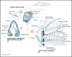
how do fish ventilate their gills?
|
Long, thin structures called gill filaments extend from each gill arch -Each filament composed of hundreds–thousands of gill lamellae – sheet-like structures-Small capillaries run through the lamellae -H2O must flow over the gills in order for the gases to be exchanged |
|
|
diagram of how fish ventilate their gills
|
|
|
|
expain how fish gill is a countercurrent system
|
the one-way flow of water through the gill lamelle has a profound impact on the gill function: the flow of blood through the capillaries in each lamelle is in the opposite direction to the flow of water. as a result, each lamella functions as a countercurrent exchanger -Flow of blood through capillaries is in opposite direction to flow of H2O over gill surface -Countercurrent exchange system in each lamellaCreates a large partial pressure of O2 in H2O over blood -Result is efficient exchange of gases over gills countercurrent flow makes fish gills extemely efficient at extracting oxygen from water because it ensures that a difference in the partial pressure of oxygen in water versus blood is maintained over the entire gas exchange surface |
|
|
how do insect trachea work?
|
insects have an extensive system of air filled tubes called trachea located within the body. - these tubes connect to the exterior through openings in the exoskeleton called Spriacles, which can be closed to minimize the loss of water due to evaporation. -Insects have tracheae (air-filled tubes) -Open to outside through spiracles -Air moves into trachea O2 diffuses into cells-For small insects, diffusion is sufficent -Larger insects = trachea alternately open & close as wing muscles around them contract & relax -As a result, V of tracheal system changes -As V changes, so does P -P and V are inversely related -Low P = air moves in; high P = air moves out |
|
|
trachea:
|
in insects,any of the small, air filled tubes that extend throughout the body and function in gas exchange
|
|
|
spiracles:
|
in insects, a small opening that connects air filled trachea to the external enviro, allowing for gas exchange
|
|
|
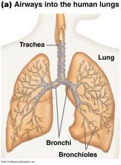
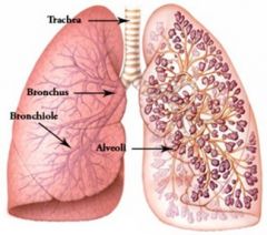
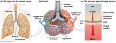
how do lungs work?
|
- in terrestrial animals, air enters through the mouth and or/nose - Trachea carries inhaled air to narrow tubes called Bronchi. *Bronchi branch into narrower bronchioles... branch into cup-like alveoli. - Lung = organ for gas exchange * lung encloses alveoli, bronchioles & portions of bronchi -animals with lungs include some fishes, repitles, amphibians, reptiles birds, mammals. |
|
|
|
|
|
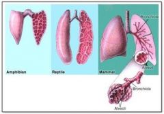
what is the difference in lung structure betw diff animal groups?
|
-In amphibians, lung is a simple sac lined with blood vessels -Mammalian lungs are divided into tiny sacs (alveoli), which greatly increase the SA for gas exchange -Humans have ~150 million alveoli per lung (70 m2 surface area) -Lung structure can vary among species and animal groups |
|
|
|
|
|
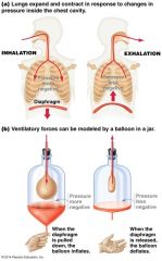
what is the difference betw positive and negative pressure ventilation and which organisms carry out each type?
|
Vertebrates actively ventilate their lungs by pumping air via muscular contractions -There are two mechanisms for pumping air:-Positive pressure ventilation: frogs -Negative pressure ventilation: mammals |
|
|
positive pressure ventilation
|
ventillation of the lungs by using increased pressure in the mouth to push air into lungs: fish
|
|
|
negative pressure ventilation
|
ventillation of the lungs by expanding the rib cage so as to pull air into the lungs: mammals
|
|
|
bronchiles
|
any of the small tubes in mammalalian lungs that carry air from the bronchi to the alveoli
|
|
|
alveoli
|
any of the tiny air filled sacs of a mammalian lung
|
|
|
diaphram
|
an elastic, sheetlike structure. in mammals, the muscular sheet that seperates the chest and abdominal cavities. it contracts and moves downward during inhalation, expanding the chest cavity |
|
|
Lung
|
any respiratory organ used for gas exchange betw blood or hemolymph and air
|
|
|
Human lung ventilation
|
Air inside human lung is under negative pressure (5 mm Hg < than atmosphere = keeps lungs expanded) -Pumping action in mammals is achieved by diaphragm (muscle) Two steps are involved: -When diaphragm moves down, pressure in chest cavity is lowered and air moves in -As diaphragm relaxes, chest cavity decreases and air is exhaled *This is a passive process driven by the elastic recoil of lungs and chest wall as diaphragm and rib muscles relax |
|
|
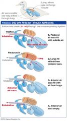
bird lung ventilation
|
Birds are able to extract enough O2 for extremely long flights & flights at high elevations 4 general steps to ventilation in birds: 1.During inhalation, air flows through the trachea into two large posterior air sacs 2.During exhalation, air flows from posterior air sacs into lung (parabronchi) 3.During 2nd inhalation, air moves to anterior air sacs 4.During 2nd exhalation, air moves out to atmosphere *Air flow through avian lung is unidirectional |
|
|
human lung ventilation diagram
|
|
|
|
bird lung ventilation diagram
|
|
|
|
diagram of different lung ventilations in animals
|
|
|
|
What is the function of blood?
|
blood is a connective tissue consisting of cell & H2O -transport of O2 & CO2 -transports nutrients to cells from the digestive system - conveys hormones to target tissues and organs -delivers cells of immune system - distributes heat **the H2O portion of blood is called Plasma |
|
|
what are the cellular components of blood?
|
~50–65% of blood is plasma Blood has cellular components: 1.Platelets: cell fragments that minimize blood loss 2.White blood cells (WBCs): immune function3.Red blood cells (RBCs): transport O2 from lungs to body tissues & CO2 from tissues to lungs *In humans, RBCs make up 99.9% of blood cells |
|
|
platelets:
|
a small membrane bound cell fragment in vertebrate blood that functions in blood clotting. derived from large cells in the bone marrow.
|
|
|
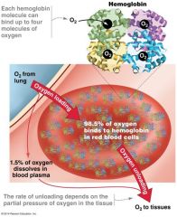
what is the structure and function of hemoglobin? |
RBC's contain oxygen carrying molecules called hemoglobin (Hb) - Consists of four polypeptide chains, each of which binds to a non-protein group called a heme -Each heme contains an Fe2+ that can bind to an O2 molecule -Each Hb molecule can thus bind up to 4 O2 molecules -In blood, 98.5% of O2 is bound to Hb -Other 1.5% is dissolved in plasma |
|
|
diagram og the structure of hemoglobin
|
|
|
|
**understand cooperative binding and why it is important in times of low oxygen in tissues (i.e. exercise) |
tendency of protein subunits of Hb to affect each other's O2 binding such that each bound O2 molecule increases the likelihood of further binding
|
|
|
how does the Bohr shift work and what is it?
|
Hb is sensitive to changes in pH and T - decreases in pH & increases in T alter Hb's conformation such that it is more likely to release O2 at all values of Po2 (partial pressure) *this is know as the Bohr shift - Makes Hb more likely to release O2 during exercise in which Pco2 is high (=pH is low) and tissues are under O2 stress |
|
|
Bohr Shift |
decreased affinity of hemoglobin for oxygen caused by an increase of carbon dioxide; the oxyhemoglobin dissociation curve is displaced to the right because of higher partial pressure of carbon dioxide and lower pH.
|
|
|
how do fetal and maternal Hb differ?
|
Hb molecules from different individuals or species may vary in ways that affect fitness. -Fetal Hb has a higher O2 affinity than adult Hb -Fetal Hb is encoded differently than adult Hb - As a result, O2 is transferred from mother's blood to fetus's blood, thus ensuring an adequate supply of O2 as fetus developes |
|
|
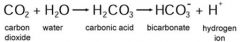
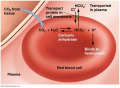
describe the buffering capacity of blood
|
CO2 produced by cellular respiration enters RBCs -Catalyzes formation of H2CO3 (carbonic acid) from CO2 & H2O -Quickly converted to bicarbonate (HCO3-) ions & protons (H+) in a reaction catalyzed by carbonic anhydrase -Most CO2 is transported in blood plasma in the form of HCO3 -Protons stay in RBCs |
|
|
|
|
|
Two reasons why Carbonic Anhydrase activity in red blood cells is important:
|
1. Protons produced by the carbonic anhydrase reaction induce the Bohr shift, which makes Hb more likely to release O2 2.The PCO2 in RBCs drops when CO2 is converted to HCO3-, maintaining a strong partial-pressure gradient favoring the entry of CO2 into RBCs |
|
|
why is Co2 released when blood returns to the lungs?
|
Many of the H+ produced by dissociation of H2CO3 are taken up by Hb -Hb also acts as a buffer, minimizing pH changes -In alveoli, a partial-pressure gradient favors the diffusion of CO2 from plasma and RBCs to atmosphere -Hb releases protons, which combine with HCO3- to form CO2 (and H2O) -Which then diffuses into alveoli and is exhaled-Hb picks up O2 during inhalation, and cycle repeats |
|
|
how does circulation differ in small vs large animals?
|
Animals maximize SA available for diffusion of gasses & other key solutes in a variety of ways. - very small animals have a small enough V that diffusion over body surface is adequate to keep them alive. - JF with a highly folded gastro vascular cavity that offers a large SA for molecular exchange - Tapeworms & flatworms have a high SA:V ratio - rotifers & tarigrades are small enough that diffusion of O2 is adequate -roundworms take up O2 directly across body wall (diffusion) *Large animals require a circulatory system in order to achieve a large SA:V ration |
|
|
What is an open circulatory system. What animals have them?
|
- Hemolyphm is pumped throughout the body in open vessels - H. comes in direct contact with body tissues - Characteristic of many invertebrates - H. is pumped by the heart through open-ended vessels LIMITATIONS: - w/o discret conti vessels, flow of h. cant be directed toward tissues that have a high O2 demand and CO2 buildup (crustaceans are an important exception) |
|
|
what is a closed circulatory system and what animals have them?
|
In closed system, blood flows in a continuous circuit of closed vessels - pressure provided by pumping action of heart - closed systems are characteristic of vertebrates & some invertebrates (annelids & cephalopods) -can be shunted in times of need e.g. legs during exercise, intestines after a meal. |
|
|
What are the three types of blood Vessels
|
1. Arteries & Aretioles 2. Capillaries 3. Veins and Venules |
|
|
Arteries and Arterioles |
though thick- walled vessels that carry blood away from the heart under high pressure - always carry oxygenated blood (except pulmonary artery!) - walls composed of smooth muscle tissue that allows for vessel elasticity |
|
|
Capillaries |
smallest vessels - walls are one cell thick, allowing gases and other molecules to exchange with tissues in a network called Capillary Beds - gases, nutrients, waste exchange betw blood and other tissues - extremely thin - form an extremely dense network throughout the body |
|
|
Veins and Venules |
- return blood to the heart under low pressure - always carry oxygenated blood (except pulmonary vein!) - thin walls and much larger interior diameter than arteries *because blood is under relatively low pressure by the time it exits the tissues *larger veins also contain valves, thin flaps of tissue that prevent any blood backflow |
|
|
what is the function of the lymphatic system in animals?
|
|
|
|
Aorta |
large vessel, generally receives blood from the heart and delivers it to body - had elastic walls allowing it to expand when blood enters it under high pressure from heart |
|
|
intestinal fluid |
the are between cells - build up because there is an outward directed hydrostatic force in capillaries created by the pressure generated by the heart |
|
|
Lymphatic System: |
intestinal fluid that is not reclaimed in the capillaries is collected here. - a collection of thin-walled, branching tubules - intestinal fluid that enters lymphatic ducts is called Lymph Three actions of the Lymphatic Duct: 1. permeate all tissues 2. eventually join with one another to form larger vessels 3. return excess intestinal fluid (lymph) to major veins entering the heart |
|
|
what are two of the chambers of the heart?
|
1. Atrium; receives blood returning from circulation 2. Ventricle: generates force to propel blood through the body |
|
|
Atrioventricular Valves |
atria are separated from ventricles by this *two distict trends as vertebrates diversified: 1. # of distinct heart chambers increased 2. in some animals, one circuit serves entire body; in more complex vertebrates, there are two circuits |
|
|
Fish have a __________ heart and a _______ circulatory circuit to both gills and rest of the body. |
1. two-chambered 2. single |
|
|
Land vertebrates evolved two separate pumping circuits:
|
1. pulmonary circulation: is a low pressure circuit to and from lungs 2. Systemic circulation: a higher-pressure circuit to and from rest of body *pulmonary and systemic circuits are completely separated in the 4 chambered hearts of crocs, birds and mammals - only partial separated in 3 chambered hearts of amphibians, lizards and turtles |
|
|
Some animals have a _______________ running from right ventricle directly into systemic circuit. What does this do? |
1. Foramen of Panizza 2. shunts blood from pulmonary to systemic circuit when an animal is underwater - greatly reduces blood flow to lungs when animal is submerged. -shunt the blood away from the lungs when they are submerged in water and not breathing. |
|
|
what is the path of blood through the human heart?
|
1. blood enters right atrium on return from body 2. blood enters the right atrium 3. blood is pumped to lungs from right ventricle 4, blood returns to left atrium from lungs 5. blood enters the left ventricle 6. blood is pumped to body from left ventricle...back to step one. |
|
|
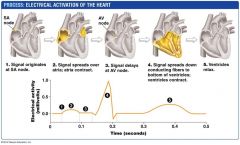
Electrical Activation of the Heart
|
Pacemaker cells that initiate contraction in the heart are located in a region of the right atrium called the sinoatrial node (SA)\ - The SA node and heart muscle cells receive input from nervous system and from chemical messengers in blood *these inputs are important for regulating heart rate. |
|
|
Electrical Activation of the Heart
|
|
|
|
AV node:
|
is a part of the electrical conduction system of the heart that coordinates the top of the heart. It electrically connects atrial and ventricular chambers.[1] The AV node is an area of specialized tissue between the atria and the ventricles of the heart, specifically in the posteroinferior region of the interatrial septum near the opening of the coronary sinus, which conducts the normal electrical impulse from the atria to the ventricles.
|
|
|
AV Valve:
|
either of two heart valves through which blood flows from the atria to the ventricles; prevents return of blood to the atrium
|
|
|
SA Node:
|
generates electrical impulses and conducts them throughout the muscle of the heart, stimulating the heart to contract and pump blood. - |