![]()
![]()
![]()
Use LEFT and RIGHT arrow keys to navigate between flashcards;
Use UP and DOWN arrow keys to flip the card;
H to show hint;
A reads text to speech;
65 Cards in this Set
- Front
- Back
|
3 main functions of the circulatory system |
1) it tansports gases (from the respiratory system), nutrient molecules and waste materials (from the digestive system) 2) it regulates internal temperature and transports chemical substances that are vital to health from one part of the body to the other 3) it protects against blood loss from injury and against disease-causing microbes or toxic substances introduced into the body |
|
|
Major components of the circulatory system |
Heart Blood vessels Blood |
|
|
Heart |
Muscular organ that continuosly pumps blood via the circulatory system to the lungs and body and generates blood flow Located slightly to the left of the middle of the chest (size of fist) Walls of heart lined with cardiac muscle Cardiac muscle cells are arranged in a network that allows the heart to contract and relax rhymically and involuntarily without fatigue Ensures blood keeps following 1 direction Ensures oxygenated blood is kept separate from deoxygenated blood Pumps blood |
|
|
Blood vessels |
A system of hollow tubes that carries blood to and from body tissues. (where blood moves) |
|
|
Cardiovascular system is comprised of |
Heart and blood vessels |
|
|
Blood |
Fluid that transports nutrients, oxygen, carbon dioxide and many other material through the body Bodily fluid in which blood cells are suspended |
|
|
Open circulatory system |
Called "open" because blood flows freely within the body cavity and makes direct contact with organs and tissues No distinction between blood and the interstitial fluid Include many invertabrates |
|
|
Mixture of blood and fluids that surround the cells |
Hemolymph |
|
|
How open circulatory system works |
IN GRASSHOPPER Hemolymph is pumped through single vessel that runs from the head to the abdomen
In the abdomen the vessel divides into chambers that function as the insect's heart
Tiny holes in the heart wall, known as ostia allow hemolymph to enter the heart chambers from the body cavity
The hemolymph is pushed from on chambe to the next by muscle contractions
Nutrients and waste are exchanged between hemolymph and the cells in the heart chambers before the hemolymph passes back into the transporting vessel to be eliminated from the insect's body |
|
|
Closed circulatory system |
Circulatory system in which the circulating blood is contained within vessels and kept separate from the interstitial fluid System that circulates blood, keeps it under pressure, pump it at a speed sufficient to supply the metabolic needs of all parts of the body Keeps blood physically contained within vessels and separate from other body tissues Blood follows a continuous fixed path of circulation and is confined to a network of vessels that keeps blood separate from the interstitial fluid |
|
|
2 top chambers of the heart |
Atria (singular= atrium) |
|
|
Atria fill with blood |
Returning from the body or the lungs |
|
|
2 chambers at the bottom |
Ventricles |
|
|
Ventricles receive |
Blood from the atria and pump it out to body or the lungs |
|
|
Atria and ventricles are separated by |
Thick muscular wall called the septum
Two valves called atrioventricular valves and semilunar valves |
|
|
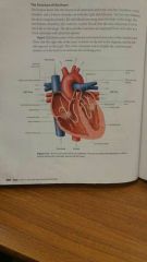
Diagram of heart |
|
|
|
Flow of blood through the heart 11 |
1) Right side of heart receives blood that is coming back from the body and then pumps this blood out to the lungs 2) The vena cava opens into the right atrium 3) Surperior vena cava collects oxygen-poor blood coming from the tissues in the head, chest and arms 4) Interior vena cava collects oxygen-poor blood coming from the tissues elsewhere in the body 5) The oxygen poor blood flows from the right atrium into the right ventriclebans the out into the pulmonary trunk 6) from there, it enters the left and right pulmonary arteries 7) it then continues to the left and right lungs for gas exchange 8) The left side of the heart does 27th reverse... Receives oxygen-rich blood from the left and right lungs and pumps this blood out to the body 9) The oxygen- rich blood flows from the lungs through the pulmonary veins to the left atrium 10) The left atrium pumps blood into the left ventricle where all the blood going to the body tissues leaves through the largest vessel in the body, the aorta 11) The 4 cables ensure the blood flows in the correct direction |
|
|
Pulmonary artery |
Large blood vessel that carries blood from the heart to the lungs |
|
|
Pulmonary vein |
Blood vessel that carries blood from the lungs to the heart |
|
|
Aorta |
An artery that carries blood directly from the heart to other arteries |
|
|
Atrioventricular valve |
A valve in the heart between the ventricle and the atrium Atrioventricular valve on the right side is called the tricupsid (made of 3 flaps) Atrioventricular valve on left side is called bicuspid or mitral valve (2 flaps) |
|
|
Semilunar valve |
A valve between the ventricle and the large arteries; it carries bloodnaway from the heart (half-moon shape) |
|
|
3 main blood vessels |
Arteries Veins Capillaries |
|
|
Arteries |
Carry oxygen-rich blood away from the heart under high pressure No valves Thick walls (tissue layers) Smaller-diameter arteries are called arterioles |
|
|
Veins |
Carry oxygen-poor blood towards the heart under low pressure Thin walls Smaller-diameter veins are called venules |
|
|
Capillaries |
One-cell-wall thick Site where gases, nutrients and other materials are tansferred from blood to tissue cells and from tissue cells to blood A tiny blood vessel that carries oxygenated blood from the arteries to the veins under low pressure Link arteries to veins (arterioles to venules) |
|
|
Structure of arteries |
Narrow lumen helps maintain blood pressure Thick walls able to withstand high blood pressure Elastic tissue gives walls flexibility to accomodate sports of blood Elasticity allows the artery to expand and contract Expansion and contraction of walls keep blood following in right direction and provides an additional pumping motion Smooth muscle allows for control over blood delivery to tissue
|
|
|
Structure of veins |
Large lumen diameter allows blood to flow easily with little resistance under low pressure Valves prevent backflow, keeping blood flowing towards the heart Contractions of nearby skeletal muscles, squeeze veins helping them push blood along |
|
|
Structure of capillaries |
Microscopic vessels whose walls are only one cell wall thick which allows materials to be exchanged with body cells, nutrients, O2 Hormones diffuse out of blood plasma into the ECF and into cells |
|
|
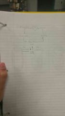
Diagram of blood flow through vessels |
|
|
|
Pulmonary circulation |
The path that blood follows from the heart to the lungs and back to the heart Movement of blood from the heart to the lungs and the from the lungs back to the heart again Blood that flows from the heart to the lungs carries waste carbon dioxide gas. As this blood passes through the respiratory surfaces of the lungs, gas exchange takes place--carbon dioxide leaves the blood and oxygen moves into the blood. The freshly rich oxygenated blood goes back to the heart and is pumped from the heart into the a second circuit that transports it throughout the rest of the body |
|
|
Systemic circulation |
The path that blood follows from the heart to the body and back to the heart Takes oxygenated blood from the heart to other tissues and organs throughout the body. After circulating throughout the body, the blood returns to the heart carrying waste carbon dioxide from the body's tissue . the blood then reenters the pulomary circulation |
|
|
Cardiac circulation |
The movement of blood through the heart tissue |
|
|
Plasma consists of |
Water + dissolved gases Proteins Sugars Vitamins Minerals Waste products 55% of blood volume |
|
|
Formed portion of blood |
Red blood cells White blood cells Platelets 45% of blood volume |
|
|
Plasma |
Clear, yellowish fluid composed of 92 percent water, 7 percent dissolved blood proteins, 1 percent other organic substances and inorganic substances such as sodium, potassoum, chloride, bicarbonate Contain electrolytes, nutrients, vitamins, hormones, clotting factors and proteins 55% of our blood |
|
|
Main proteins in blood are |
Albumin, globulins, fibrinogen |
|
|
Component of plasma: WATER FUNCTION |
dissolved and transports other substances |
|
|
Component of plasma: PLASMA PROTEINS FUNCTION |
-maintain fluid balance in plasma, in cells, in spaces between cells -help maintain slightly alkaline oH -fibrinogen helps with blood clotting -globulins (antibodies) strengthen immunity |
|
|
Component of plasma: SALTS FUNCTIONS |
-maintain fluid balance in plasma, in cells, in spaces between cells -help maintain slightly alkaline pH -assist in nerve and muscle function |
|
|
Example of salts (ions) |
Bicarbonate Calcium Chloride Magnesium Potassium Sodium |
|
|
Red blood cells are also called |
Erythrocytes
|
|
|
Red blood cells take up |
45 percent of the total volume of blood |
|
|
Red blood cells are specialized forb |
Oxygen transport |
|
|
The oxygen carrying-capacity of the blood is dependant on the |
Number of erythrocytes that are present and the amount of hemoglobin that each red blood cell contains |
|
|
A muture mammalian erythrocyte |
Disk-shaped cell= provides Moe surface area for gas exchange No nucleus Pack with 280 million iron-containing molecules of the respiratory protein hemoglobin
|
|
|
Hemoglobin |
Protein Hemoglobin has special properties that allow it to pick up or chemically bind with oxygen
Releases oxygen in the presence of cells that need it
Transports some of the carbon dioxide waste from cells
After carbon dioxide diffused into the blood it enters the red blood cells where a small amount binds to hemoglobin |
|
|
Red blood cells |
Most abundant cells in our blood, they are produced in the bone marrow and contain protein called hemoglobin that carries oxygen to our cells |
|
|
White blood cells are also called |
Leukocytes |
|
|
White blood cells |
Part of our immune system and destroy infection agents called pathogens Response to infection 1% of total blood volume but may increase to more than double normals levels when your body is fighting infection |
|
|
Characteristics of white blood cells |
Have nuclei Appear colorless |
|
|
5 types of white blood cells |
Neutrophil Eosinophil Basophil Lymphocyte Monocyte |
|
|
Phagocytosis |
Process that engolf and destroy pathogens |
|
|
Cells that carry out phagocytosis are called |
Phagocytes |
|
|
Neutrophils |
Most abundant leukocytes Found in body tissues of an animal as well as blood |
|
|
Eosinophils |
Found in the mucous lining of the digestive and respiratory tracts |
|
|
Basophil |
Adi in immunity by secreting substances that attract phagocytes to destroy pathogens |
|
|
Lymphocytes |
Produce proteins called antibodies that incapacitate pathogens and allow them to be easily detected and destroyed |
|
|
Monocytes |
Circulate in the bloodstream for only a few days before they become specialized as macrophages which destroy bacteria |
|
|
Platelets are also called |
Thrombocytes |
|
|
Platelets |
Clotting factors that are carried in the plasma; they clot together in a process called coagulation to seal a wound and prevent a loss of blood |
|
|
How platelets are formed |
Membrane-bound fragments of cells that form when larger cells in bone marrow break apart |
|
|
Characteristics of platelets |
Contain nuclei Break down blood in blood within 7-10 days after they are formed |
|
|
Steps in blood clotting |
1. When a blood vessel is broken due to injury it releases chemicals that attract platelets to the site of injury 2. The platelets rupture releasing chemicals that combined with other chemicals in the plasma to produce the enzyme thromboplatin 3. As long as calcium ions are present thromboplatin with prothrombin to produce amother enzyme called thrombin 4.thrombin reacts with fibrogen to produce fibrin 5. Fibrin is an insoluble protein that forms a fibrous mesh over the site of injury 6.This mesh prevents the loss of blood cells and eventually solidifies to form a clot |
|
|
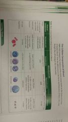
Comparing cellular components chart |
|