![]()
![]()
![]()
Use LEFT and RIGHT arrow keys to navigate between flashcards;
Use UP and DOWN arrow keys to flip the card;
H to show hint;
A reads text to speech;
68 Cards in this Set
- Front
- Back
|
Squamous |
flat, many-sided cells with centrally located nucleus |
|
|
Cuboidal |
squarish cells with centrally located nucleus |
|
|
Columnar |
cells composed of two or more layers of cells |
|
|
Simple |
composed of one layer of cells |
|
|
Stratified |
composed of two or more layers of cells |
|
|
Glandular Epithelium |
are cells that invaginated into the underlying lamina propria |
|
|
Acini |
spherical group, specialized for secretion |
|
|
Unicellular gland |
one-celled gland that secretes mucous, solitary cell which appears cup-shaped resulting from accumulation of mucoid secretion |
|
|
Multicellular glands |
this gland is made up of many cells |
|
|
Serous |
These cells are columnar, cytoplasm is acidophilic and granular |
|
|
Mucous |
These cells are cuboidal, cytoplasm is basophilic |
|
|
Neuro-epithelium |
composed of pseudo-stratified epithelium specialized to receive sensations |
|
|
Retina |
design for vision |
|
|
Organ for Corti |
design for hearing |
|
|
Ampulla Crista Ampullaris |
function for equilibration |
|
|
Olfactory cells |
design for smell |
|
|
Surface epithelium |
This structure is for surface modification of the epithelium |
|
|
Microvilli |
submicroscopic filaments projecting from the superficial layer of the cytoplasm |
|
|
Striated border |
composed of many extremely thin, short, uniform and closely packed protoplasmic projections |
|
|
Brush border |
short processes which are irregulary arranged as cuboidal cells seen in the kidneys |
|
|
stereo cilia |
very elongated, non-motile hair like processes |
|
|
cilia |
hairlike processes with basal corpuscles |
|
|
Gap junctions |
small channels that allow materials to cross the epithelial layers |
|
|
Connective tissues |
serve to connect, give support and anchor part to the body and organs; developed from mesoderm where cells unite to make a network. |
|
|
Hyaluronic acid |
present in many connective tissues; has the capacity to bind water and is an important factor for the changes in viscosity and permeability of the ground substance in the tissue |
|
|
Supporting Tissue |
includes bones and cartilages |
|
|
Vascular Tissue |
includes blood and lymph |
|
|
Fibroblast |
most common and numerous in all types of connective tissues; large, flat, branching with many processes; are active during tissue injury and repair forming fibers |
|
|
Plasma Cells |
small round or irregular in shape; nucleus is eccentric with coarse chromatin forming "spoke of the wheel" pattern; actual formers of circulating antibodies. |
|
|
Mast Cells |
large, polyhedral or flattened cells with small nuclei, numerous along the blood vessel beds; motile but slow-moving and forms heparin and serotonin |
|
|
Macrophages |
or Histlocytes, irregulary shaped cells with short processes and smaller nuclei; phagocytic in inflammatory conditions |
|
|
Pigment cells |
elongated cells with short irregular outgrowths which cytoplasm contained granules of melanin; formed from melanoblasts |
|
|
Undifferentiated mesenchyme |
cells are similar but smaller to fibroblasts; found along blood vessels |
|
|
Adipose cells |
large brilliant spherical cells, "fat cells" |
|
|
Blood cells |
main component found in tissue spaces; these are neutrophil, monocyte and eosinophil which move in and out of the tissue |
|
|
Collagenous |
white fibers, most common type of fiber which posses little elasticity but high tensile strength |
|
|
Fibrillae |
bundles of fine wavy fibrils which are cemented together |
|
|
Elastic |
yellow fibers, homogenous straight and stretchable, resistant to acid and composed of the protein called elastin |
|
|
Reticular fibers |
similar to white fibers, on boiling yeilds reticulin; believed to be immature forms of white fibers |
|
|
Embryonal and Adult Tissue Proper |
Two types of connective tissue proper |
|
|
Mesenchyme |
called packing or filling tissue in the embryo from mesodermal cells migrating to spaces between germ layers. |
|
|
Mucous |
ground substance rich in mucin |
|
|
Areolar/Loose connective tissue |
irregulary arranged tissue that serves as packing tissue in adult between organs and other tissues |
|
|
Reticular tissue |
forms a network that serves as a supporting framework to lungs, liver, kidneys, marrow, bone, spleen, etc. |
|
|
Lymphoid Tissue |
common variety lf reticular tissue, for protection, hemopoeisis, and filtration of tissue fluid and lymph. |
|
|
Adipose tissue |
abundant in fat cells |
|
|
Dense Fibrous tissue |
collagenous fibers are the predominating fibers in the tissue spaces, appears silver white |
|
|
Tendons |
muscle to bone |
|
|
Ligaments |
helps hold bones together at joints |
|
|
Aponeurosis |
connects muscle or the periosteum to the bones |
|
|
Membranes |
investing and protecting organs all over the body |
|
|
Fasciae |
bands that wraps around muscles and hold them in place |
|
|
Superficial and Deep Fasciae |
Two types of Fasciae |
|
|
Dense elastic tissue |
elastic fiber that predominates in the tissue space which runs parallel to each other |
|
|
Cartilage |
sometimes called gristle, firm and tough also flexible, called chindriocytes |
|
|
Hyaline Cartilage |
type of cartilage, referred as skeletal or embryonic cartilage, bluish to white appearance, forms a matrix where most bones are developed |
|
|
Fibrous Cartilage |
type of cartilage is pervaded with heavy collagenous fibers |
|
|
Elastic Cartilage |
type of tissue wherein the intercellular matrix is pervaded with elastic fibers together woth cartilage cells |
|
|
Bones |
their cells are called osteocytes |
|
|
Long, short, flat, irregular bones |
Shape and size of bones |
|
|
Compact or Spongy |
Histologic texture of bones |
|
|
Cancellous |
or spongy, this has bigger cavities and is interior of the bone tissue |
|
|
Dense |
or compact, appears as a continous hard mass, has fewer spaces and is exterior to the bone tissue |
|
|
Membrane |
origin: develops directly from mesenchyme of the embryo forming membrane e.g flat bone of skull |
|
|
Cartilage |
Origin: develops from mesenchyme passing through a performed cartilage model e.g long bones |
|
|
Intra-membranous Ossification |
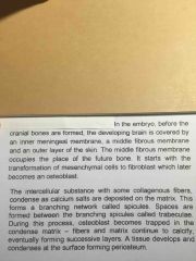
Back (Definition) |
|
|
Intracartilagenous Ossification |
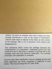
Back (Definition) |
|
|
so ******* tired just review this **** |
Back (Definition) |