![]()
![]()
![]()
Use LEFT and RIGHT arrow keys to navigate between flashcards;
Use UP and DOWN arrow keys to flip the card;
H to show hint;
A reads text to speech;
109 Cards in this Set
- Front
- Back
|
What 3 things can occur after injury?
|
Progression (further injury developing into chronic inflammation),
Healing (fibrosis and loss of function), and Resolution (return to normal function after clearance of injurous stimuli, mediators, and inflammatory cells) |
|
|
What are the cardinal signs of acute inflammation?
|
Red (rubor),
Swelling (tumor), Warm (calor), tender (dolor), loss of function (functio laesa) |
|
|
In what order do the inflammatory cells come to the site of injury?
|
After edema comes the neutrophils (about 1 day) and then the monocytes/macrophages (about 2 days)
|
|
|
What is a PMN?
|
It is a polymorphonuclear lekocyte aka a neutrophil
|
|
|
How does a neutrophil get into a site of injury?
|
Margination (pavementing) and adhesion: so rolling, activation with chemokines, stable adhesion, then migration through endothelium.
|
|
|
What are the mediators of emigration and chemotaxis?
|
Bacterial products (LPS), Complement derivatives (C5a), arachidonic acid derivatives (leukotriene B4), and cytokines (IL-8)
|
|
|
What comes from WBC defects?
|
Chediak-higashi syndrome and diabetes mellitus
|
|
|
What happens with chemotactic factor defects?
|
C5 or immunoglobulin deficiency.
|
|
|
What happens when there is a defect with the serum chemotaxis inhibitors?
|
C5 inactivator defects cause an increase in cirrhosis, sarcoidosis, and other diseases.
|
|
|
What happens when WBC locomotion inhibitors are defective?
|
Chloroquine, cancer, and other chronic diseases can occur.
|
|
|
What are the 3 steps of phagocytosis?
|
Recognition, attachment, engulfment
|
|
|
How does a cell recognize what its going to phagocytose?
|
With opsoninzation (attachment of antibodies) and opsonins (Rc fragment of IgG, C3b, mannose-binding protein from liver) that cover the surface that allow it to be recognized.
|
|
|
What happens after a macrophage attaches a bacteria it will phagocyize?
|
It engulfs it and then merges it with a lysosome to degrade it with enzymes, NO, and reactive oxygen species.
|
|
|
What is oxygen dependent killing?
|
It is the use of reactive oxygen species such as HOCl and OH free radicals that is used to destroy a microorganism.
|
|
|
How does a cell produce HOCl and OH free radicals?
|
Myeloperoxidase uses hydrogen peroxide and halide (Cl-) to make HOCl, and Hydrogen peroxide and ferrous ion will generate OH free radical if myeloperoxidase is absent.
|
|
|
What role does the liver play in the immune system?
|
It produces the essential elements of coagulation (Factor XII – Hagerman factor) and inflammation (Complement activation – e.g. memgrane attack complex)
|
|
|
What chemical mediators of inflammation are stored in secretory granules of mast cells, basophils, platelets, neutrophils, and macrophages?
|
Histamine, serotonin, and lysosomal enzymes.
|
|
|
Which chemical mediators of inflammation are newly synthesized?
|
Prostaglandins, leukotrines, platelet-activating factors, activated oxygen specises, nitric oxide, and cytokines.
|
|
|
Where does the arachidonic acid come from?
|
Cell membranes
|
|
|
What are some things that can be produced from arachidonic acids?
|
Prostaglandins, leukotrines, lipotoxins, and other inflammatory mediators. They affect vasoconstriction/dialation, chemotaxis, edema, etc.
|
|
|
What is transudate?
|
Fluid with low protein content and a specific gravity <1.012
|
|
|
What is Exudate?
|
Inflammatory extracellular fluid composed of plasma proteins, principally albumin, cells and cellular debris, and a specific gravity >1.020.
|
|
|
What is Anasarca?
|
Edema which is severe and generalized
|
|
|
What is an abcess?
|
Localized collections of pus caused by suppuration buried in a tissue, organ, or confined space.
|
|
|
What is cellulitis?
|
Spreading suppurations in subcutaneous tissue.
|
|
|
Name the types of exudates and an example of each.
|
Serous – blister;
Fibrinous – uremic pericarditis, viral/chemical pneumonitis; Purulent – meningococcal meningitis; Eosinophilic – asthma and parasitic infections (IgE mediated); Hemorrhagic – rickettsial; pseudomembranous – diptheria and pseudomembranous enterocolitis. |
|
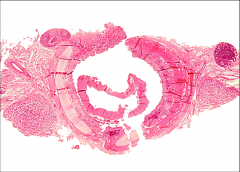
What does this picture show? |
Pulmonary edema with transudate. |
|
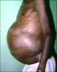
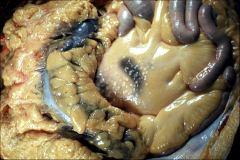
What does this image show? |
Ascites: the accumulation of fluid in the peritoneal cavity, causing abdominal swelling. |
|
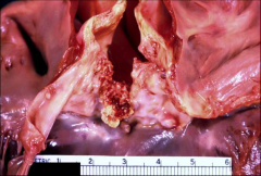
This is an example of: |
Fibrinous pericarditis. |
|
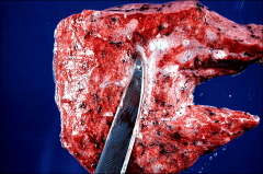
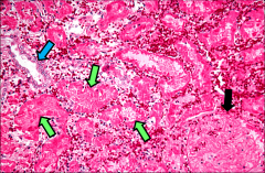
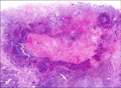
What type of acute inflammation is this? |
Purulent: you can see its filled with cells and the remains of cells - pus = dead white blood cells. |
|
What type of exudate is this? |
Pseudomembranous, specifically from diphtheria. |
|
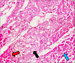
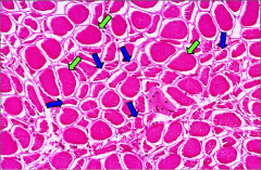
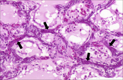
What is this? What are the arrows pointing to? |
This is hydropic degeneration: The accumulation of water in a cell that usually leads to cell death. Red arrow: dead heart cells Black arrow: Hydropic degeneration, reversible swelling. Still alive, but not healthy. Blue arrow: Normal heart cells. |
|
What type of necrosis is this? |
Coagulative Necrosis. |
|
What type of necrosis is this? |
Liquefaction necrosis. |
|
What type of necrosis is this? |
Liquefaction. |
|
What are the tiny bumps? |
Pancreatic fat necrosis |
|
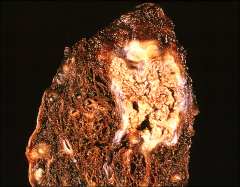
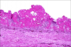
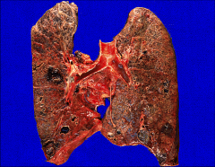
What type of necrosis is this? |
This is caseous necrosis caused by TB. |
|
What is this? |
Caseous necrosis |
|
What type of necrosis is this? |
fibrinoid necrosis |
|
What type of necrosis is this? |
Gangrenous necrosis. |
|
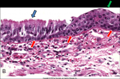
What is this an example of? |
squamous metaplasia. |
|
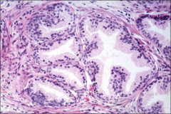
What is this an example of? |
Prostatic hyperplasia |
|
What is this? |
This is disuse atrophy. |
|
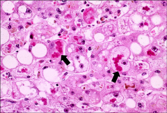
What is this? |
This is fatty change in the liver stained with Oil Red O stain. |
|
What is this? |
Alcoholic hyalin |
|
What is this? |
Anthracosis |
|
What is this? |
Metastatic calcification |
|
What is this? |
dystrophic calcification. |
|
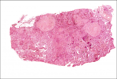
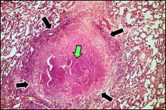
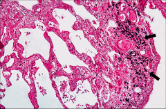
What is this? What could have caused it? |
Granulomatous Inflammation that could be caused by TB. |
|
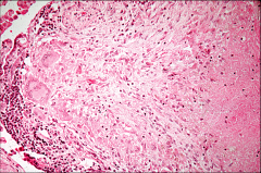
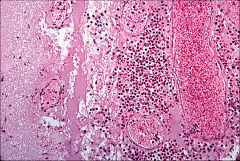
What is this? |
A closeup of granulomatous inflammation. Note the epithelioid macrophages. |
|
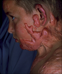
What is this? |
This is an excessive wound repair known as a keloid. It is made of fibrinous tissue. |
|
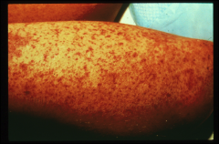
What is this? What disease is it associated with? |
This is a petechiae rash and is associated with Meningococcal Meningitis. |
|
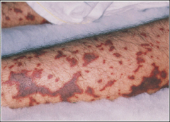
What is this? What disease is it associated with? |
This is a purpura rash (formed when the splotches from the petechiae accumulate) and is associated with Meningococcal Meningitis. |
|
|
What are the 4 main types of cell adaptation? |
Atrophy, hypertrophy, hyperplasia, metaplasia. |
|
|
What are the 2 possible outcomes from an irreversible injury? What is the main difference between them? |
Necrosis and apoptosis. Necrosis involves inflammation whereas apoptosis does not. |
|
|
Define Hypoxia and Ischemia. |
Hypoxia: decreased availability of oxygen (pneumonia) and/or loss of oxygen carrying capacity of blood (anemia). Ischemia: Insufficient blood supply, often caused by occlusion of artery or vein. |
|
|
What is troponin I? |
It is a protein released by dying heart cells. |
|
|
What are the results of oxygen depravation? |
The mitochondria cannot use oxidative phosphorylation which leads to: ER swelling, cellular swelling, loss of microvilli, blebs, clumping of nuclear chromatin (due to low pH from buildup of lactic acid), and lipid deposition. |
|
|
What are the signs of a reversible injury? |
Cell swelling leading to hydropic change or vacuolar degeneration. |
|
|
What happens with an irreversible injury? |
Cell death leading to necrosis. Also, nuclear pyknosis followed by karyorrhexis and karyolysis. |
|
|
What are pyknosis, karyorrhexis, and karyolysis? |
They are the steps in the destruction of genetic material. Pyknosis: shrinking of chromosomes Karyorrhexis: breaking up of nucleus Karyolysis: all pieces of nucleus dissolve away. |
|
|
Why do dead cells appear so bright on histology slides? |
The build up of lactic acid as the cells died causes the degeneration and coagulation of proteins which allows the stain to bind really well. |
|
|
How can ROS be generated? |
Redox reactions, absorption of radiant energy, in activated leukocytes during inflammation, enzymatic metabolism of exogenous chemicals/drugs, transition metals (iron and copper), nitric oxide and peroxynitrite anion. |
|
|
How do cells remove ROS? |
Antioxidants (vitamins E, A, C, and glutathione) Iron and copper binding proteins (transferrin, ferritin, lactoferrin, and ceruloplasmin) Enzymes (catalase, superoxide dismutases, glutathione peroxidase) |
|
|
What are the pathologic effects of ROS? |
Lipid peroxidation in membranes (they become stiff and break), oxidative modification of proteins, DNA damage. |
|
|
Define coagulative necrosis. |
Dissolution of nucleus with preservation of cellular shape and tissue architecture, also coagulation (denaturation) of cell proteins. It appears to leave behind a "ghost" of the cell. |
|
|
Define liquefaction necrosis. |
Hydrolytic enzymes cause autolysis and heterolysis (liquefaction) of cells/tissues. Begins as coagulation, but then the cells break apart into mush. It is the complete degradation of cells down to the molecular level. |
|
|
What are the components of pus? |
neutrophils, bacteria, and dead tissue. |
|
|
What is fat necrosis? |
Destruction of adipose tissue due to the action of lipases: happens especially in liver and pancreas. The lipase reacts to lipids, releases free fatty acids that react with calcium in the blood to make salts: saponification. |
|
|
What is caseous necrosis? |
A combination of coagulative and liquefaction necrosis that is primarily found in the center of tubercles. It is the result of an inability to digest and remove material from center of granuloma. |
|
|
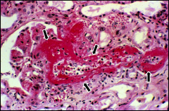
What is fibrinoid necrosis? |
Necrotic tissue due to immunologic reaction. It is usually seen in blood vessels with deposition of complement and antibodies in vessel wall (e.g. kidney) |
|
|
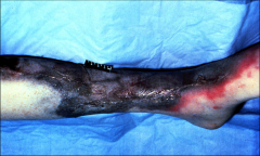
What is gangrenous necrosis? |
It is coagulative necrosis with a secondary bacteria infection that leads to liquefaction. |
|
|
What are the 2 forms of gangrenous necrosis? |
Dry gangrene: coagulative necrosis is predominant pattern. Usually when there isn't enough blood for lots of liquefaction. Wet gangrene: liquefactive process is the dominant pattern. Usually when there is plenty of blood available. |
|
|
What are the 2 types of apoptosis? |
Physiologic apoptosis: embryogenesis and hormone-dependent involution (e.g. menstrual cycle) Pathologic apoptosis: viral diseases and injurious agents leading to cell death. |
|
|
What are the mechanisms for apoptosis? |
Activation of endonuclease, cytoskeleton disruption by proteases, cytoplasmic protein cross-linking by transglutaminase, and cell surface changes leading to phagocytosis. |
|
|
What are the morphologic characteristics of apoptosis? |
General cell shrinkage, chromatin condensation, bleb formation and apoptotic bodies, phagocytosis, and lack of an inflammatory reaction. |
|
|
Define atrophy. |
Shrinkage in the size of the cell by loss of structural components. This may be due to decreased work load, loss of innervation, diminished blood supply, inadequate nutrition, loss of endocrine stimulation. |
|
|
Define hypertrophy. |
Increased size of cells and the organ. Can be physiologic (hormonal stimulation like uterus) or pathologic (increased functional demand e.g. left ventricular hypertrophy (LVH)) |
|
|
Define hyperplasia. |
Increase in the number of cells in an organ or tissue. Can be physiologic (hormone induced like breast in pregnancy) or pathologic (viral induced or excessive hormonal stimulation). |
|
|
Define metaplasia. |
Reversible change in which one differentiated cell type is replaced by another cell type. |
|
|
True/False: An example of metaplasia is ciliated columnar epithelium turning directly into squamous epithelium. |
False! The ciliated columnar do not turn into squamous epithelium. It is the stem cells that will begin forming the squamous to replace the columnar as they die. A ciliated columnar epithelium cannot change directly into a squamous epithelium. |
|
|
Give an example of when metaplasia would occur. |
If a kidney stone continually rubbed up against the columnar epithelium of the kidney, then the kidney would develop squamous epithelium to prevent/mitigate the damage. |
|
|
True/False: Metaplasia is reversible. |
True. |
|
|
Name some normal and abnormal cellular constituents than can abnormally accumulate in a cell. |
Normal: lipids, proteins, glycogen, carbohydrates. Abnormal: carbon, silica, asbestos, bacteria. |
|
|
What is fatty change? |
It is when the amount or distribution of fat changes. So too much lipid going in or not enough coming out. Lipid in macrophages: foam cells - atherosclerosis Lipid in parenchyma cells: alcoholic fatty liver. |
|
|
What are hyaline droplets? |
Accumulation of intracellular proteins, especially in the kidney proximal tubules. |
|
|
Where can you find Russell bodies? |
In plasma cells. |
|
|
What is alcoholic hyaline? |
A poorly defined accumulation of eosinophilic material in the cytoplasm of damaged liver cells in certain forms of cirrhosis. Looks like tangled skeins of cytokeratin intermediate filaments and other proteins. Also called Mallory Alcoholic hyaline. |
|
|
What are some exogenous pigments? |
Carbon (anthracosis) tattooing natural substances (beta carotene) poisons (Lead: Pica) |
|
|
What are some endogenous pigments? |
Lipofuscin, melanin, and hemosiderin. |
|
|
What is hemosiderosis and hemochromatosis? |
Hemosiderosis: iron overload in phagocytic cells, no tissue damage. Hemochromatosis: iron overload in parenchymal (functional, not structural) cells, tissue damage occurs. |
|
|
What is metastatic calcification? |
Deposition of calcium in normal tissues due to hypercalcemia. Often found in interstitial tissues of blood vessels, kidneys, lungs, and gastric mucosa. |
|
|
What is dystrophic calcification? |
Deposition of calcium salts in necrotic tissues. Can be intracellular, extracellular or both. Heterotopic bone may form with time. |
|
|
What are the different types of fixed macrophages and where are they found? |
Kupffer cells - liver histiocytes - connective tissue microglia - brain osteoclasts - bone Langerhans cells - epidermis. |
|
|
What is granulomatous inflammation? |
Focal accumulations of activated macrophages with an epithelial-like (epithelioid) appearance. |
|
|
What is a granuloma? |
A microscopic aggregation of epithelioid macrophages surrounded by a collar of lymphocytes and occasionally plasma cells. |
|
|
What is granulation tissue? |
It is the proliferation of new blood vessels and fibroblasts. Fibroblasts turn into myofibroblasts. |
|
|
In general terms, what is the order of healing? |
Inflammation, formation of granulation tissue (when inflammation ends, collagen accumulation begins), and then finally wound contraction. |
|
|
What is primary union wound healing? |
Healing by first intention: The healing of a clean surgical incision. Very little tissue damage is present. |
|
|
What is secondary union wound healing? |
Healing by second intention: The healing of a large tissue defect such as road rash. |
|
|
Which is quicker, primary union or secondary union and why? |
Primary union is quicker because it is easier to pull the small incision together than it is with a large scrape. |
|
|
What are some of the causes of wound healing abberations? |
Inadequate granulation tissue/healing, excessive repair (hypertropic scar or keloid), contractures (shortening and hardening of muscles). |
|
|
What are some conditions that would inhibit or delay wound healing? |
Poor local vascular supply (poor oxygenation, decreased bacterial resistance, low temperature, and acidic pH) Deficiencies of vitamin C, protein, zinc Infecton Tissue necrosis (foreign materal delay healing and acts as a nidus for bacterial infection Excessive movement. |
|
|
What is the main component of the extracellular matrix? |
Collagen. Specifically extra cellular which is after the procollagen has been cleaved and the lysine oxidized to promote cross-linking. It has very high tensile strength. |
|
|
What will allow the maximal tensile strength of 70 to 80% of normal? |
The replacement of Type III collagen with Type I in wound healing. |
|
|
What will lead to fibrosis (tissue scar)? |
Persistent tissue damage such as chronic inflammatory diseases (cirrhosis, chronic pancreatitis, pulmonary fibrosis) |
|
|
What is associated with acute injury? |
Vasoconstriction/dilation, fluid and protein leakage into interstitium (exudate), neutrophils (transudate), monocytes/macrophages cleaning up mess (phagocytosis) |
|
|
What is associated with chronic injury? |
Macrophages (cleaning up and recruiting more/proliferating), lymphocytes (regulating macrophages, orchestrating healing), granulomatous inflammation and/or granulomas. |
|
|
What is associated with healing? |
Macrophages (still cleaning up, becoming mature for that tissue), early granulation tissue (fibroblasts), early fibrosis (Type III collagen, myofibroblasts), collagen maturation (to Type I), and scar (with 70% of tensile strength). |