![]()
![]()
![]()
Use LEFT and RIGHT arrow keys to navigate between flashcards;
Use UP and DOWN arrow keys to flip the card;
H to show hint;
A reads text to speech;
77 Cards in this Set
- Front
- Back
|
Chapter 14
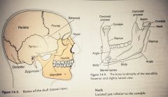
Name and label the bones and bony prominences of the skull: Page 199
|
|
|
|
Actions of the TMJ joint: |
-Depression -Elevation -Protraction -Retraction -Right and left lateral deviation |
|
|
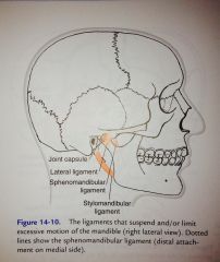
Ligaments of the TMJ:
Page 202 Fig. 14-10 and 14-11
(Next 6 cards include attachment sites for ligaments and other structures.) |
|
|
|
Lateral ligament (temporomandibular ligament): |
-Attaches anteriorly onto the neck of the mandibular condyle and disk, and then runs superiorly to the articular tubercle of the temporal bone.
-Limits downward, posterior, and lateral motions of the mandible.
|
|
|
Sphenomandibular ligament: |
-Attaches to the spine of the sphenoid bone and runs to the middle of the ramus on the internal surface of the mandible.
-Suspends the mandible and limits excessive anterior motion. |
|
|
Stylomandibular ligament |
-Runs from the styloid process of the temporal bone to the posterior inferior border of the mandible's ramus.
-It lies between the masseter and medial pterygoid muscles and plays a role in limiting excessive anterior motion. |
|
|
Stylohyoid ligament: |
-Attaches from the styloid process of the temporal bone to the hyoid bone.
-Holds the hyoid bone in place. |
|
|
Joint capsule: |
-Envelops the TMJ by attaching superiorly to the articular tubercle and borders of the fossa of the temporal bone. Inferiorly, it attaches to the neck of the condyle of the mandible. |
|
|
Articular Disk:
Figure 14-12 on page 202 |
(Similar to the articular disk of the sternoclavicular joint.)
-Divides the joint space into upper and lower spaces. |
|
|
The 4 major muscles of the TMJ:
Next 4 cards on each muscle. |
1. Temporalis
2. Masseter
3. Medial pterygoid
4. Lateral pterygoid |
|
|
Temporalis:
Figure 14-15 page 203 |
O: Temporal fossa
I: Coronoid process and ramus of mandible
A: Elevation, retraction, deviation
N: Trigeminal V |
|
|
Masseter:
Figure 14-16 page 204 |
O: Zygomatic arch or temporal bone and zygomatic process of maxilla
I: Angle of the ramus and coronoid process of mandible
A: Elevation, deviation
N: Trigeminal V |
|
|
Medial pterygoid:
FIgure 14-17 page 204 |
O: Lateral pterygoid plate of the sphenoid bone and tuberosity of the maxilla
I: Ramus and angle of the mandible
A: Elevation, protraction, deviation
N: Trigeminal V |
|
|
Lateral pterygoid:
Figure 14-17 page 204 |
O: Lateral pterygoid plate and greater wing of the sphenoid
I: Mandibular condyle and articular disk
A: Depression, protraction, deviation
N: Trigeminal V |
|
|
Suprahyoid muscles:
(Located above the hyoid bone and connect it to the skull, primarily the mandible.) |
-Mylohyoid
-Geniohyoid
-Stylohyoid
-Digastric |
|
|
Infrahyoid muscles:
(Located below the hyoid bone and serve to depress it.) |
-Sternohyoid
-Sternothyroid
-Thyrohyoid
-Omohyoid |
|
|
Chapter 15
|
:D |
|
|
Cervical vertebrae form what type of curve?
(Concave or convex?) |
Convex |
|
|
Thoracic vertebrae form what type of curve? |
Concave |
|
|
Lumbar vertebrae form what type of curve? |
Convex |
|
|
Sacral vertebrae form what type of curve? |
Concave |
|
|
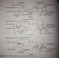
Parts of vertebrae and how they articulate on each level:
Table 15-2 and photos - page 216 |
|
|
|
Ligaments of the spine:
(Next 5 cards on ligaments.) |
-Anterior longitudinal ligament
-Posterior longitudinal ligament
-Supraspinal ligament
-Interspinal ligament
-Ligamentum flavum |
|
|
Anterior longitudinal ligament: |
- Runs down the vertebral column on the anterior surface of the bodies. - Prevents excessive hyperextension. |
|
|
Posterior longitudinal ligament: |
- Runs along the vertebral bodies posteriorly, inside the vertebral foramen.
- Prevents excessive flexion. |
|
|
Supraspinal ligament: |
- Extends from the seventh cervical vertebra distally to the sacrum posteriorly along the tips of the spinous processes.
** In the cervical vertebrae the nuchal ligament takes the place of the supraspinal and interspinal ligaments that are located in the thoracic and lumbar vertebrae. |
|
|
Interspinal ligament: |
- Runs between successive spinous processes in thoracic and lumbar vertebrae.
|
|
|
What is the name of the ligament that takes the place of the supra spinal and interspinal ligaments in the cervical region?
This ligament also becomes the supra spinal ligament at C7. |
Ligamentum nuchae (Figure 15-15 page 219) |
|
|
Ligamentum flavum: |
- Connects adjacent laminae anteriorly. |
|
|
Muscles of the neck:
(Next several cards will be on these.) |
Anterior: -Sternocleidomastoid -Scalenes (3) -Prevertebral (4)
Posterior: -Erector spinae group (3) -Splinius capitus -Splenius cervicus -Suboccipital group (4) |
|
|
Sternocleidomastoid: |
O: sternum and clavicle
I: Mastoid process
A: Flexes neck, hyperextends head, laterally bends neck, cervical rotation
N: Accessory nerve XI; second and third cervical nerves |
|
|
Scalene: |
O: Transverse processes of the cervical vertebrae
I: First and second ribs
A: neck flexion, lateral neck bending
N: Lower cervical nerves |
|
|
Splenius capitis: |
O: Lower half of nuchal ligament, spinous processes C7-T3
I: Lateral occipital bone, mastoid process
A: Extend the head and neck, rotate and laterally bend neck
N: Middle and lower cervical nerves |
|
|
Splenius cervicus: |
O: Spinous processes of T3-T6
I: Transverse processes of C1-C3
A: extend neck, rotate and laterally bend neck
N: middle and lower cervical nerves |
|
|
Prevertebral muscles: (4) |
-Longus colli
-Longus capitis
-Rectus capitis anterior
-Rectus capitis lateralis |
|
|
Longus Colli: |
O: Bodies and transverse processes of C3-T12
I: Transverse processes and bodies of C1-C6
A: Flex neck |
|
|
Longus capitis: |
O: Transverse processes of C3-C6
I: Occipital bone
A: flex head |
|
|
Rectus capitis anterior: |
O: ATLAS (C2)
I: Occipital bone
A: flex head |
|
|
Rectus capitis lateralis: |
O: transverse process of atlas
I: Occipital bone
A: laterally bend head |
|
|
Suboccipital muscles: (4) |
-Obliquus capitis superior -Obliquus captifs inferior -Rectus capitis posterior minor -Rectus capitis posterior major
**ALL are located posterior and extend the neck**
|
|
|
Trunk muscles: |
Anterior: -Rectus abdominus -External oblique -Internal oblique -Transverse abdominus
Posterior: -Erector spinae group (3) -Transversospinalis group (3) -Interspinales -Intertransversarii
Lateral: -Quadratus lumborum |
|
|
-Rectus abdominus: |
O: Pubis
I: Xiphoid process and costal cartilage of ribs 5-7
A: Trunk flexion, compression of abdomen
N: 7-12 intercostal nerves |
|
|
External oblique: |
O: lower 8 ribs
I: iliac crest and linea alba
A: trunk flexion, compression of abdomen, lateral bending, trunk rotation
N: 8-12 intercostal, iliohypogastric, and ilioinguinal nerves |
|
|
Internal oblique: |
O: inguinal ligament, iliac crest, thoracolumbar fascia
I: 10-12 ribs, abdominal aponeurosis
A: trunk flexion, compression of abdomen, lateral bending, trunk rotation
N: 8-12 intercostal, iliohypogastric, and ilioinguinal nerves |
|
|
Transverse abdominis: |
O: inguinal ligament, iliac crest, thoracolumbar fascia, last 6 ribs
I: abdominal aponeurosis and linea alba
A: compression of abdomen
N: 7-12 intercostal, iliohypogastric, and ilioinguinal nerves |
|
|
Erector spinae: |
O: spinous processes, transverse processes, and posterior ribs from the occiput to the sacrum and ilium
I: Spinous processes, transverse processes, posterior ribs from the occiput to the sacrum
A: extend neck and trunk, laterally bend neck and trunk
N: spinal nerves |
|
|
Transversospinalis muscles: |
O: Transverse processes
I: spinous processes of vetebrae above
A: extend neck and trunk, rotate neck and trunk
N: spinal nerves |
|
|
Interspinales muscles: |
O: spinous process below
I: spinous process above
A: neck and trunk extension
N: spinal nerves |
|
|
Intertransversarii muscles: |
O: transverse process below
I: transverse process above
A: neck and trunk lateral bending
N: spinal nerves |
|
|
Quadratus lumborum: |
O: iliac crest
A: trunk lateral bending
N: 12th thoracic and 1st lumbar nerves |
|
|
Chapter 16 |
:-| |
|
|
What is the difference between primary and accessory muscles for breathing? |
Primary muscles: diaphragm and intercostals - they move the diaphragm, thus changing thoracic volume and pressure, moving the ribs, and lowering the diaphragm
Accessory muscles: come into play during forced respiration |
|
|
Diaphragm muscle: |
O: xiphoid process, ribs, lumbar vertebrae
I: central tendon
A: inspiration
N: phrenic nerve (C3, C4, C5)
|
|
|
External intercostal muscles: |
O: rib above
I: rib below
A: elevate ribs
N: intercostal nerve (T2-T6) |
|
|
Internal intercostal muscles: |
O: rib below
I: rib above
A: depress ribs
N: intercostal nerve (T2-T6) |
|
|
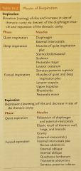
Phases of respiration: Table 16-2 page 243 |
|
|
|
What is Valsalva's maneuver? |
Occurs when people hold their breath and attempt to exhale.
Prolonged breath-holding and straining forces exhalation against the closed glottis. This increases intrathoracic pressure, which traps blood in veins and prevents it from entering the heart. When the breath is released, intrathoracic pressure drops and the trapped blood is quickly propelled through the heart, increasing the heart rate and blood pressure. Immediately, a reflex bradycardia follows. This event can have no consequences, or it can lead to cardiac arrest. |
|
|
What is the difference between diaphragmatic breathing and chest breathing? |
Diaphragmatic breathing is the most efficient method of breathing and requires the least amount of energy.
Chest breathing requires greater effort and is much less efficient than diaphragmatic breathing.
Page 244 |
|
|
Chapter 17: |
:-) |
|
|
What bones make up the pelvic girdle? |
-Sacrum
-Coccyx
-Ilium
-Ischium
-Pubis |
|
|
What are the three joints found in the pelvic girdle? |
1. Right and left sacroiliac joints (SI joints)
2. Symphysis pubis
3. Lumbosacral joint |
|
|
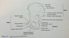
Bony landmarks of the pelvic girdle: Figure 17-7, page 251 |
|
|
|
Ligaments of the pelvic girdle:
(The next several cards will cover ligaments.) |
-Anterior sacroiliac ligament -Interosseous sacroiliac ligament -Short posterior sacroiliac ligament -Long posterior sacroiliac ligament -Sacrotuberous ligament -Sacrospinous ligament -Iliolumbar ligament -Superior pubic ligament -Inferior pubic ligament |
|
|
Anterior sacroiliac ligament: |
-Connects the pelvic surface of the sacrum to the articular surface of the ilium. Holds together the anterior surface. |
|
|
Interosseous sacroiliac ligament: |
-The deepest, shortest, and strongest of the SI ligaments.
-Connects the tuberosities of the ilium to the sacrum. |
|
|
Short posterior sacroiliac ligament: |
-Runs obliquely between the ilium and upper portion of the sacrum on the dorsal surface.
-Prevents forward movement of the sacrum. |
|
|
Long posterior sacroiliac ligament: |
-Runs vertically between the posterior superior iliac spine and the lower portion of the sacrum.
-Prevents downward movement of the sacrum. |
|
|
Sacrotuberous ligament: |
-Runs between the PSIS and PIIS of the ilium, from the posterior and lateral side of the sacrum inferior to the articular surface and to the coccyx.
|
|
|
Sacrospinous ligament: |
-Lies deep to the sacrotuberous ligament
-Attaches from the lower lateral sacrum and coccyx on the posterior side to the spine of the ischium. |
|
|
What two ligaments convert the greater sciatic notch into a foramen through which the sciatic nerve passes? |
Sacrospinous and sacrotuberous ligaments |
|
|
Iliolumbar ligament: |
-Connects the transverse process of L5 with the ala of the sacrum. |
|
|
The pubic symphysis is held together by these two ligaments: |
-Superior pubic ligament
-Inferior pubic ligament |
|
|
Pelvic girdle motions:
(The next 4 cards will cover the motions.)
Pictures can of each can be found on pages 254-255, figures 17-13 thru 17-17. |
-Anterior tilt
-Posterior tilt
-Lateral tilt
-Pelvic rotation |
|
|
Anterior tilt: |
-Occurs when the pelvis tilts forward, moving the ASIS anterior to the pubic symphysis |
|
|
Posterior tilt: |
-Occurs when the pelvis tilts backward, moving the ASIS posterior to the pubic symphysis. |
|
|
Lateral tilt: |
-Occurs when the two iliac crests are not level. |
|
|
Pelvic rotation: |
-Occurs in the transverse plane around a vertical axis when one side of the pelvis moves forward or backward in relation to the other side.
**Pelvic rotation occurs because the pelvis moves on the weight bearing hip joint. If there is right forward rotation of the pelvis, there is left medial hip rotation..Remember that medial rotation occurs because the pelvis moves on the femoral head rather than the other way around.** |