![]()
![]()
![]()
Use LEFT and RIGHT arrow keys to navigate between flashcards;
Use UP and DOWN arrow keys to flip the card;
H to show hint;
A reads text to speech;
33 Cards in this Set
- Front
- Back
|
Intrinsic Muscles |
- muscles which don't cross a major muscle
- found entirely in the area the muscle is acting on |
|
|
Extrinsic Muscles |
- muscle crosses a major joint
- found in one area, acts on another |
|
|
Compartments of the Lower Leg |
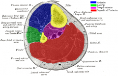
- anterior
- lateral
- superficial posterior
- deep posterior |
|
|
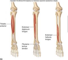
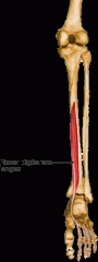
Anterior Compartment |
- primarily dorsiflexors of the ankle & extensors of the toes
- Muscles include: - anterior tibialis - extensor hallucis longus - extensor digitorum longus - fibularis tertius - deep fibular nerve |
|
|
Retinaculums |
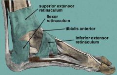
- strong bands which cross the anterior aspect of the ankle and binds the extensor tendons
- Superior Extensor Retinaculum & Inferior Extensor Retinaculum |
|
|
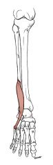
Tibialis Anterior (Origin & Insertion) |
Origin: Lateral condyle & superior 1/2 of the lateral surface of the tibia & interosseus membrane
Insertion: Medial & inferior surfaces of medial cuneiform & base of the 1st metatarsal |
|
|
Tibialis Anterior (Action, Innervation, Blood Supply, Strengthen/Stretching) |
Action: dorsiflex the ankle & invert the foot
Innervation: deep fibular nerve
Blood Supply: anterior tibial artery
Strengthen: dorsiflex & invert against band or resist again hand
Stretch: plantarflex & evert |
|
|
Extensor Digitorum Longus (Origin & Insertion) |
Origin: Lateral condyle & superior 3/4 medial surface of fibula & interosseus membrane
Insertion: Middle & distal phalanges of the lateral 4 digits |
|
|
Extensor Digitorum Longus (Action, Innervation, Blood Supply, Strengthen & Stretching) |
Action: extends lateral 4 digits & dorsiflexes the ankle
Innervation: deep fibular nerve
Blood Supply: anterior tibial nerve
Strengthen: dorsiflex & extend digits against resistance
Stretch: plantarflex & flex digits |
|
|
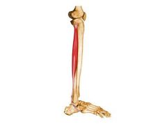
Extensor Hallucis Longus (Origin & Insertion) |
Origin: middle part of the anterior surface of the fibula & interosseus membrane
Insertion: dorsal aspect of the base of the distal phalanx of the great toe |
|
|
Extensor Hallucis Longus (Action, Innervation, Blood Supply, Strengthen & Stretch) |
Action: extends the great toe & dorsiflexes the ankle
Innervation: deep fibular nerve
Blood Supply: anterior tibial artery
Strengthen: extend great toe & dorsifex against resistance
Stretch: flex great toe & plantarflex |
|
|
Fibularis Tertius (Origin & Insertion) |
Origin: inferior 1/3 of anterior surface of the fibula & interosseus membrane
Insertion: dorsum of the base of the 5th metatarsal |
|
|
Fibularis Tertius (Action, Innervation, Blood Supply, Strengthen & Stretch) |
Action: dorsiflex ankle & evert foot
Innervation: deep fibular nerve
Blood Supply: anterior tibial artery
Strengthen: dorsiflex & evert against resistance
Stretch: plantarflex & invert |
|
|
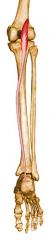
Lateral Compartment |
- primarily everters
- fibula (laterally)
- fascias anteriorly & posteriorly
Includes: - fibularis (peroneal) longus & brevis - superficial fibular nerve |
|
|
Fibular Retinaculum |

Held in place by the superior & inferior fibular retinaculum |
|
|
Fibularis Longus (Origin & Insertion) |
Origin: head & superior 2/3 of lateral surface of the fibula
Insertion: base of 1st metatarsal & medial cuneiform |
|
|
Fibularis Brevis (Origin & Insertion) |
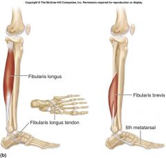
Origin: inferior 2/3 of lateral surface of the fibula
Insertion: dorsal surface of the tuberosity on the lateral side of the base of the 5th metatarsal |
|
|
Fibularis Longus & Brevis (Action, Innervation, Blood Supply, Strengthen & Stretch) |
Action: everts foot & plantarflexes ankle (weakly)
Innervation: superficial fibular nerve
Blood Supply: anterior tibial artery & fibular artery
Strengthen: everted toe raises & plantarflex and evert against reistance
Stretch: dorsiflex & invert |
|
|
Superficial Posterior Compartment |
- primarily ankle plantarflexors - triceps surae
- divided from deep posterior compartment from a fascia
- includes: - grastrocnmeius - soleus - plantaris |
|
|
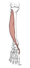
Gastrocnmeius (Origin & Insertion) |
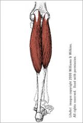
Origin: Lateral head: lateral aspect of the lateral condyle of the femur Medial head: popliteal surface of the femur, superior to the medial condyle
Insertion: posterior calcaneal tubercle via the calcaneal tendon |
|
|
Gastrocnmeius (Action, Innervation, Blood Supply, Strengthen & Stretch) |
Action: plantarflexion of the ankle when knee is extended; raises heel when walking; flexes knee
Innervation: tibial nerve
Blood Supply: popliteal artery/posterior tibial artery
Strengthen: |
|
|
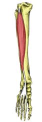
Soleus (Origin & Insertion) |
Origin: posterior aspect of the head of the fibula, superior 1/4 of the posterior surface of the tibial soleal line and the medial border of the tibia
Insertion: posterior calcaneal tubercle via calcaneal tendon |
|
|
Soleus (Action, Innervation, Blood Supply, Strengthen & Stretch) |
Action: plantarflexion of the ankle
Innervation: tibial nerve
Blood Supply: popliteal artery, posterior tibial, fibular artery
Strengthen: plantarflex with knee flexed
Stretch: dorsiflexion with knee flexed |
|
|
Plantaris (Origin & Insertion) |
Origin: inferior end of the lateral supracondylar line of the femur and oblique popliteal ligament of the knee
Insertion: medial aspect of the posterior calcaneus via the calcaneal tendon |
|
|
Plantaris (Action, Innveration, Blood Supply, Strengthen & Stretch) |
Action: weakly assist gastrocnemius -plantarflexion of ankle - flexion of knee
Innervation: tibial nerve
Blood Supply: popliteal artery
Strengthen: plantarflex with knee extended
Stretch: dorsiflex with knee extended |
|
|
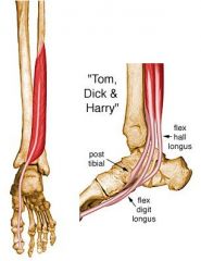
Deep Posterior Compartment |
Includes: - popliteus - posterior tibialis - flexors - posterior tibial artery/fibular artery - tibial nerve |
|
|
Tibialis Posterior (Origin & Insertion) |
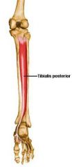
Origin: interosseus membrane, posterior surface of the tibia inferior to the soleal line & posterior surface of the fibula
Insertion: navicular tuberosity, plantar surface of the cuneiforms, plantar surface of the cuboid and bases of 2nd, 3rd, and 4th metatarsals |
|
|
Tibialis Posterior (Action, Innervation, Blood Supply, Strengthen & Stretch) |
Action: plantarflexion & inversion of ankle
Innervation: tibial nerve
Blood Supply: posterior tibial artery
Strengthen: plantarflex & invert against resistance
Stretch: dorsiflex & evert |
|
|
Flexor Digitorum Longus (Origin & Insertion) |
Origin: medial part of the posterior surface of the tibia inferior to the soleal line
Insertion: base of the distal phalanges of the lateral 4 digits |
|
|
Flexor Digitorum Longus (Action, Innervation, Blood Supply, Strengthen & Stretch) |
Action: flexion of lateral 4 digits, plantarflexion of ankle, supports longitudinal arch
Innervation: tibial nerve
Blood Supply: posterior tibial
Strengthen: flex toes by pulling towel/flex toes & plantarflex against resistance
Stretch: extend toes & dorsiflex |
|
|
Flexor Hallucis Longus (Origin & Insertion) |
Origin: inferior 2/3 of posterior surface of the fibula & inferior part of the interosseus membrane
Insertion: base of the distal phalanx of the great toe |
|
|
Flexor Hallucis Longus (Action, Innervation, Blood Supply, Strengthen & Stretch) |
Action: flexes great toe, weakly plantarflexes ankle, supports the medial longitudinal arch
Innervation: tibial artery
Blood Supply: posterior tibial artery
Strengthen: flex great toe against resistance
Stretch: extend great toe & dorsiflex |
|
|
Intrinsic Muscles of the Foot |
Include: - abductor hallucis: abducts great toe - flexor digitorum brevis: flexes lateral 4 digits - abductor digiti minimi: abducts 5th digit - quadratus plantae: supports arch - lumbricals: help flexes lateral 4 digits - flexor hallucis brevis: flexes great toe - flexor digiti minimi brevis: flexes 5th digit - plantar interossei (3) - PAD (adducts metatarsals) - dorsal interossei (4) - DAB (abducts metatarsals) |