Reading...
![]()
Play button
![]()
Play button
![]()
Use LEFT and RIGHT arrow keys to navigate between flashcards;
Use UP and DOWN arrow keys to flip the card;
H to show hint;
A reads text to speech;
56 Cards in this Set
- Front
- Back
- 3rd side (hint)
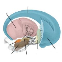
Label these parts of the Basal Ganglia
|
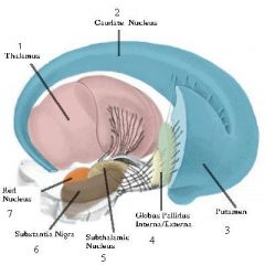
1. Thalamus 2. Caudate Nucleus 3. Putamen 4. Globus Pallidus Interna/Externa 5. Subthalamic Nucleus 6. Substantia Nigra 7. Red Nucleus
|
|
|
|
Virtually all inputs to the basal ganglia arrive via ________. Outputs leave the basal ganglia via the ________ ________ ________ and the ________ ________ ________ ________.
|
Inputs arrive via the STRIATUM and outputs exit via the GLOBUS PALLIDUS INTERNA and SUBSTANTIA NIGRA PARS RETICULATA
|
Inputs enter STRaight through the STRiatum and outputs Go out via the Globus Pallidus and Substantia Nigra
|
|
|
Which two structures comprise the "striatum" in the basal ganglia?
|
Caudate Nucleus and the Putamen.
|
|
|
|
Which two structures comprise the "lentiform" nucleus?
|
Putamen and the Globus Pallidus.
|
It's called lentiform because it looks like a lentil
|
|
|
What are the two parts that comprise the globus pallidus?
|
Internal segment and external segment.
|
|
|
|
What is the internal capsule and what is its relation to the basal ganglia?
|
The internal capsule is a continuation of the corona radiata as it funnels down into more compact tracts.
Location: Passes through the gaps between the cellular bridges. |
Pages 692-693 in Blumenfeld
|
|
|
The head of the caudate connects primarily to the ________ lobe, the body of the caudate to the ________ lobe, and the tail to the ________ lobe.
|
Frontal lobe, Parietal lobe, and Temporal lobe.
|
Remember by visualizing what these structures are closest to within the brain.
|
|
|
What are the four "channels" of the basal ganglia.
|
Cognition, Occularmotor, Motor, and Emotion.
|
Remember the word "COME"
|
|
|
What do the terms "bradykinesia" and "bradyphrenia" mean?
|
Slow moving and slow thinking.
|
"brady"=slow (as in bradycardia=slow heart rate)
"phrenia"=thinking (as in schizophrenia=split thinking) "kinesia"=movement |
|
|
The ________ pathway travels from the striatum and has a net excititory effect on behavior and the ________ pathway has a net inhibitory effect on behavior
|
Direct Pathway and Indirect Pathway.
|
DIrect=Disinhibitory
INdirect=Inhibitory |
|
|
In Parkinson's Disease, the ________ pathway is impaired, causing a reduction in excitation of movement and thinking.
|
Direct Pathway
|
DIrect=Disinhibitory pathway activity is reduced, causing bradykensia and bradyphrenia.
|
|
|
In Huntington's Disease, the ________ pathway is impaired, causing a net increase in movement.
|
Indirect Pathway
|
INdirect=Inhibitory pathway activity is reduced, causing ballismus.
|
|
|
In addition to the better recognized "prototype" disorders associated with the basal ganglia (Parkinson's & Huntinton's) what are two other disorders associated with basal ganglia abnormalities?
|
Obsessive-Compulsive Disorder and Tourette's Syndrom.
|
|
|
|
Clumsy stiff movements and hyperreflexia caused by corticospinal & upper motor neuronal damage are known as ________, while abnormal movements caused by basal ganglia dysfunction are known as ________, while jerky, uncoordinated movements associated with cerebellum damage is known as ________.
|
Spasticity, Dyskinesia, Ataxia.
|
|
|

Label these parts of the Cerebellum
|

1. Horizontal Fissure 2. Primary Fissure 3. Vermis 4. Anterior Lobe 5. Posterior Lobe.
|
|
|
|
The primary fissure of the cerebellum divides the cerebellum in an ________ lobe and ________ lobe.
|
Anterior & Posterior.
|
Primary fissure is a very deep fold which runs HORIZONTALLY.
|
|
|
The cerebellum forms the roof of the ____ ventricle.
|
Fourth Ventricle.
|
|
|
|
In the cortex, ridges are referred to ________ while cerebellum ridges are called ________.
|
Gyri & Folia
|
Think Foliage because they look like leaves (Folia=leaves)
|
|
|
The thick, durable shealth which looks much like the dura matter and separates the cerebellum from the inferior occipital lobes is called ________ ________.
|
Tentorium Cerebelli
|
|
|
|
The three main functional regions of the cerebellum are
_____ & ________ lobe, ________ hemispheres, and ________ hemispheres. |
Vermis and Flocculonodular lobe, Intermediate hemisphere, and Lateral hemisphere.
|
Remember the mnemonic "The Volleyball Flew Into the Lake."
|
|
|
The Vermis is important in control of ________ muscles.
|
Proximal and trunk muscles.
|
Lesion cause truncal ataxia and vertigo.
|
|
|
The flocculonodular lobes are important in ________-_____ control.
|
Vestibulo-occular control (balance and related eye-movements).
|
Recall that when someone is very dizzy/drunk, their eye movements tend to be affected as well.
|
|
|
The intermediate hemispheres is mainly involved in control of more distal (distant) parts of the appendicular muscles in the _____ and _____
|
Arms and Legs.
|
"Appendicular" refers to the word "Appendage." Arms and legs are both appendages.
|
|
|
The largest functional unit of the cerebellum is the ________ hemispheres and is involved in ______ ________ for the extremities.
|
Lateral Hemisphere, motor planning.
|
It's also involved in cognitive functioning.
|
|
|
Ataxia always occurs on the ________ side of a lesion on the cerebellum or nearby structures.
|
Ipsilateral.
|
|
|
|
Ataxia consists of two different components or aspects:________ & ________.
|
Dysmetria and Dysrhythmia.
|
DYSMETRIA=Difficulty judging the required effort to move muscles through a given distance--thus overshooting or undershooting a traget.
DYSRHYTHMIA=Deficit in the timing or sequencing of movement. |
|
|
The walls of the forth ventricle are formed by the ________ ________.
|
Cerebellar Peduncles.
|
There are three peduncles: Superior, Middle, and Inferior.
|
|
|
All cerebellar axons projecting upward are ________ while all axons projecting downward are ________.
|
Excitatory & Inhibitory.
|
Upward projecting axons are from mossy & climbing fibers, downward projecting axons are from purkinje, stelate, basket, and golgi cells.
|
|
|
The Superior peduncle contains primarily ________ from the cerebellum, while the middle and inferior contain primarily ________ from the cerebellum.
|
Outputs & Inputs.
|
Mnemonic: the highest peduncle (superior) is closest to the higher levels of processing--therefore it makes sense that it contains outputs. Conversely, it makes sense that the two lower peduncles contain inputs.
|
|
|
The Tentorium Cerebelli, which has sharp, stiff edges can cause damage if the brain ________ during swelling.
|
Herniates.
|
|
|
|
Cerebellar lesions are associated with ________ _____ in contrast to basal ganglia lesions which are associated with ________ _____.
|
Intentional tremor & resting tremor.
|
|
|
|
What are two ways cerebellar and basal ganglia functions are similar?
|
1. smoothing motor movement 2. involvment in higher cognitive processing.
|
|
|
|
The "prototypical disease" of the cerebellum and it's symptoms are what?
|
Alcohol Intoxication. Sx: "whirlies" (vertigo, poor balance); unstable gait (truncal ataxia); awkward arm and leg movements (appendicular ataxia); slurred articulation; slowed thinking and poor judgment.
|
|
|

Label the main structures of the the brain stem and surrounding structures.
|

1. Medulla Oblongata 2. Pons 3. Midbrain 4. hypthalamus 5. Thalamus 6. Diencephalan 7. Corpus Callosum 8. Cerebral Acqueduct 9. Reticular Formation 10. Spinal Cord
|
|
|
|
The somatic nervous systems consisits of _______ nerves that convey messages from sense organs to the CNS and ________ nerves that carry motor signals from the CNS to muscles.
|
Afferent & Efferent
|
Afferent for Away from the periphery and Efferent for Exit from the central.
|
|
|
The central nervous system is covered by three protective layers Meninges. These three layers are, from superior to inferior.
|
1. Dura 2. Arachnoid 3. Pia
|
Thinking PAD for padding.
|
|
|
The inner surface of the skull has ridges that divide the cranial cavity into several areas called ________.
|
Fossae.
|
|
|
|
The three fossae are ________, ________, & ________.
|
Anterior Fossa, Middle Fossa, & Posterior Fossa.
|
Think MAP
|
|
|
The meninges form three spaces called?
|
Epidural Space, subdural space, & Subarachnoid space.
|
|
|
|
When referring to orientation below the midbrain, the term equivalent to anterior is ________, posterior is ________, superior is ________, & inferior is ________.
|
Ventral, Dorsal, Rostral, & Caudal.
|
|
|
|
When referring to orientation above the midbrain, the term equivalent to anterior is ________, posterior is ________, superior is ________, & inferior is ________.
|
Rostral, Caudal, Dorsal, & Ventral
|
|
|
|
CSF is produced by the ________ ________.
|
Choroid Plexus.
|
|
|
|
________ are holes spread throughout the skull. The largest is known as the ________ ________.
|
Foramina & Foramen magnum.
|
Latin translation means big hole.
|
|
|
When the ventricles become enlarged, is condition is called what?
|
Hydrocephalus.
|
|
|
|
A dissection which cuts through the brain from top to bottom, so that the plane is parallel to the face is called the ________ plane of dissection.
|
Coronal
|
|
|
|
The plane which divides the two hemispheres and is derived from the Latin word for "archer" is call the ________ plane of dissection.
|
Sagittal
|
|
|
|
The plane of dissection which is parallel to the ground is called the ________ plane of dissection.
|
Horizontal.
|
|
|
|
The four lobes of the brain are:________ lobe, ________ lobe, ________ lobe, ________ lobe.
|
Frontal lobe, Parietal lobe, Temporal lobe, & Occipital lobe.
|
|
|
|
Which lobe is most closely assocaited with vision?
|
Occipital lobe.
|
|
|
|
Which lobe is most closely associated with hearing and language?
|
Temporal lobe.
|
|
|
|
Which lobe is most closely associated with "executive" functions?
|
Frontal lobe
|
|
|
|
The deep fold which divides the brain into left and right hemispheres is called the ________ ________.
|
Longitudinal Fissure.
|
|
|
|
Primary functions of the medulla include: ________ & ________
|
Respiration & Blood Pressure.
|
|
|
|
The "folds" in the brain are called ________, while the "bumps" are called ________.
|
Sulci (sulcus) & Gyri (gyrus).
|
|
|
|
The words "tract", "commissure", "association fibers", & "projection fibers" all refer to a collection of ______ ______, which represent the part of a neuron called the ______.
|
White matter & Axon.
|
|
|
|
the ________ is especially important because it is a sensory relay station for many different senses.
|
Thalamus.
|
|