![]()
![]()
![]()
Use LEFT and RIGHT arrow keys to navigate between flashcards;
Use UP and DOWN arrow keys to flip the card;
H to show hint;
A reads text to speech;
19 Cards in this Set
- Front
- Back
|
vulva |
|
|
Labia Minora
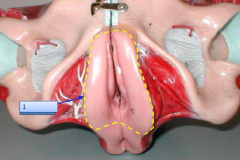
Vaginal Orifice Labia Majora Anus Obstetric Perineum |
|
|
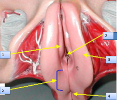
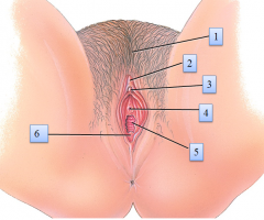
Mons Pubis
Prepuce Clitoris External Urethral Orifice Vaginal Orifice Hymen |
|
|
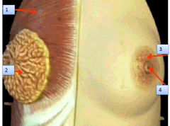
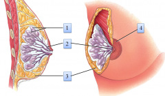
Pectoralis Major Muscle
Mammary Gland AreolaNipple |
|
|
Lobule with Alveoli
Lactiferous Sinus Adipose Tissue Areola |
|
|
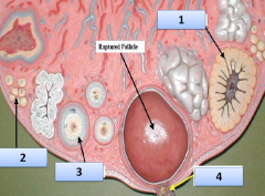
Corpus Luteum
Primordial Follicle Primary follicle Secondary Oocyte |
|
|
Follicular Cells
Zona Pellucida Primary Oocyte |
|
|
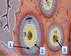
Primordial Follicle
Ovarian Medulla Primary Follicle Ovarian Cortex |
|
|
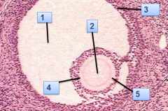
Antrum
Oocyte Granulosa Cells Corona Radiata Zona Pellucida |
|
|
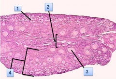
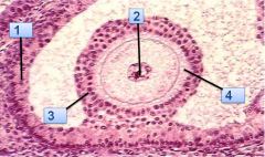
Granulosa Cells
Oocyte Corona Radiata Zona Pellucida |
|
|
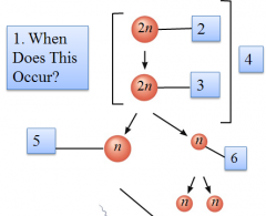
Prenatally
Oogonium Primary Oocyte Mitosis Secondary Oocyte First Polar Body |
|
|
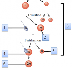
Spermatozoon
Secondary Oocyte Meiosis II Ovum Second Polar Body Zygote |
|
|
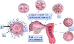
Corona Radiata
Zona Pellucida Secondary Oocyte Morula Blastocyst Cleavage Mitosis |
|
|
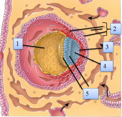
Yolk Sac
Chorion Amnion Amnionic Cavity Bilaminar Embryonic Disc |
|
|
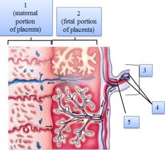
Decidua Basilis
Chorion Umbilical Cord Umbilical Arteries Umbilical Vein |
|
|
Draw a line below to indicate the end of the embryonic phase/beginning of the fetal phase.How did you know where to draw the line?
|
The line is drawn after the 8th week of development. The embryonic period is where the structures are being made – fetal period is where they are enlarging.
|
|
|
sperm cells rely on __________ (an enzyme found in the acrosome of spermatozoa) to disperse the corona radiata from the zona pellucida of the secondary (ovulated) oocyte
|
hyaluronidase |
|
|
exist during pregnancy between the developing fetus and mother |
chorion |
|
|
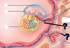
Connecting stalk Chorionic villus |