![]()
![]()
![]()
Use LEFT and RIGHT arrow keys to navigate between flashcards;
Use UP and DOWN arrow keys to flip the card;
H to show hint;
A reads text to speech;
25 Cards in this Set
- Front
- Back
|
Select all that are true about the histology and general organization of the esophagus a. The epithelium consists of thousands of beating cilia which are collectively known as the brush border. b. The muscularis mucosae is responsible for peristaltic contractions. c. A lacteal (that absorbs and transports nutrients) is found in the Submucosa. d. The submucosa is richly supplied with blood vessels, nerves, and glands. e. The most superficial layer is the adventitia. f. The epithelium consists of stratified squamous epithelium. g. The muscularis consists of two layers of smooth muscle. running at right angles to each other. h. The most superficial layer is the serosa. |
d e f g |
|
|
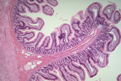
Select all that are true about the histology and general organization of the small intestine a. The most superficial layer is the serosa. b. A lacteal (that absorbs and transports nutrients) is found in the Submucosa. c. The epithelium consists of thousands of beating cilia which are collectively known as the brush border. d. The most superficial layer is the adventitia. e. The mucosa contains numerous microvilli on its apical surface. f. The submucosa is richly supplied with blood vessels, nerves, and glands. g. The muscularis consists of two layers of smooth muscle running at right angles to each other. h. The epithelium consists of stratified squamous epithelium. |
a e f g |
|
Which of the following are true: a. These are pancreatic acini. b. These are Islets of Langerhans (pancreatic islets). c. They release their secretions directly into the blood stream via capillaries. d. These are the exocrine cells of the pancreas. e. They produce digestive enzymes. f. These are pancreatic ducts. g. They use a series of ducts to transport their secretions into the small intestine. h. They produce insulin and glucagon. i. These are the endocrine cells of the pancreas. |
b c h i |
|
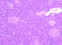
a. These are pancreatic acini.
b. They release their secretions directly into the blood stream via capillaries. c. These are Islets of Langerhans (pancreatic islets). d. These are pancreatic ducts. e. These are the endocrine cells of the pancreas. f. These are the exocrine cells of the pancreas. g. They produce digestive enzymes. h. They produce insulin and glucagon. i. They use a series of ducts to transport their secretions into the small intestine |
a f g i |
|
Which tissue layer (tunic) of the small intestine is the most superficial? |
serosa |
|
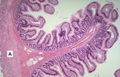
Which tissue layer (tunic) of the small intestine is represented here by the letter 'A' |
muscularis |
|
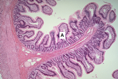
Which tissue layer (tunic) of the small intestine is represented here by the letter 'A' |
mucosa |
|
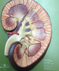
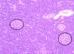
Which of the following statements are true about highlighted area of this image? Select one or more: a. It is called the renal pyramid b. Its apex is in the cortex and its base is the renal papillae c. Its base is in the cortex and its apex is the renal papillae d. You would be able to find the entire nephron in this area e. It is called the renal lobule f. It just contains blood vessels and connective tissue g. This area along with the calyces make up the renal medulla h. You would be able to find the loop of henle and collecting ducts in this area i. This area along with the renal columns make up the renal medulla j. It contains blood vessels, connective tissue, and some of the functional tissue of the kidney |
a c h i j |
|
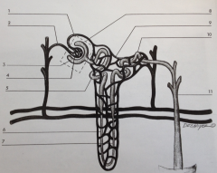
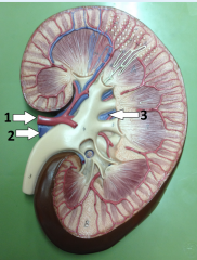
Label |
1 – Efferent arteriole, 2 – Afferent Arteriole, 3 – Bowman's Capsule, 4 – Glomerulus, 8 – Renal corpuscle |
|
Label |
1 – Podocytes, 2 – Endothelium, 4 – Bowman's capsule, 3 – Bowman's space |
|
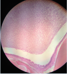
What is the feature seen here and what is its function? |
It is a renal papillae and its purpose is to allow urine to pass out of the renal pyramid and eventually onto the ureter |
|
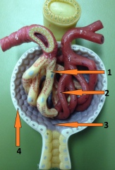
Label |
1 – Renal artery, 2 – Renal vein, 3 – Renal sinus, The indent where 1 and 2 are entering/exiting the kidney – Renal hilus |
|
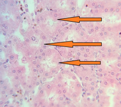
Which of the following statements are true about the feature at the end of the pointers? a. They are the location in the nephron where most of the filtration occurs. b. They are collectively referred to as the bush border and they decrease the sruface area of cells c. In the nephron they are only found in distal convoluted tubules d. They finger like projections of the basal cellular membrane known as villi e. They finger like projections of the apical cellular membrane known as microvilli f. In the nephron they are only found in proximal convoluted tubules g. They are collectively referred to as the brush border and they increase the surface area of cells h. They are the location in the nephron where most of the selective reabsorption occurs. |
e f g h |
|
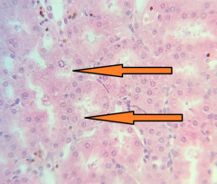
What is the name of the structures at the end of the pointers and what type of epithelium lines their lumen? |
They are proximal convoluted tubules and their lumen is lined by simple cuboidal epithelium that posess microvilli. |
|
|
Which beverage is the control beverage? |
Distilled Water |
|
|
What time point was the control for your beverage? |
30 minutes |
|
|
Why do people with diabetes mellitus have glucose in their urine? |
There is too much glucose in the filtrate for it to be completely reabsorbed into the blood |
|
|
What is water going to have an effect on? |
volume and specific gravity |
|
|
What is v8 going to have an effect on? |
sodium concentration and sodium excretion rate |
|
|
what is tums going to have an effect on? |
pH |
|
|
What is cranberry juice going to have an effect on? |
ph and specific gravity |
|
|
what is beer going to have an effect on? |
volume and specific gravity |
|
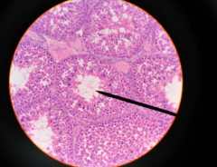
What anatomical structure is shown in this image? |
seminiferous tubules |
|
|
the Ph of the vagina is : Select one or more: a. between 7.4-8.2 due to the presence of potassium hydroxide b. acidic because of glycogen fermentation caused by bacteria c. between 3.5-4.0 due to lactic acid d. high in order to be naturally spermicidal e. between 8.5-11.0 due to lactic acid f. acidic because of glycogen oxidation caused by viruses |
b and c |
|
|
what is the procedure called when the prepuce, which covers the glans, is surgically removed? |
circumcision |