![]()
![]()
![]()
Use LEFT and RIGHT arrow keys to navigate between flashcards;
Use UP and DOWN arrow keys to flip the card;
H to show hint;
A reads text to speech;
90 Cards in this Set
- Front
- Back
|
Exencephaly |
Early manifestation of anencephaly with neutral tissue still present that eventually degenerates |
|
|
Recurrence risk of anencephaly and prevention strategy |
2-5%, folic acid 4mg daily |
|
|
Imaging findings with anencephaly |
No calvarium with no neural tissue above orbits |
|
|
When in gestation can you diagnose exencephaly? |
First trimester |
|
|
Ultrasound findings of exencephaly |
Neural tissue present, abnormal head contour, CRL < dates |
|
13 weeks Identify diagnosis |
Exencephaly |
|
|
Why is amniotic fluid echogenic in anencephalic pregnancies? |
Dissolved neural tissue |
|
|
Differential diagnosis for fetus with anencephalic appearance. Explain differences |
Amniotic band syndrome (slash defects, fetus may appear stuck) Encephalocele (cranium present) Severe microcephaly (cranium present, cerebrum present) Atelencephaly or aprosencephaly (severe craniofacial defects, severe microcephaly plus limb abnormalities) |
|
|
What races have higher risk of anencephaly |
White, Hispanic (not in Hispanic) |
|
|
Definition of cephalocele |
Defect in skull and dura with protrusion of intracranial structures |
|

MRI |
Encephalocele |
|
|
Most common genetic disorder associated with cephalocele |
Meckel Gruber (Encephalocele, polydactyly, polycystic kidneys) |
|
|
Top Differential diagnoses for appearance of cephalocele |
Cystic hygroma Amniotic band syndrome |
|
|
What is cyst within cyst of fetus with a cephalocele? |
Prolapsed forth ventricle |
|
|
Iniencephaly |
Hyperextension neck Encephalocele Cervical spina bifida |
|
|
Majority of cephaloceles are in what portion of the head? |
Occipital |
|

|
Occipital cephalocele with target sign |
|
|
What is an atretic cephalocele? |
Cystic scalp mass that contains durable, fibrous tissue, neutral cells Involuted true cephalocele |
|
|
DDx for scalp mass |
Atretic cephalocele (can be difficult to see small bony defect) , hemangioma, lymphagioma, lipoma, epidermoid cyst |
|
|
Fetal facial mass + hypertelorism = |
Frontal encephalocele |
|
|
Percentage of fetuses with cephalocele that have an additional major anomaly? |
65% |
|

Suspected diagnosis |
Cephalocele |
|
|
Management of fetus with cephalocele |
Detailed anatomy scan, fetal ECHO. Genetic counseling possible amnio Peds surg/neurosurg consult Repeat ultrasound 3rd trimester |
|
|
Mortality rate with cephalocele diagnosed prenatally |
80% |
|
|
Imaging features of agenesis of corpus callosum (ACC) |
Absent CSP Colpocephaly (enlarged occipital horn of lateral ventricle) Abnormal pericallosal artery Teardrop lateral ventricle |
|
|
Percentage of fetuses with ACC with other (1) CNS anomalies and (2) non CNS anomalies |
1) 85% 2) 65% |
|

|
Absent CSP, concern for ACC, colpocephaly with tear drop shape ventricle |
|
|
What can you mistake on ultrasound for a corpus callosum? |
Paired fornices |
|
|
Pregnancy management for ACC |
Detailed US, fetal ECHO genetics consult, amnio (with karyotype and microarray) Third trimester us to reassess hydrocephaly Consider MRI |
|
|
Syndromes associated with ACC |

Dandy Walker (common) Chiari 2 Walker Warburg Aicardi Meckel Gruber Apert |
|
|
What should be in your differential for absent CSP? |
ACC SOD (fused frontal horns) Lobar holoprosencephaly Open lip schizencephaly Isolated absent CSP (Dx of exclusion) |
|
|
AVID anomaly |
Asymmetric Ventriculomegaly with Interhemispheric cyst and dysgenesis of the corpus callosum |
|
|
DDx for interhemispheric cystic mass |
Porencephalic cyst, arachnoid cyst, schizencephaly |
|
|
When would you consider AVID? |
Markedly asymmetric ventriculomegaly Normal appearing adjacent brain |
|

26 wks |
AVID |
|
|
Pregnancy management of AVID |
Follow for hydrocephalus Amnio not necessary if no other findings |
|
|
Aprosencephaly |
Failed development of prosencephalon |
|
|
DDx of aprosencephaly |
Anencephaly Holoprosencephaly Hydrancephaly |
|
|
How to differentiate between aprosencephaly and anencephaly |
Similar cranial contour Calvarium present in aprosencephaly |
|
|
Findings in aprosencephaly |
Severe microcephaly Limb abnormalities may be present |
|
|
Incidence of CPCs |
1-3% |
|
|
Size of choroid plexus cyst to meet criteria |
2mm |
|

Diagnosis |
Bilateral choroid plexus cysts (CPCs) |
|
|
Chromosomal anomaly seen with CPCs |
T18 |
|
|
Pregnancy management for CPCs |
Detailed ultrasound (ensure open hands, normal cardiac anatomy) Fetal ECHO only if can't clear cardiac views Consider NIPT Amnio if other anomalies No other ultrasound f/u indicated if everything else normal |
|
|
3 forms of holoprosencephaly |
1. Alobar (most severe, complete lack of division and no midline structures) 2. Semilobar (intermediate form, posterior portion of the brain does divide) 3. Lobar (ventral neocortex fused, ACC, ventricles divided) |
|
|
Major ddx for appearance of lobar holoprosencephaly |
SOD |
|
|
Mild Interhemispheric Variant (MIHV) |
Form of holoprosencephaly 1. failure of separation of posterior frontal and parietal cortex 2. Incomplete separation thalami 3. Absent body of CC |
|
|
Imaging findings with alobar holoprosencephaly... What can you see in first trimester? |
Mono-ventricle No falx No butterfly appearance of choroid plexus in first trimester |
|
|
Imaging findings in semilobar holoprosencephaly |
Absence of interhemispheric separation but some separation posteriorly Fusion of anterior horns of lateral ventricles Partial separation of thalami Hypotelorism |
|
|
Imaging findings with Lobar holoprosencephaly |
Absent CSP, CC |
|
|
Imaging findings MIHV |
Incomplete separation of the thalami and caudate nucleus, absent body of the corpus callosum Brain heterotopias ("out of place") |
|
|
Percent of newborns with holoprosencephaly who have chromosomal anomaly? What types |
25-45% T18, T13, triploidy |
|

14 weeks gestation Diagnosis |
Alobar holoprosencephaly |
|
|
Management of pregnancies with holoprosencephaly |
Detailed imaging Genetic counseling, amnio Offer termination Peds consult Consider fetal MRI if lobar form Repeat imaging 3rd trimester |
|
|
Porencephalic cyst |
Fluid filled cavity that takes up space where normal brain tissue used to be, occurs after ischemic stroke, hemorrhage or infection Not a true cyst |
|
|
DDx when you see large cystic space in brain |
Porencephalic cyst Arachnoid cyst Interhemispheric cyst Hydrancephaly Schizencephaly |
|
|
Will you see mass effect with porencephalic cyst? |
No, the space is caused by degenerating tissue so no mass effect |
|
|
Thrombophilias associated with increased risk of fetal perinatal stroke |
FVL, Protein C def, APLAS |
|

Suspected diagnosis |
Porencephalic cyst |
|
|
Management of pregnancy for fetus with porencephalic cyst |
Detailed US and MRI Generic counseling Thrombophilia work up both parents TORCH titers Peds neurosurgery consult Offer termination Third trimester scan to monitor hydrocephaly |
|
|
Leading cause of CP |
Porencephaly |
|
|
Ventriculomegaly vs hydrocephaly |
Often used interchangeably but technically hydrocephaly is when ventricle > 15mm and ventriculomegaly just mild enlarged |
|
|
Most common cause of ventriculomegaly |
Aqueductal stenosis |
|
|
Imaging findings with aqueductal stenosis |
Hydrocephalus Macrocephaly Brain mantle thinned May not see corpus callosum Posterior structures normal |
|
|
What hands finding do you see with aqueductal stenosis |
Bilateral abducted thumbs (>50%!) |
|
|
Management of pregnancy with fetal hydrocephaly |
Detailed US and MRI Genetic counseling/ amnio TORCH work up Serial US to monitor hydrocephaly Delivery planning Peds neurosurg consult |
|
|
DDx appearance holoprosencephaly |
Aprosencephaly Hydrancephaly (no vertebral tissue, normal face) Aqueductal stenosis (falx present, thalami not fused) |
|
|
Risk factors for holoprosencephaly |
Diabetes (1%risk) Retinoic acid Alcohol |
|

32 weeks |
Lobar holoprosencephaly |
|
|
SOD definition (3 parts) |
Optic nerve hypoplasia Midline brain malformation (absent CSP) Hypopituitarism |
|
|
Imaging findings in SOD |
Absent CSP frontal horns in communication across midline |
|

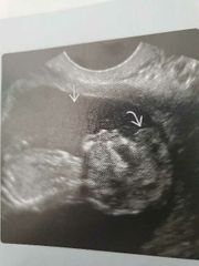
28 weeks Findings and possible diagnosis |
SOD Straight arrow =fused frontal horns Curved arrows= fornices |
|
|
Findings with Walker Warburg |
Lissencephaly, hydrocephalus, encephalocele, micropthalmia, cataracts, kinking of mesencephalic-pontine junction Most severe congenital muscular dystrophy(Smooth brain, eye problems, weakness) |
|
|
Rhomboencephalosynapsis |
Fusion of cerebellar hemispheres and vermian agenesis Associated with aqueductal stenosis |
|

|
Aqueductal stenosis with rhomboencephalosynapsis See cerebellum is small and abnormally shaped with no vermis |
|

Diagnosis and what you see in each picture |
Walker warburg You can see hydrocephalus, kinking of brainstem, cerebellar hypoplasia, eye malformation, lissencephaly |
|
|
Arnold Chiari cause |
Open NTD leading to CSF leakage and lack of development of posterior fossa |
|
|
DDx borderline ventriculomegaly |
Normal variant NTD Early hydrocephaly ACC TORCH |
|
|
Classic fruit findings with Arnold Chiari |
Lemon and banana |
|
|
Percent cases of borderline ventriculomegaly with associated structural or chromosomal anomalies |
~40% |
|
|
Percent spina bifida associated with chromosomal anomalies (which ones? ) |
10% T13/18 rarely 22q11 |
|
|
Most common chromosomal anomaly associated with borderline ventriculomegaly |
T21 |
|
|
Pregnancy management borderline ventriculomegaly |
Detailed US, fetal ECHO TORCH titers Genetics/amnio MRI brain Serial US |
|
|
Dandy Walker findings |
Enlarged posterior fossa Elevated cerebellar tentorium Dilation 4th ventricle |
|
|
Ddx enlarged cisterna magna |
Dandy Walker Posterior fossa Arachnoid cyst Blake pouch cyst Mega cisterna magna Vermian hypoplasia |
|
|
US findings Dandy Walker |
Large cisterna magna that communicates with 4th ventricle Absent vermis with splayed cerebellar hemispheres Elevated tentorium VM |
|

Likely diagnosis |
DWM |
|
|
Pregnancy mgmt when ultrasound findings concerning for DWM |
Detailed US, ECHO Genetics, amnio with karyotype and microarray MRI Peds NS, neuro consults Serial US |
|
|
IUFD risk with DWM |
~15% |