![]()
![]()
![]()
Use LEFT and RIGHT arrow keys to navigate between flashcards;
Use UP and DOWN arrow keys to flip the card;
H to show hint;
A reads text to speech;
77 Cards in this Set
- Front
- Back
|
Vuvla disorders:
What is Lichen sclerosis? What is Lichen simplex chronicus? |
Lichen sclerosis: postmenopausal women, thinning of the epidermis, small risk for SCC.
Lichen simplex chronicus: leukoplakia due to squamous hyperplasia, small risk for SCC. |
|
|
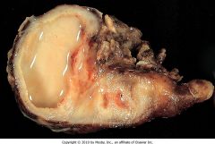
Diagnose: painful nodule on the labia majora; benign tumor of the apocrine sweat gland.
|
Papillary hidradenoma.
|
|
|
Name the important risk factors for SCC on the vulva.
|
HPV type 16, smoking cigarettes, immunodeficiency.
|
|
|
This vulvar tumor is an intraepithelial adenocarcinoma which spreads along the epithelium but rarely invades the dermis. It can also be located on the nipple. What test can be used to identify the malignant cells?
|
Extramammary Paget's disease - red, crusted vulvar lesion. Malignant Paget's cells contain mucin. Mucin is periodic acid-Schiff (PAS) positive.
|
|
|
Sexually active female presents with creeping, raised sores on her genitalia. Some have healed and are now scars. Biopsy reveals a gram-negative infection resulting in granuloma formation.
|
Calymmatobacterium granulomatis of the Klebsiella family is an STD that causes granuloma inguinale (donovanosis). Organism phagocytized by macrophages (Donovan bodies).
|
|
|
This microbe is the second most common cause of vaginitis in the US. It presents as a pruritic vaginitis with a white discharge and fiery red mucosa. Diagnosis?
|
Candida albicans - yeast and pseudohyphae. It can be part of the normal vaginal flora. Risk factors: diabetes, antibiotics, pregnancy, OCP.
|
|
|
Whats the pathogenesis of Chlamydia trachomatis?
|
STD: incubation period 7-12 days after exposure; red inclusions (reticulate bodies) in infected metaplastic squamous cells; reticulate bodies divide to form elementary bodies, which are the infective bodies.
|
|
|
What is Fitz-Hugh-Curtis syndrome?
|
Perihepatitis. Scar tissue between peritoneum and surface of liver from pus from pelvic inflammatory disease. Can be caused by chlamydia trachomatis.
|
|
|
What disease do the subtypes L1, L2, and L3 of Chlamydia trachomatis cause?
|
Lymphogranuloma venereum. Papules with no ulceration; inguinal lymphadenitis with granulomatous microabscesses and draining sinuses. Lymphedema of scrotum or vulva. RF: rectal strictures.
|
|
|
Most common cause of vaginitis. Malodorous vaginal discharge. Vaginal pH > 4.5. What cells would be present?
|
Gardenerella vaginalis:
Clue cells - organisms adhere to squamous cells. RF: preterm delivery and low-birth weight newborns. Tx: metronidazole. |
|
|
Scrotal chancroid lesion in an HIV-positive patient. The patient also had suppurative inguinal nodes and perianal ulcers. What is the gram-negative organism that causes this?
|
Haemophilus ducreyi. Diagnosis with Gram stain (school of fish appearance).
|
|
|
What histological changes occur in HPV infection?
|
HPV 16 and 18 have the highest association with dysplasia and squamous cancer. Virus produces koilocytic changes in squamous epithelium. Cells have wrinkled pyknotic nuclei surrounded by clear halo.
|
|
|
What are some complications of Neisseria gonorrhoeae STD infection?
|
Ectopic pregnancy, male sterility, disseminated gonococcemia (C6-C9 deficiency), septic arthritis, Fitz-Hugh-Curtis syndrome.
|
|
|
Patient presents with painless chancre on the penis. What will his next symptoms be if he is infected by a spirochete?
|
Treponemal disease.
Secondary: maculopapular rash on trunk, palms, soles; generalized lymphadenopathy; condylomata lata (flat lesions in same area as condylomata acuminata (venereal warts)). Tertiary: neurosyphilis, aortitis, gummas. |
|
|
Women presents with greenish, frothy discharge from her strawberry colored cervix and fiery red vaginal mucosa. Diagnosis?
|
Trichonomas vaginalis - protozoan with jerky motility.
|
|
|
What is Rokitansky-Kuster-Hauser syndrome?
|
Absence of the upper vagina and uterus. Anatomic cause of primary amenorrhea.
|
|
|
Gartner's duct cyst is due to the remnant of what duct?
|
Wolffian (mesonephric) duct. Cyst is on the lateral wall of the vagina.
|
|
|
Necrotic, grape-like mass protrudes from the vagina. Diagnosis?
|
Embryonal rhabdomyosarcoma.
|
|
|
What is associated with clear cell adenocarcinoma of the vagina/cervix?
|
Diethylstilbestrol (DES) exposure and adenosis precursor lesion (mullerian gland remnants).
|
|
|
What is the differential for acute cervicitis (inflammation in the transformation zone of the cervix)? What are the clinical findings?
|
Chlamydia trachomatis, N. gonorrhoeae, Trichomonas vaginalis, Candida, HSV-2, HPV. CF: vaginal discharge (most common complaint), pelvic pain, dyspareunia, painful on palpation, bleeds easily when obtaining cultures, exudate on cervical os.
|
|
|
How do you diagnose Trichomonas acute cervicitis?
|
Clinical findings (discharge, pelvic pain, easy bleed...) and wet mount.
|
|
|
What cells must be present on a PAP to indicate proper sampling?
|
Metaplastic squamous cells (in the transformation zone) or mucus-secreting columnar cells (from endocervix).
|
|
|
In a pap smear what are superficial squamous cells a sign of? What do intermediate squamous cells indicate? Parabasal?
|
Superficial - adequate estrogen.
Intermediate - adequate progesterone. Parabasal - lack of estrogen and progesterone. Normal nonpregnant - 70% superficial 30% intermediate. |
|
|
What are the clinical findings associated with cervical polyps?
|
Postcoital bleeding, vaginal discharge. Non-neoplastic polyp that protrudes from the cervical os. They arise from the endocervix and are most common in multigravida women and perimenopausal women 30-50. Not precancerous.
|
|
|
Cervical intraepithelial neoplasia is the precursor to cervical cancer. How long does it take to progress from CIN I (out of III) to invasive cancer?
|
~20 years. Average age for cervical cancer is ~45 years old.
|
|
|
45 year old women presents with malodorous discharge and postcoital bleeding. History reveals she has had multiple partners, she smoked, and she use to be on OCPs. Blood work up reveals an increase in BUN. Its also confirmed that she does not have an infection. What should you be concerned about?
|
Cervical cancer: 80% are SCC; other - small cell cancer and adenocarcinoma. Postrenal azotemia leading to renal failure is common.
|
|
|
What decreases the amount of clotting during menstruation?
|
Plasmin. Increased clotting is a sign of menorrhagia.
|
|
|
What factors decrease sex hormone-binding globulin (SHBG) and what is the affect?
|
SHBG is increased by estrogen and decreased by androgens, obesity, and hypothyroidism. Decreased SHBG increased free testosterone level which can cause hirsutism in women.
|
|
|
How is plasma volume and RBC mass altered in normal pregnancy?
|
Plasma volume and RBC mass increases, as well as Plasma volume/RBC mass ratio.
This decrease Hb (dilutional effect). |
|
|
Why are pregnant women slightly alkalotic in pregnancy?
|
Respiratory alkalosis is due to the effects of estrogen and progesterone on the respiratory center. A decrease in PCO2 causes a corresponding increase in PO2.
|
|
|
How are thyroid binding globulin, total thyroxine, and free thyroxine levels altered in pregnancy?
|
TBG and total thyroxine are increased, but the free thyroxine remains normal.
|
|
|
Define menopause.
|
No menses for 1 year after age 40.
|
|
|
What is the best serum marker to indicate menopause?
|
FSH.
|
|
|
Where is testosterone in a women produced? Where is DHEA-sulfate?
|
Testosterone - mostly synthesized from the ovary.
DHEA--sulfate - 95% from the adrenal cortex. |
|
|
Name the most common causes of hirsutism/virilization in women.
|
Polycystic ovary syndrome is the most common cause.
Idiopathic. Adrenogenital (21 or 11 hydroxylase deficiency). Drugs: phenytoin, cyclosporin, minoxidil. Leydig cell tumor. Adrenal tumor (Cushing's). Obesity. Hypothyroidism. |
|
|
Why is there an increase risk in endometrial cancer in polycystic ovary syndrome?
|
Patho: increased pituitary LH, decreased FSH. Increased LH increases androgen (hirsutism). Androgens aromatize to estrogen in adipose cells thus increasing endometrial hyperplasia.
|
|
|
What is the most common symptom/complaint of a patient with polycystic ovary syndrome?
|
Oligomenorrhea.
|
|
|
What is the pathology of primary dysmenorrhea?
What is the most common cause of secondary dysmenorrhea? |
Increased PGF - increases uterine contractions.
Endometriosis. |
|
|
What is dysfunctional uterine bleeding?
What is anovulatory DUB? |
Abnormal uterine bleeding unrelated to an anatomic cause (e.g. NOT cancer NOT a polyp).
Anovulatory DUB - excessive estrogen stimulation and a reduced secretory phase. |
|
|
What are three general causes of amenorrhea? (Both primary and secondary causes).
|
1. Hypothalamic/pituitary disorder: decreased FSH, LH, E, PE (hypogonadotropic hypogonadism).
2. Ovarian disorder: increased FSH,LH, but decreased E, PE (hypergonadotropic hypogonadism). 3. End organ defect: prevents the egress of blood. Normal FSH, LH, E, PE. |
|
|
What are some causes of primary amenorrhea?
|
Turners (45, X), imperforate hymen, Rokitansky-Kuster-Hauser syndrome (no vagina), Asherman syndrome (removal of stratum basalis owing to repeat curettage).
|
|
|
1. Do Turners have a Barr body?
2. How can a newborn with Turners by diagnosed at birth? 3. What heart defect is present in Turners? 4. Whats the genetic make up of a Turner with mosaic? 5. Why are Turners patient infertile? |
1. No
2. Lymphedema feet/hands. Nuckle, nuckle, dimple, nuckle. Webbed neck. 3. Preductal coarctation. 4. XO, XX. 5. Menopause before menarche by 2 years - all follicles are gone - streak gonad. Susceptible to dysgerminomas. |
|
|
What is the difference between adenomyosis and endometriosis?
|
Adenomyosis - glands and stroma within the myometrium.
Endometriosis - functioning glands and stroma outside the uterus. |
|
|
Postpartum women presents with lochia, what is the most common cause?
|
Acute endometritis is due to bacterial infection following delivery or abortion. Most common pathogen: Strep agalactiae (other - strep pyogenes, staph, bacteroides fragilis,....). Lochia - purulent or foul vaginal discharge following pregnancy. Fever, uterine tenderness, and abdominal pain.
|
|
|
Women presents with fever, uterine tenderness, and abdominal pain. History reveals that she has an intrauterine device. Swab of the vagina reveals plasma cells. Diagnosis?
|
Chronic endometritis with an actinomyces israelli infection. Other causes: retained placenta and gonorrhea.
|
|
|
26 year old female presents with dysmenorrhea, painful stooling during menses, and infertility complaints. What is the pathogenesis of her complaint?
|
Endometriosis in the pouch of Douglas. Postulated pathogenesis: reverse menses through fallopian tubes, coelomic metaplasia, or vascular/lymphatic spread. Other common sites: ovary (most common), fallopian tubes, intestines.
|
|
|
What test can be done to exclude the diagnosis of endometriosis?
|
Serum cancer antigen 125. High sensitivity, low specificity.
|
|
|
Endometrial polyps are a common cause of menorrhagia in 20-40 year olds. Does it progress to endometrial carcinoma?
|
No.
|
|
|
What are the risks for endometrial hyperplasia?
|
Prolonged estrogen stimulation: early menarche, late menopause, nulliparity, obesity, POS, anovulatory menstrual cycles, Lynch syndrome.
|
|
|
What ages groups are affected by the three most common female gynecologic tumors?
|
Most to least common.
1. Endometrial - 55 2. Ovarian - 65 3. Cervical - 45 |
|
|
What are the three types of endometrial cancer?
|
Well-differentiated: 1) adenoacanthoma (contains foci of benign tissue), 2) adenosquamous (contains foci of malignant tissue).
Papillary adenocarcinoma: 3) highly aggressive. |
|
|
What is a hydatid cysts? Whats the risk of a hydatid cysts?
|
Cystic mullerian remnants. Located around the fimbriated end of the fallopian tube. Risk of torsion - abdominal pain.
|
|
|
Female, 26, presents with fever, abdominal pain, uterine bleeding and a mucopurulent vaginal discharge. You tell the patient she's at risk of infertility and ectopic pregnancies. What are the non-STD causes of her disease?
|
PID - most common cause of female infertility and ectopic pregnancy. STD causes: gonorrhoeae, Chlamydia. Non-STD causes: B. fragilis, streptococci, Clostridium perfringens, Mycobacteria tuberculosis, CMV.
|
|

|
PID - fallopian tubes are filled with pus. Most common cause of hydrosalpinx (pus resorbs, leaving a clear fluid distending the tube).
|
|
|
What is salpingitis isthmica nodosa?
|
Tubal diverticulosis.
|
|
|
What are the most common implantation sights of an ectopic pregnancy?
|
Majority occur within the tubes - broad ampullary portion below the fimbriae.
Ovaries. Abdominal cavity. |
|
|
Female, 30, presents with abdominal pain and tenderness. She also has rebound tenderness in her lower right quadrant. She also has tachycardia and low BP. History reveals its been 6 weeks since her last normal menstrual period.
|
Ectopic pregnancy. Complications: rupture with intra-abdominal bleed - most common cause of death in early pregnancy.
|
|
|
What is the most common ovarian mass (non-neoplastic)?
What is the most common ovarian mass in pregnancy (non-neoplastic)? |
Follicular cyst. Can cause peritonitis. Most regress.
Corpus luteum cysts. |
|
|
What is stromal hyperthecosis?
|
Postmenopausal women with hypercellular ovarian stroma that is producing androgens - hirsutism/virilization. Associated with acanthosis nigricans and insulin resistance.
|
|
|
What genetic factors are linked to ovarian tumors?
|
Mutations of BRCA1, BRCA2.
Lynch syndrome. Turner's syndrome. Peutz-Jeghers syndrome. |
|
|
What is the most common benign ovarian tumor?
What is the most common malignant, bilateral ovarian tumor? |
Both are surface derived.
Serous cystadenoma. Serous cystadenocarcinoma. |
|
|
What ovarian tumor has psammoma bodies?
|
Serous cystadenocarcinoma - psammoma bodies are dystrophically calcified tumor cells. Bilateral.
|
|
|
What is the most common germ cell tumor in a female?
|
Cystic teratoma - a germ cell ovarian tumor. Usually benign.
Struma ovarii type has functioning thyroid tissue. |
|
|
Female, 70, presents with a mass in her ovaries. LDH levels are also increased. You inform her that she has what malignant tumor?
|
Dysgerminoma. Malignant germ cell tumor of the ovary.
|
|
|
Women presents with ascites, right-sided pleural effusion. The tumor on her ovary is removed and the effusion regresses. Diagnosis?
|
Sex-cord stromal tumor of the ovary: Thecoma-fibroma with Meigs' syndrome.
|
|
|
Ovary has signet-ring cells. Is this a metastasis or primary malignancy?
|
Metastasis from hematogenous spread of a gastric cancer - Krukenberg tumor.
|
|
|
1. Most common cause of late pregnancy bleeding?
2. What is the greatest risk factor for abruptio placentae? 3. Most common cause of late pregnancy painless bleeding? |
1. Abruptio placentae.
2. Hypertension greatest risk factor. 3. Placenta previa - implantation over the cervical os. |
|
|
What are the pathological findings of the placenta in a pregnant women with preeclampsia?
|
Premature aging of the placenta.
Multiple placenta infarctions. Spiral arteries show intimal atherosclerosis. Pathogenesis of preeclampsia: 1) abnormal placentation (obstruction of spiral arteries), 2) vasoconstrictions > vasodilators, 3) Net effect is placental hypoperfusion. |
|
|
Patient presents with vaginal bleeding in her fourth month of pregnancy. She reports vomiting. An ultrasound reveals a snowstorm appearance. Her hCG levels are increased and her uterus is too large for the gestational age. What is your diagnosis?
|
Hydatidiform mole (complete). No embryo is present. Mole has 46, XX - both X chromosomes are of paternal origin (androgenesis).
|
|
|
Whats the genetic make up of a partial hydatidiform mole?
|
69 chormosomes (triploid, XY). Egg with 23, X is fertilized by 23, X and 23, Y sperm.
|
|
|
1. What carries a higher risk for choriocarcinoma: complete or partial hydatidiform mole?
2. What is a choriocarcinoma? 3. What are the common metastatic sights of choriocarcinoma? 4. Treatment of choice? |
1. Complete.
2. Malignant tumor composed o trophoblast tissue. 3. Metastasizes to lung and vagina. 4. MTX - great response. |
|
|
Most common cause of:
1. Galactorrhea? 2. Bloody nipple? 3. Purulent nipple discharge? 4. Greenish brown nipple discharge? |
1. Mechanical stimulation of the nipple. (Other: antipyschotics, methyldopa, hypothyroidism).
2. Intraductal papilloma. 3. Acute mastitis - due Staph aureus. 4. Mammary duct ectasia - plasma cell mastitis. |
|
|
Name the benign breast tumors:
1. Develop in 50% of women who receive cyclosporine after renal transplantation. 2. Most common breast tumor in women under 40. 3. Potential to be malignant. 4. Most common cause of bloody nipple discharge in women < 50. |
1. Fibroadenoma (stromal)
2. Fibroadenoma 3. Phyllodes tumor (stromal) 4. Intraductal papilloma (lactiferous ducts or sinuses) |
|
|
1. Most common breast tumor in a women under 35?
2. What is the only fibrocystic change that carries the risk of developing into cancer? |
1. Fibroadenoma (estrogen-sensitive)
2. Atypical ductal hyperplasia (other FCC: fibrosis, sclerosing adenosis). |
|
|
How does Paget's disease of the breast look?
|
Paget's disease is extension of DCIS into lactiferous ducts and skin of nipple producing a rash with or without nipple retraction.
|
|
|
1. What genetic factors increase the risk of breast cancer?
2. What factors decrease the risk for breast cancer? 3. What are the clinical findings in breast cancer? |
1. Mother, sister, BRCA1 or 2, Li-Fraumeni, Ras, ERBB2, RB
2. Breast-feeding, vigorous physical training, health body weight. 3. Painless mass, skin or nipple retraction, painless axillary lymphadenopathy, bone pain with metastasis. |
|
|
What drugs can cause gynocomastia?
|
Durgs that displace estrogen from SHBG: spironolactone, ketoconazole.
Drugs with estrogen activity: DES, digoxin. Drugs that block androgen-R: spironolactone, flutamide. Drugs that decrease androgen production: leuprolide. |

