![]()
![]()
![]()
Use LEFT and RIGHT arrow keys to navigate between flashcards;
Use UP and DOWN arrow keys to flip the card;
H to show hint;
A reads text to speech;
21 Cards in this Set
- Front
- Back
- 3rd side (hint)
|
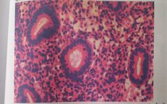
Proliferative Endometrium Comment: endometrial glands: -Round or tubular -uniform in size, shape and distribution -lined by one layer of cubical epithelial cells with oval nuclei. The stroma : - spindle cells -scanty cytoplasm. |
|
|
|
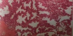
Secretory Endometrium Comment: -the gland show cork-screw appearance with decidualized stroma |
|
|
|
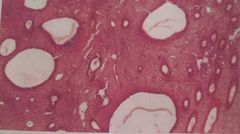
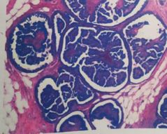
Simple Hyperplasia (Cystic Hyperplasia) Comment: endometrial giands: - Increased number -variable in size and shape, -lined by multiple layers of columnar epithelial cells. -cystic dilatation forming swiss- cheese appearance. The stroma hypercellular |
|
|
|
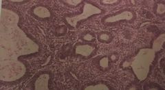
Adenomatous Hyperplasia Comment: the endometrial glands : -variable in size and iregular in shape -lined by multiple layers of columnar epithelium -arranged back to back |
|
|
|
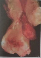
Endometrial Polyp Comment: opened uterus, the uterine cavity shows -a single polyp 3x2 cm with short pedide - smooth greyish surface |
|
|
|
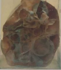
Multiple intramural and submucous leiomyoma Comment: Opened enlarged uterus. The myometrium shows multiple variable-sized masses (intramural leiomyomata), greyish white whorly
The uterine cavity shows a large submucous lelomyoma10x7cm |
|
|
|
Puerperal sepsis Comment: opened enlarged (sublinvoluted) uterus. The uterine cavity is lined by shredded yellowish necrotic tissue The uterine wall (myometrium) is thickened and shows dilated veins with yellowish septic thrombl inside |
|
|
|
Carcinoms of cervix (Stage 4) Comment: A sagittal section of a female pelvis. The cervix shows infiltration of the posterior lip by greyish white tumor which infiltrates the upper vagina, the recto-vaginal septum & the rectal wall |
|
|
|
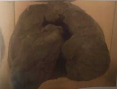
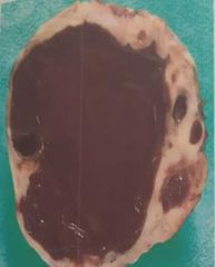
Ovarian Endometriosis Cyst (Chocolate Cyst) Comment:Cut section of ovary shows a large cyst filled with haemorhagic brownish material The wall is iregular and thick |
|
|
|
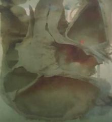
Papillary serous cystadenoma of the ovary. Comment: Half of an oval -unilocular ovarian cyst -smooth outer surface -The inner surface shows many small papillae, projecting in the cyst cavity. |
|
|
|
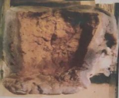
Mucinous cystadenoma of the ovary Comment: Half of a large -multilocular ovarian cyst -thin wall -smooth surface - The locules contain gelatinous material. - No papillae were detected |
|
|
|
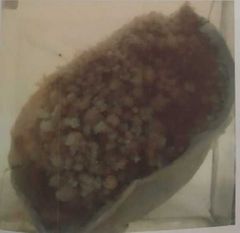
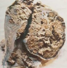
Vesicular mole Comment: placenta show : numerous transparent variable-sized vesicles, less than 1cm. These vesicles are chorionic villi with hydropic degeneration (bunch of grapes like) |
|
|
. |
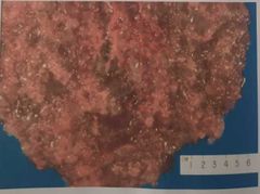
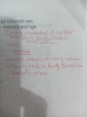
Choriocarcinoma Comment: Half of an enlarged uterus
infiltrated by a large brownish mass (non capsulated ) with ill-defined borders , extensive necrosis and hge |
|
|
|
Intraductal papillomatosis,Breast Comment: Multiple branching papillae inside duct of breast formed of : -vascular core -Columnar cells |
|
|
|
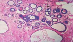
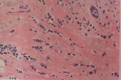
Fibrocystic disease of the breast Comment: Adenosis, epitheliosis,lobular hyperplasia, dilated cysts, fibrosis and calcification(arrow) |
|
|
|
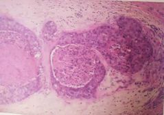
Intra-ductal Carcinoma (Comedo Type) Comment: The ducts - dilated -lined by several layers of malignant cells without invasion of basement membrane - filled with central eosinophilic areas of necrosis |
|
|
|
Invasive Duct Carcinoma Comment: -Infilitrating the breast tissue with chalky white foci -Ill-defined firm -gritty greyish white mass -iregular border -Infilitrating the breast tissue with chalky white foci |
|
|
|
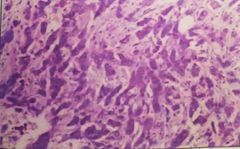
Invasive Duct Carcinoma Comment: Section in breast showing malignant tumor formed of infiltrating sheets of malignant epithelial cells separated by dense fibrous stroma. The malignant cells are rounded or polygonal and variable in size. Their nuclei are large, pleomorphic, hyperchromatic |
|
|
|
Invasive Lobular Carcinoma Comment: Section in breast
mallignant tumor formed of small malignant cells invading the stroma in linear cords (Indian File appearance) |
|
|
|
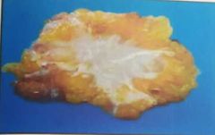
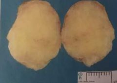
Fibroadenoma Comment: -A bisected breast mass -oval in shape 5x3 cm -rubbery to firm, -greyish white - capsulated. |
|
|
|
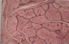
Intracanalicular Fibroadenoma Comment: Proliferating ducts -lined by cubical epithelium -separated by dense fibrosis which compress and even close the lumen forming slit like appearance |
|