![]()
![]()
![]()
Use LEFT and RIGHT arrow keys to navigate between flashcards;
Use UP and DOWN arrow keys to flip the card;
H to show hint;
A reads text to speech;
74 Cards in this Set
- Front
- Back
- 3rd side (hint)

|
Fibrous Tunic Outermost tunic made up of sclera and cornea |
|
|

|
Vascular Tunic (Uveal Tract) |
|
|

|
Nervous Tunic |
|
|

|
Fibrous Tunic Outermost tunic made up of sclera and cornea |
|
|
|
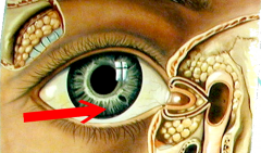
Conjunctiva Outer surface of the eye, mucous membrane Fxn: mechanical protection, attaches skeletal muscles (extrinsic muscles) |
|
|

|
Palpebral Conjunctiva Attaches to interior surface of eyelid |
|
|
|
Palpebral Conjunctiva Attaches to interior surface of eyelid |
|
|
|
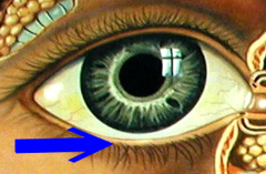
Bulbar Conjunctiva Surface of the eyeball |
|
|
|
Bulbar Conjunctiva Surface of the eyeball |
Surface of the eyeball |
|
|
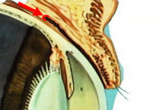
Conjunctival Sac (Feature) The fold where palpebral and bulbar conjunctivameet |
|
|
|
Conjunctival Sac (Feature) The fold where palpebral and bulbar conjunctivameet |
(Feature) The fold where palpebral and bulbar conjunctiva meet |
|
|
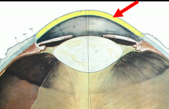
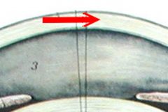
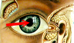
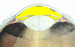
Cornea Anterior surface of the eye, transparent layer, avascularFxn: light refraction |
|
|
|
Corneal Epithelium (nonkeratinized stratified squamous ET) |
|
|
|
Corneal Stroma (Dense Irregular CT) |
|
|
|
Corneal Endothelium (Simple Squamous Epithelium) |
|
|
|
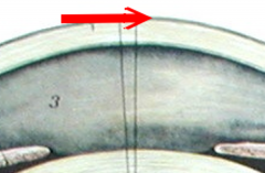
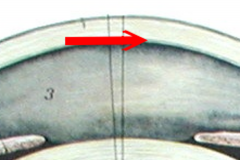
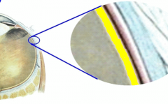
Sclera The white of the eyeFxn: Protection, gives shape to eye |
|
|
|
Sclera The white of the eyeFxn: Protection, gives shape to eye |
|
|

|
Sclera The white of the eyeFxn: Protection, gives shape to eye |
The white of the eye Fxn: Protection, gives shape to eye |
|
|
Vascular Tunic (Uveal Tract) |
2nd tunic AKA uvea |
|

Name the layer |
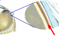
Choroid Layer of blood vesselsFxn: nourishes posterior eye structures and increases melanin to absorb stray light |
|
|
|
Choroid Layer of blood vesselsFxn: nourishes posterior eye structures and increases melanin to absorb stray light |
Layer of blood vessels Fxn: nourishes posterior eye structures and increases melanin to absorb stray light |
|
|
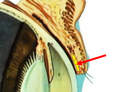
Ciliary Muscle Fxn: constricts to accommodate lens, produces aqueous humor |
|
|
|
Ciliary Processes Attach to zonular fibers, secrete aqueous humor |
|
|
|
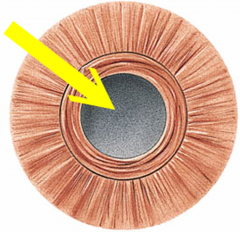
Iris Contains melanocytes, pigments Fxn: regulates amount of light that enters the eye |
|
|
|
Iris Contains melanocytes, pigments Fxn: regulates amount of light that enters the eye |
Contains melanocytes, pigments Fxn: regulates amount of light that enters the eye |
|
Which muscles are contracting? What are they responsible for? |
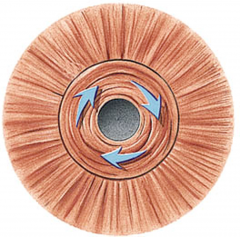
Circular Muscles Constriction |
|
|
Which muscles are contracting? What are they responsible for? |
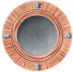
Radial Muscles Dilation |
|
|
|
Pupil An opening that lets light into the eye |
|
|
|
Pupil An opening that lets light into the eye |
An opening that lets light into the eye |
|
|
Nervous Tunic |
Deepest Tunic |
|

Name the Layer |
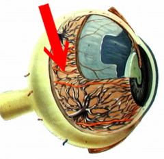
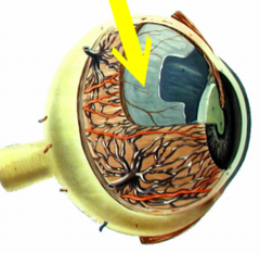
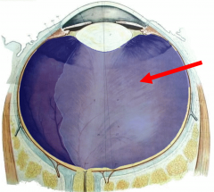
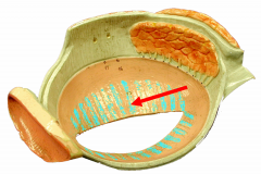
Retina Fxn: Converts light into nerve impulses |
|
|

|
Retina Fxn: Converts light into nerve impulses |
Fxn: Converts light into nerve impulses |
|

|
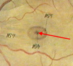
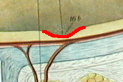
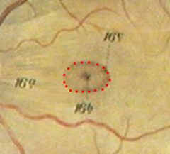
Fovea Centralis Aka central fovea: area of sharpest vision |
|
|
|
Fovea Centralis Aka central fovea: area of sharpest vision |
Aka central fovea: area of sharpest vision |
|
|
Macula Lutea Where the central fovea is located |
|
|
|
Macula Lutea Where the central fovea is located |
Where the central fovea is located |
|
|
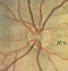
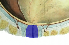
Optic Disc Moves visual sensory information to the brain |
|
|
|
Optic Disc Moves visual sensory information to the brain |
Moves visual sensory information to the brain |
|
|
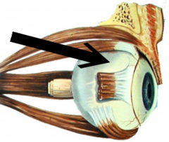
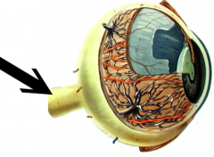
CN 2 Optic Nerve Fxn: Sensation of vision |
|
|
|
CN 2 Optic Nerve Fxn: Sensation of vision |
Fxn: Sensation of vision |
|

|
Pigmented Epithelium (Melanin) absorbs stray light and stores vitamin A |
|
|

Name the Cavity |
Anterior Cavity |
|
|
Name the Chamber |
Anterior Chamber |
|
|
|
Posterior Chamber |
|
|
Name the fluid in this cavity |
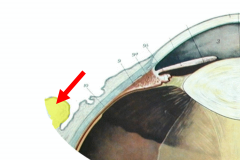
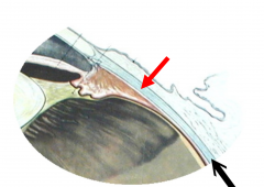
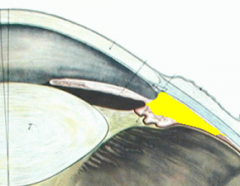
Aqueous Humor Fluid in the anterior cavity Fxn: nourishes the cornea and lens |
|
|
|
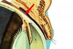
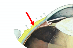
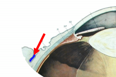
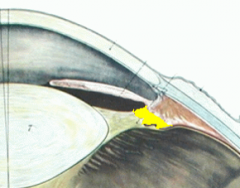
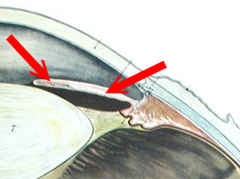
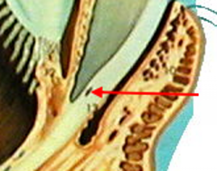
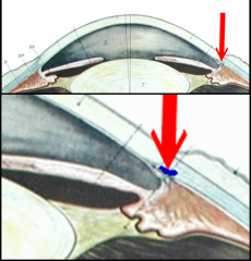
Canal of Schlemm Drains aqueous humor |
|
|
|
Canal of Schlemm Drains aqueous humor |
|
|

|
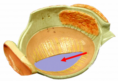
Posterior Cavity |
|
|
Name the Fluid in this Cavity |
Vitreous Humor Holds the retina against the choroid and gives the eyeball shape |
|
|
Name the structure |
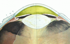
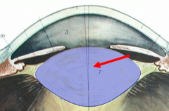
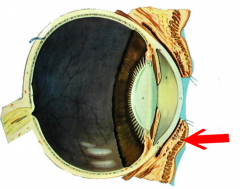
Lens |
|
|

Name the protein found here |
Crystallins A protein that functions in the refraction of light |
|
|
|
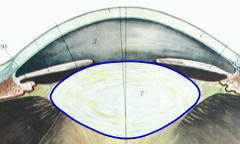
Lens Capsule Fxn: protects the lens |
|
|
|
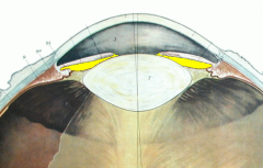
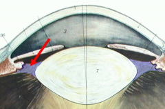
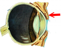
Suspensory Ligaments Attach the lens to the ciliary processes |
|
|
|
Upper Palpebrae |
Upper eyelid |
|
|
Lower Palpebrae |
Lower eyelid |
|
|
Tarsal glands Secrete watery secretion and prevent eyelids from sticking |
|
|
|
Tarsal glands Secrete watery secretion and prevent eyelids from sticking |
|
|
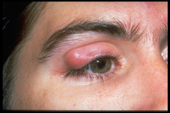
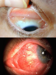
Chalazion |
Infection of the tarsal gland |
|
|
|
Ciliary Glands Open the eyelash follicle Fxn: lubrication; secrete an oily secretion |
|
|
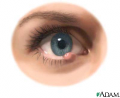
Stye |
Infection of the ciliary gland |
|
|
|
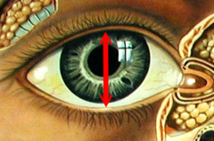
Palpebral Fissure Opening between upper and lower palebrae |
|
|
|
Palpebral Fissure Opening between upper and lower palebrae |
Opening between upper and lower palebrae |
|
|
Medial and Lateral Commissure Corners of the eyes |
|
|
|
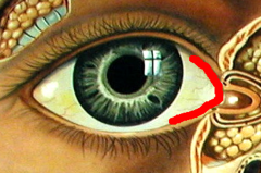
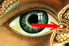
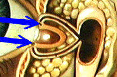
Caruncle Mucous membrane that produces eye goobers |
|
|
|
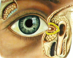
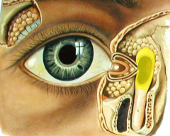
Lacrimal Gland Produces/drains tears |
|
|
|
Lacrimal Punctum Produces/drains tears |
|
|
|
Lacrimal Canal Produces/drains tears |
Produces/drains tears |
|
|
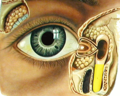
Lacrimal Sac Produces/drains tears |
Produces/drains tears |
|
|
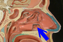
Nasolacrimal Duct Produces/drains tears |
Produces/drains tears |
|
|
Nasolacrimal Duct Produces/drains tears |
Produces/drains tears |
|
|
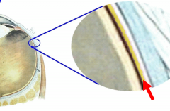
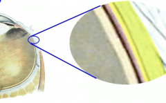
Retina Fxn: Converts light into nerve impulses |
|
|
|
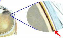
Pigmented Epithelium (Melanin) absorbs stray light and stores vitamin A |
|
|
|
Choroid Layer of blood vesselsFxn: nourishes posterior eye structures and increases melanin to absorb stray light |
|
|
|
Sclera The white of the eyeFxn: Protection, gives shape to eye |
|