Reading...
![]()
Play button
![]()
Play button
![]()
Use LEFT and RIGHT arrow keys to navigate between flashcards;
Use UP and DOWN arrow keys to flip the card;
H to show hint;
A reads text to speech;
383 Cards in this Set
- Front
- Back
|
How many components make up the nervous system, and what are they?
|
Two. Central and Peripheral.
|
|
|
The Central Nervous System includes...
|
Brain and spinal cord
|
|
|
The Peripheral Nervous System includes...
|
The entire nervous system EXCEPT the brain and spinal cord
|
|
|
How many kinds of cells make up the nervous system, and what are they?
|
Two. Neurons and glia.
|
|
|
What is the popular name for neurons, and what do they do?
|
Nerves. They transmit information in the body.
|
|
|
How are glia cells different from neurons?
|
They don't transmit information.
|
|
|
What is the function of glia cells?
|
Support the neurons in many ways.
|
|
|
What are the most important cells of the nervous system?
|
Neurons
|
|
|
What are the three major types of neurons?
|
Sensory, motor, interneurons
|
|
|
What is the function of sensory neurons?
|
Detect changes in the environment (light, sound, odor, taste, touch, etc.)
|
|
|
What is the function of motor neurons?
|
They are responsible for movement
|
|
|
What is the function of interneurons?
|
They analyze and relay information throughout the body
|
|
|
The basic structure of neurons is made up of how many regions?
|
Four
|
|
|
What is the "central headquarters" of the neuron?
|
The soma
|
|
|
What part of the neuron contains the nucleus?
|
The soma
|
|
|
What part of the neuron receives messages from other neurons?
|
Dendrites
|
|
|
What part of the neuron has a surface lined with receptors?
|
Dendrites
|
|
|
What part of the neuron carries messages from one major part of the neuron to another, and what are these parts?
|
The axon carries messages from the soma to the terminal buttons.
|
|
|
What is an important feature of the geometry of the axon?
|
Its diameter is constant
|
|
|
What is the term for the message carried by the axon?
|
Action potential
|
|
|
What major classes of chemicals make up the myelin sheath, and in what proportions?
|
Lipid (80%) and protein (20%)
|
|
|
What are the gaps in the myelin sheath called?
|
Nodes of Ranvier
|
|
|
Presynaptic terminals are another name for...
|
Terminal buttons
|
|
|
What happens when an action potential reaches a terminal button?
|
The nerve secretes neurotransmitters
|
|
|
The membrane of the neuron is also called the...
|
Plasma membrane
|
|
|
Describe the chemical structure of the neuron's membrane
|
Made up of two layers of lipid molecules and protein channels
|
|
|
What kinds of molecules can pass freely through the neuron's membrane?
|
Small, uncharged molecules
|
|
|
What are three examples of small, uncharged molecules?
|
H₂0, 0₂, CO₂ (water, oxygen, carbon dioxide)
|
|
|
By what means may some ions pass through the cell membrane?
|
Protein channels
|
|
|
What are four examples of ions that can pass through the cell membrane by a special route?
|
Na, K, Ca, Cl
|
|
|
O==O
O==O O==O What does the above ASCII picture resemble? |
Phospholipid molecules in a cell membrane
|
|
|
What is the function of the nucleolus?
|
Makes ribosomes
|
|
|
What is the function of ribosomes?
|
Production of proteins
|
|
|
What are the major functions of proteins?
|
Provide physical structure and act as enzymes
|
|
|
What are enzymes?
|
Catalysts for chemical reactions in the body
|
|
|
What are chromosomes?
|
Long strands of DNA that contain genetic information
|
|
|
What is the jelly-like substance that fills most of the space within the membrane?
|
Cytoplasm
|
|
|
What is the function of mitochondria?
|
Extracts energy from nutrients
|
|
|
What is the chemical that the mitochondria use in its role in cell metabolism?
|
Adenosine Triphosphate (ATP)
|
|
|
What is the function of the endoplasmic reticulum?
|
Separation, storage, and transportation of proteins
|
|

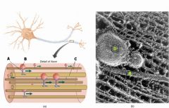
Label the following:
|
A: Cytoplasm
B: Nucleus C: Dendritic spines D: Dendrite E: Membrane F: Microtubules G: Myelin sheath H: Mitochondria |
|
|
The process of getting items back and forth between the nucleus/soma and the terminal buttons
|
Axoplasmic transport
|
|
|
Cytoplasm in the axon is called...
|
Axoplasm
|
|
|
What are the proteins that walk down the microtubules, carrying stuff?
|
Kinesin and dynein
|
|
|
What protein is responsible for anterograde transport?
|
Kinesin
|
|
|
What protein is responsible for retrograde transport?
|
Dynein
|
|
|
If you're picking stuff up at the soma and dropping it off at the terminal buttons:
1) what are you called? 2) what is your activity called? |
1) Kinesin
2) Anterograde transport |
|
|
If you are picking stuff up at the terminal buttons and dropping it off at the soma:
1) what are you called? 2) what is your activity called? |
1) Dynein
2) Retrograde transport |
|
|
A: Kinesin
B: Vesicle C: Microtubule D: Vesicle E: Microtubule |
|
|
Five supporting functions of glia
|
1) Surround neurons
2) Hold neurons in place 3) Control neurons' nutrient supply 4) Insulate neurons from each other so messages don't get scrambled; and 5) Destroy and remove dead neurons |
|
|
Into what chemical do astrocytes break down glucose?
|
Lactate
|
|
|
How do astrocytes support neurons?
|
1) Provide physical support
2) Clean up debris via phagocytosis 3) Provide some nutrients |
|
|
How do astrocytes provide some nutrients to neurons?
|
Obtain and break down glucose from blood vessels
|
|
|
What, physically, do the arms of the astrocytes do, and why?
|
1) wrap around blood vessels to obtain nutrients
2) wrap around neuron to pass processed nutrients to cell |
|
|
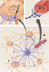
A: Lactate
B: Lactate C: Energy D: Glucose E: Glucose F: Glycogen (storage) G: Lactate H: Blood vessel I: Astrocyte J: Neuron |
|
|
What do oligodendrocytes do?
|
Create myelin sheaths and repair myelin
|
|
|
How are oligodendrocytes and Schwann cells different?
|
Oligodendrocytes do their job in the central nervous system; Schwann cells do their job in the peripheral nervous system
|
|
|
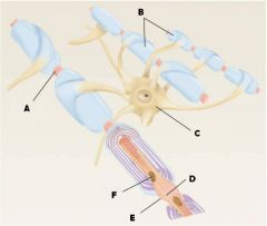
A: Node of Ranvier
B: Myelinated Axons C: Soma of oligodendrocyte D: Microtubule E: Node of Ranvier F: Mitochondrion in Axoplasm |
|
|
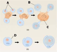
A: Axons
B: Myelin sheath C: Oligodendrocyte D: Schwann cell |
|
|
What is the function of microglia that makes it part of the immune system?
|
Protect brain from invading microorganisms by causing an inflammatory reaction in response to brain damage
|
|
|
What is the blood-brain barrier?
|
A semipermeable barrier between the blood and the brain, made up of the walls of the capillaries in the brain
|
|
|
What are capillary walls made of?
|
Endothelial cells
|
|
|
What are special about endothelial cells in the blood-brain barrier?
|
They are very close together
|
|
|
What does it mean to say the blood-brain barrier is semipermeable?
|
Some things can pass from the blood to the fluid of the brain and some things cannot
|
|
|
What is the purpose of the blood-brain barrier?
|
To protect the fluid of the brain from blood chemicals that would be dangerous
|
|
|
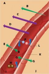
A: Brain tissue
B: Fat-soluble molecule C: Glucose transport D: Amino-acid transport E: CO₂ F: Charged molecules G: O₂ H: Large molecule I: Blood vessel J: Brain tissue K: Endothelial cell L: Cell wall tight junction |
|
|
What is the primary energy source for the brain?
|
Glucose
|
|
|
What organ converts other nutrients into glucose?
|
Liver
|
|
|
What element is needed to metabolize glucose?
|
Oxygen
|
|
|
What nutrient is necessary for the use of glucose?
|
Thiamine
|
|
|
What is another name for Thiamine?
|
Vitamin B1
|
|
|
What impairments characterize Korsokoff's Syndrome?
|
Severe memory impairments
|
|
|
Prolonged deficiency of what nutrient leads to Korsokoff's Syndrome?
|
Thiamine
|
|
|
Prolonged deficiency of Thiamine can lead to two things:
|
1) Death of neurons
2) Koroskoff's Syndrome |
|
|
Many alcoholics have a diet deficient in...
|
Thiamine
|
|
|
How fast are action potentials transmitted from the soma down the axon to the terminal buttons?
|
1 to 100 meters per second
|
|
|
A resting potential is also called...
|
Electrical gradient
|
|
|
What kinds of axons have some baseline level of potential electrical power?
|
All axons.
|
|
|
The axon's baseline level of potential electrical power is called...
|
Resting potential (or electrical gradient)
|
|
|
The resting potential can be described as...
|
The difference in voltage or electrical charge between the inside and outside of a neuron.
|
|
|
How many millivolts exist between the inside and the outside of neurons?
|
-70mV
|
|
|
At its baseline level of electrical power, the neuron contains more of what kinds of ions on the outside than inside?
|
Na+ (positive sodium ions) and Cl- (negative chlorine ions)
|
|
|
At the neuron's baseline level of electrical power, what is the charge of the interior of the neuron?
|
Negative (more electrons than protons)
|
|
|
At the neuron's baseline line level of electrical power, what is the charge of the exterior of the neuron?
|
Positive (more protons than electrons)
|
|
|
How long, in relative terms, does an action potential last?
|
A brief time.
|
|
|
An action potential is a ______ of voltage.
|
reversal
|
|
|
The threshold of excitation is the ________ voltage level that must be reached for the neuron to produce an action potential
|
minimum
|
|
|
The neuron, in order to produce an action potential, must possess a voltage level that does what?
|
Crosses the threshold of excitation
|
|
|
Increase in the negative charge inside of the neuron is called:
|
hyperpolarization
|
|
|
Decrease in the negative charge inside of the neuron is called:
|
depolarization
|
|
|
Rapid depolarization and slight reversal of the usual polarization inside of a neuron is called
|
an action potential
|
|
|
When dissolved in water, ________ split into two electrically opposed ions.
|
Electrolytes
("-lyte" is Greek for "loose," so the ions get loosened up by the water) |
|
|
Salt, dissolved in water, is an example of _______
|
an electrolyte
|
|
|
Ions that have a positive charge are called:
|
cations
|
|
|
Ions that have a negative charge are called:
|
anions
|
|
|
Sodium and potassium ions are two examples of:
|
cations
|
|
|
Two examples of common anions
|
Chloride (Cl-)
Acetate (CH3COO-) [Acetate is a "polyatomic" ion] |
|
|
Diffusion is...
|
Movement of molecules (like ions) from regions of high concentration to regions of low concentration
|
|
|
The force resulting from opposite charges attracting and like charges repelling...
|
Electrostatic pressure
|
|
|
Na+ and K+ ions get into the neuron by way of...
|
Protein channels
|
|
|
When the neuron membrane is at rest, _____ channels are closed, and ____ channels are almost closed.
|
Sodium (Na), Potassium (K)
|
|
|
When the neuron membrane is at rest, almost all ___ flow is stopped.
|
Sodium (Na)
|
|
|
When the neuron membrane is at rest, ___ flows through very slowly
|
Potassium (K)
|
|
|
The (A) / (B) pump lets 3 (C) ions out while allowing 2 (D) ions in.
|
A) Na
B) K C) Na D) K |
|
|
The Na/K pump doesn't stop some ___ from leaking back in through the channels...
|
Potassium (K)
|
|
|
All else being equal, given the relative concentration of Na+ inside and outside the neuron at resting potential, what would the effect of diffusion be?
|
Diffusion would push Na+ inside (Na+ is more dense on the outside)
|
|
|
All else being equal, given the charge of the inside of the neuron at resting potential, what would you expect to have happen to Na+ on the outside of the neuron?
|
Na+ would be attracted to the inside, which is negatively charged
|
|
|
Why doesn't Na+ just stream into the negatively charged interior of the neuron all the time?
|
Two reasons:
1) The cell membrane isn't very permeable 2) The Na+/K+ pump continually pushes Na+ out and K+ in. |
|
|
What must happen to the membrane of the neuron for Na+ to stream in and create an action potential?
|
Sodium channels must open up
|
|
|
What causes the change of permeability in the neuron membrane that leads to Na+ rushing in?
|
Sodium ion channels open
|
|
|
When enough electrical charge is added to the inside of the cell to cross the threshold of excitation, what happens?
|
Sodium channels open
|
|
|
What does it mean to say that sodium ion channels are voltage dependent?
|
They open in response to electrical charge inside the cell crossing the threshold of excitation
|
|
|
There are two forces that cause Na+ to rush into the neuron, causing an action potential. What are these?
|
1) Diffusion
2) Electrostatic pressure |
|
|
At resting potential, the neuron has a charge of __ mV. When an action potential occurs, this charge changes to about __ mV.
|
1) -70
2) +40 |
|
|
After Na+ channels have become refractory, what must happen for them to open again?
|
The membrane must return to its resting potential
|
|

|
A: +40
B: -70 C: Threshold of excitation D: Na+ channels open; Na+ begins to enter cell E: K+ channels open; K+ begins to leave cell F: Na+ channels become refractory; no more Na+ enters cell G: K+ continues to leave cell, causes membrane to return to resting level H: K+ channels close; Na+ channels reset I: Extra K+ outside diffuses away |
|

|
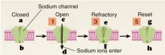
A: Closed
B: Open C: Refractory D: Reset E: Sodium channel F: Sodium ions enter |
|
|
a: +
b: - c: + d: - e: - f: + g: + h: - |
|
|
Ion movement during action potentials: step 1
|
An electrical impulse strong enough to reach the threshold of excitation occurs.
|
|
|
What causes the axon membrane's Na+ channels to open?
|
An electrical impulse strong enough to reach the threshold of excitation
|
|
|
Ion movement during action potentials: step 2
|
The axon membrane's K+ channels begin to open and K+ begins to leave the cell
|
|
|
Ion movement during action potentials: step 3
|
Action potential reaches its peak and Na+ channels become refractory
|
|
|
Ion movement during action potentials: step 4
|
But, the K+ channels are still open and K+ is still leaving, so the inside of the neuron begins to return to being negatively charged. As the inside charge returns to normal, the K+ channels close, too
|
|
|
Ion movement during action potentials: step 5
|
Everything resets so it’s ready for the next action potential. Na+/K+ pumps remove anything leftover that’s in the wrong place
|
|
|
Where does the action potential start?
|
Axon hillock, a small bump in the soma where it meets the axon
|
|
|
What is important about the size of the action potential as it travels?
|
The voltage remains constant
|
|
|
What is the relationship between the size, velocity, and amplitude of an action potential and the intensity of the stimulus that initiated it?
|
There is no relationship
|
|
|
What property of action potentials determines the strength of the messages they transmit?
|
Frequency
|
|
|
What happens to the size of the action potential when it is split between two or among three or more terminal buttons?
|
The size stays the same
|
|
|
There are two laws governing action potentials that enable nerve signals to trigger behaviors that are continuous, like movement. What are these laws?
|
Rate law
All-or-none law |
|
|
Do any electrical currents travel down the axon that are too small to create an action potential?
|
Yes
|
|
|
Electrical currents traveling down the axon that are too small to create an action potential are said to have _____ properties.
|
Cable
|
|
|
Electrical currents traveling down the axon that are too small to create an action potential have the property of ____________ conduction.
|
Decremental
|
|
|
The phenomenon of electrical currents becoming smaller as they travel is called:
|
Decremental conduction
|
|
|
The phenomenon of electrical currents maintaining energy by hopping from node to node is called:
|
Saltatory conduction
|
|
|
Saltare is Latin for:
|
"To dance"
|
|
|
What is the role of the Nodes of Ranvier in facilitating saltatory conduction?
|
They generate new action potentials and "recharge" the message represented by the original action potential, sending the recharged signal further down the axon
|
|
|
Who do neurons communicate to?
|
a) other neurons
b) muscles c) sense organs |
|
|
How do neurons communicate between each other?
|
Neurotransmitters
|
|
|
Communication within neurons is...
|
electrical
|
|
|
Communication between neurons is...
|
chemical
|
|
|
The presynaptic neuron is always sending signals from what part of the neuron?
|
terminal button
|
|
|
The postsynaptic neuron can be what part of the neuron?
|
a) dendrites
b) soma c) axon |
|
|
The three kinds of synapses, corresponding to which part of the neuron is receiving the synapse, are called...
|
Axodendritic (on the dendrite)
Axosomatic (on the soma) Axoaxonic (on the axon) |
|
|
Bumps on dendrites that increase surface area to hold more receptors are called...
|
Dendritic spines
|
|
|
Membrane on the end of the terminal button is called...
|
Presynaptic membrane
|
|
|
Membrane on the neuron receiving a message from a terminal button is called...
|
Postsynaptic membrane
|
|
|
Another word for the synapse
|
Synaptic cleft
|
|
|
How wide is a synapse?
|
Usually 20 nanometers
|
|
|
What do synapses contain?
|
Extracellular fluid
|
|
|
What do vesicles contain?
|
Neurotransmitters or peptides
|
|
|
What are vesicles produced by?
|
a) Golgi apparatus of the soma
b) cisternae |
|
|
The region of the terminal button where synaptic vesicles attach and release neurotransmitters into the synaptic cleft is called...
|
the release zone
|
|
|
What are cisternae?
|
Located in the terminal buttons, cisternae recycle portions of the presynaptic membrane into synaptic vesicles
|
|
|
Which is thicker, the pre- or postsynaptic membrane, and why?
|
The postsynaptic membrane, because it has receptors on it
|
|
|
Describe how action potentials lead to getting neurotransmitters into the synapse...
|
Action potentials cause some of the synaptic vesicles in the terminal buttons to fuse with the presynaptic membrane and then break open, spilling their neurotransmitters into the synaptic cleft.
|
|
|
What is it called when proteins on the synaptic vesicle attach to proteins on the presynaptic membrane?
|
Docking
|
|
|
A type of ion channel not (ordinarily?) found on other parts of the neuron is found on the presynaptic membrane...
|
Ca²+ (calcium)
|
|
|
What opens the Ca²+ channels on the membrane of the terminal buttons?
|
The depolarization caused by an action potential
|
|
|
What happens when Ca²+ channels open?
|
Calcium ions from the extracellular fluid rush into the presynaptic neuron.
|
|
|
What happens when calcium ions from the extracellular fluid rush into the presynaptic neuron?
|
Fusion pore is opened, allowing a synaptic vesicle to fuse with the presynaptic membrane
|
|
|
Repeated fusion of synaptic vesicles with terminal button membranes would lead to too much membrane on the TB unless...
|
Little pieces of TB membrane get pinched off into the cytoplasm. This is called pinocytosis.
|
|
|
Bits of membrane in the TB pinched off by pinocytosis are picked up by what, and what is done with them?
|
Cisternae pick up the bits of membrane and recycle them into synaptic vesicles. Then the proteins and neurotransmitters are inserted
|
|
|
What do postsynaptic receptors do when neurotransmitters bind to them?
|
They open neurotransmitter-dependent ion channels
|
|
|
When ion channels on postsynaptic neurons have their own built-in receptor site, these ion channels are called:
|
ionotropic receptors
|
|
|
When metabotropic receptors are activated, what is the next thing that happens?
|
a G protein is activated
|
|
|
When a G protein is activated, what does it do?
|
Produces a chemical called a "second messenger."
|
|
|
If there's a second messenger, what is the first?
|
The neurotransmitter that activated a metabotropic receptor
|
|
|
Postsynaptic potentials can be either...
|
excitatory or inhibitory
|
|
|
This kind of postsynaptic potential increases the chances that the postsynaptic neuron will have an action potential
|
excitatory
|
|
|
This kind of postsynaptic potential decreases the chances that the postsynaptic neuron will have an action potential
|
inhibitory
|
|
|
What determines whether a postsynaptic potential will be inhibitory or excitatory?
|
The type of ion channel (Na+, K+, Cl-, or Ca2+) that is opened by the neurotransmitter that caused the postsynaptic potential
|
|
|
What ion channels, if opened in the course of a postsynaptic potential, will be inhibitory?
|
K+
|
|
|
What ion channels, if opened in the course of a postsynaptic potential, will be excitatory?
|
Na+ & Ca+
|
|
|
If Cl- ion channels are opened in the course of a postsynaptic potential, what happens?
|
Nothing, unless the cell has already been excited, in which case the Cl- ions bring the neuron back to baseline (hyperpolarize to -70mV)
|
|
|
What are the ways in which postsynaptic potentials are brought to an end?
|
Reuptake and enzymatic deactivation
|
|
|
I am the process whereby neurotransmitters are "sucked" back directly through the terminal button membrane. Who am I?
|
Reuptake
|
|
|
I am the process whereby neurotransmitters in the synapse are broken down and deactivated by enzymes. Who am I?
|
Enzymatic deactivation
|
|
|
The enzyme that breaks down acetylcholine (ACh) is...
|
Acetylcholinesterase (AChE)
|
|
|
ACh is responsible for...
|
Muscle contractions
|
|
|
I am an autoimmune disease that destroys ACh receptors. What might I be?
|
Myasthenia gravis
|
|
|
I am a poison that blocks neural transmission at muscles. What am I?
|
Curare
|
|
|
I counteract curare, and am effective at treating (but not curing) myasthenia gravis. What am I?
|
Physostigmine
|
|
|
The process by which inhibitory and excitatory postsynaptic potentials add up and control the rate of firing of a neuron
|
Neural integration
|
|
|
In the process of neural integration, if the activity of excitatory postsynaptic potentials goes up, what happens?
|
The rate of firing will go up, and vice versa
|
|
|
Neural inhibition does not always equal...
|
behavioral inhibition
|
|
|
What kinds of receptors initiate potentials in one neuron in response to neurotransmitters released by another neuron?
|
postsynaptic
|
|
|
Where are autoreceptors located?
|
Anywhere on a neuron!
|
|
|
What do autoreceptors respond to?
|
Neurotransmitters released by the autoreceptor's own neuron
|
|
|
What receptors control and regulate the neuron's internal processes?
|
Autoreceptors
|
|
|
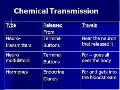
Not all chemical communication stays in the synapse. What can bring signals past the synapse?
|
Neuromodulators
|
|
|
I am a chemical that doesn't elicit postsynaptic potentials--instead, I hit receptors beyond the synapse. What am I?
|
Neuromodulator
|
|
|
I accomplish chemical transmission of neural information, but I can travel all over the body, not just in the brain. What am I?
|
Neuromodulator or hormone
|
|
|
I accomplish chemical transmission of neural information, but being small and fat-soluble, I can readily pass the blood-brain barrier and travel through the bloodstream. What am I?
|
Hormone
|
|
|
I accomplish chemical transmission of neural information. But being small and fat-soluble, I have a talent for passing through cell membranes into the nucleus, where I bind to special receptors. What am I?
|
Hormone
|
|
|
|
|
|
In order for a chemical message to be received somewhere else, there has to be a ______ for that type of chemical at the destination
|
Receptor
|
|
|
Receptors have _____ sites
|
Binding
|
|
|
A chemical that binds to a binding site is called a _____
|
Ligand
|
|
|
Neurotransmitters, neuromodulators, and hormones can all attached to binding sites, and are therefore three kinds of...
|
ligands
|
|
|
I am an imaginary line drawn down the center of the CNS. What am I?
|
Neuraxis
|
|
|
Toward the head
|
Anterior or rostral
|
|
|
Toward the tail
|
Posterior or caudal
|
|
|
Toward the back
|
Dorsal
|
|
|
Toward the belly/under the chin
|
Ventral
|
|
|
Above
|
superior
|
|
|
Below
|
inferior
|
|
|
Toward the side, away from the middle
|
lateral
|
|
|
Toward the middle, away from the sides
|
medial
|
|
|
Located on the same side of the body
|
ipsilateral
|
|
|
Located on the opposite side of the body
|
contralateral
|
|
|
If you slice up the spinal cord like a salami, and you are not a serial killer, what are you doing?
|
taking cross-sections (or coronal sections, if applied to the brain)
|
|
|
If you slice up the brain in slices parallel to the forehead, and you are not Hannibal Lecter getting ready for a picnic, what are you doing?
|
taking cross-sections, or coronal sections
|
|
|
If you slice up the brain in slices parallel to the ground, and you are not misdirecting a helicopter, what are you doing?
|
Taking horizontal, or transverse, sections
|
|
|
If you are slicing up a head (with a brain in it) in such a way that the first and last slice would slice the ears off, and you are not the subject of a future Lifetime movie, what are you doing?
|
Taking sagittal sections of the brain
|
|
|
This plane divides the brain into two symmetrical halves, left and right...
|
Midsagittal plane
|
|
|
Name the three meninges...
|
1) Dura mater
2) Arachnoid membrane 3) Pia mater |
|
|
The fluid-filled space between the arachnoid membrane and pia mater...
|
Subarachnoid space
|
|
|
Clean fluid in the subarachnoid space and other areas of the brain...
|
Cerebrospinal fluid (CSF)
|
|
|
Which of the 3 meninges only covers the CNS?
|
Arachnoid membrane
|
|
|
How heavy is 3 pounds in grams?
|
1400
|
|
|
How heavy is the brain?
|
3 pounds or 1400 grams
|
|
|
In its bath of CSF, what is the weight of the brain?
|
80 grams
|
|
|
Which ventricle is located in the middle of the brain?
|
Third
|
|
|
Which ventricle(s) is/are the largest?
|
Lateral ventricles
|
|
|
The cerebral aqueduct connects which ventricles?
|
Third and fourth
|
|
|
CSF is extracted from ____.
|
Blood
|
|
|
CSF is created by...
|
the choroid plexus
|
|
|
Blood-rich tissue that protrudes into all four ventricles...
|
choroid plexus
|
|
|
Where does the path of the CSF end?
|
Reabsorbed into bloodstream by flowing through structures called arachnoid granulations
|
|
|
Total volume of CSF in brain at any given time...
|
125 mL (about 1/2 cup)
|
|
|
Half-life of CSF
|
3 hours
|
|
|
When flow of CSF is blocked, the result is...
|
Obstructive hydrocephalus
|
|
|
The three major divisions of the brain...
|
Forebrain, midbrain, hindbrain
|
|
|
Ventricle(s) of the forebrain...
|
Laterals and third
|
|
|
Ventricle(s) of the midbrain...
|
Cerebral aqueduct
|
|
|
Ventricle(s) of the hindbrain...
|
Fourth
|
|
|
Subdivisions of the forebrain...
|
Telencephalon, diencephalon
|
|
|
Subdivisions of the midbrain...
|
Mesencephalon
|
|
|
Subdivisions of the hindbrain...
|
Metencephalon, myelencephalon
|
|
|
Principal structures of the forebrain...
|
Cerebral cortex, basal ganglia, limbic system, thalamus, hypothalamus
|
|
|
Principal structures of the midbrain...
|
Tectum, tegmentum
|
|
|
Principal structures of the hindbrain...
|
Cerebellum, pons, medulla oblongata
|
|
|
Development of the CNS begins on day __ after conception
|
18
|
|
|
Day of embryonic development that sees the formation of the neural tube...
|
21
|
|
|
Day of embryonic development that sees the rostral end of the developing CNS develop three chambers surrounded by tissue...
|
28
|
|
|
Region around the 2 lateral ventricles...
|
Telencephalon
|
|
|
Region around the 3rd ventricle...
|
Diencephalon
|
|
|
Region around the cerebral aqueduct...
|
Mesencephalon
|
|
|
|
|
|

|

|
|
|
Cells that line the inside of the neural tube and produce the cells of the CNS
|
Ventricular zone
|
|
|
What part of the developing brain do we know the most about?
|
Cerebral cortex
|
|
|
In what pattern does the cerebral cortex develop?
|
From the inside out: cells come from the neural tube and create the first layer; then new cells pass through that and build the second layer, and so on...
|
|
|
What guides new neurons from the ventricular zone to the cortex?
|
Radial glia
|
|
|
Cells of the ventricular zone that produce neurons...
|
Founder (stem) cells
|
|
|
Two ways in which founder cells produce neurons...
|
symmetrical and asymmetrical division
|
|
|
When a founder cell divides into two equal founder cells, it is called...
|
symmetrical division
|
|
|
When a founder cell divides onto one founder cell that stays put and one neuron that travels to its spot in the brain, it is called...
|
asymmetrical division
|
|
|
When a neuron in the developing brain has found its spot, what does it have to do next?
|
Find its friends by growing dendrites and axons
|
|
|
Most of the cerebral hemispheres is called the...
|
Telencephalon
|
|
|
Most of the cerebral hemispheres is covered by the...
|
Cerebral cortex
|
|
|
The subcortical region of the telencephalon contains...
|
the limbic system and the basal ganglia
|
|
|
Small grooves...
|
sulci
|
|
|
Large grooves...
|
fissures
|
|
|
Bumps between grooves...
|
gyri
|
|
|
Three areas of the cortex that receive information from sense organs...
|
1) Primary visual cortex
2) Primary auditory cortex 3) Primary somatosensory cortex |
|
|
What's on the calcarine fissure?
|
Primary visual cortex
|
|
|
Where is the primary visual cortex?
|
Back of brain on the calcarine fissure
|
|
|
What's on the lateral fissure?
|
Primary auditory cortex
|
|
|
Where is the primary auditory cortex?
|
Middle of brain on the lateral fissure
|
|
|
What's behind the central sulcus?
|
Primary somatosensory cortex
|
|
|
What kind of information does the primary somatosensory cortex receive?
|
Body sense information: touch, temperature, pressure
|
|
|
What's on the top of the brain, right in front of the somatosensory cortex?
|
Primary motor cortex
|
|
|
What part of the cortex controls skeletal muscles?
|
Primary motor cortex
|
|
|
Each primary sensory area in the brain sends information to adjacent regions. These are called...
|
Sensory association cortex
|
|
|
Some regions of the sensory association cortex (SAC) receive information from many sensory systems, and some regions of the SAC receive information from only one sensory system. What determines this?
|
Proximity--regions of SAC close to a primary sensory area get information only from that system, while regions of SAC farther away receive AND integrate information from many primary systems
|
|
|
In addition to receiving and integrating sensory information, the sensory association cortex are involved with other functions...
|
Memory and perception
|
|
|
Rule of thumb about the functions of different cortical regions...
|
Back: perceiving, learning, spatial relations
Front: movement-related activities such as planning and executing movements and executive functions |
|
|
The rule of thumb about the back region of the cortex is that it controls...
|
Perceiving, learning, and spatial relations
|
|
|
The rule of thumb about the front region of the cortex is that it controls...
|
Movement-related activities such as planning and executing movements and executive functions
|
|
|
Damage in one of the three primary sensory cortical areas results in...
|
Impairment in using the corresponding sense function(s)
|
|
|
Damage in the more general sensory association cortex results in...
|
Impairment in integrating sensory information
|
|
|
Damage in the visual association cortex may result in...
|
Being unable to recognize objects by sight
|
|
|
Damage in the auditory association cortex may result in...
|
Having trouble perceiving or producing speech
|
|
|
The motor association cortex is also known as...
|
The premotor cortex
|
|
|
What is the location of the motor association cortex?
|
Just in front of (anterior to) the primary motor cortex, in the front of the brain
|
|
|
What does the motor association cortex do?
|
Controls primary motor cortex
|
|
|
Anterior to the premotor cortex is the...
|
Prefrontal cortex
|
|
|
To a small degree, the prefrontal cortex is involved in...
|
Movement
|
|
|
The main business of the prefrontal cortex is...
|
Formulating plans and strategies (executive functioning)
|
|
|
What is underneath the neocortex?
|
Limbic cortex
|
|
|
The neocortex includes...
|
Premotor and prefrontal cortices
|
|
|
According to one rule of thumb, which half of the brain is responsible for things of a verbal nature?
|
Left
|
|
|
Which half of the brain analyzes (rule of thumb)?
|
Left
|
|
|
Which half of the brain synthesizes (rule of thumb)?
|
Right
|
|
|
Which half of the brain is good at drawing, reading maps, and spatial reasoning? (rule of thumb)
|
Right
|
|
|
What allows the two halves of the brain to communicate?
|
Corpus callosum
|
|
|
Large groove on cerebral cortex that runs from ear to ear...
|
Central sulcus
|
|
|
Position of parietal lobes...
|
On top--behind frontal lobes
|
|
|
If the right parietal lobe is damaged, you might experience...
|
Unilateral neglect
|
|
|
This lobe is responsible for space and integrates information about that space (sight and sounds)...
|
Parietal lobe (usually right lobe)
|
|
|
Primary function of the limbic system...
|
Emotion and motivation
|
|
|
Major structures of the limbic system...
|
Limbic cortex, hippocampus, amygdala
|
|
|
Portion of the limbic system most strongly associated with learning and memory...
|
Hippocampus
|
|
|
Portion of the limbic system most strongly associated with feeling and expressing emotions, recognizing others' emotions, and emotional memories...
|
Amygdala
|
|
|
Primary function of basal ganglia...
|
Control of movement
|
|
|
Major structures of the basal ganglia...
|
Caudate nucleus
Putamen Globus pallidus |
|
|
What disease is caused by degeneration of neurons going to the basal ganglia?
|
Parkinson's disease
|
|
|
Major symptoms of Parkinson's disease...
|
Weakness, tremor, rigidity, poor balance, and difficulty initiating movement
|
|
|
This division of the forebrain surrounds the third ventricle...
|
Diencephalon
|
|
|
Major structures of the diencephalon...
|
Thalamus
Hypothalamus |
|
|
Primary function of the thalamus...
|
Relay station
|
|
|
Major structures of the thalamus...
|
Lateral geniculate nucleus
Medial geniculate nucleus Ventrolateral nucleus |
|
|
Function of the lateral geniculate nucleus...
|
Receives information from the eye and sends axons to the primary visual cortex
|
|
|
Function of the medial geniculate nucleus...
|
Receives information from the ear and sends information to the primary auditory cortex
|
|
|
Function of the ventrolateral nucleus
|
Receives information from the cerebellum and sends information to the primary motor cortex
|
|
|
Primary functions of the hypothalamus...
|
Controls autonomic nervous system and endocrine system; organizes behaviors necessary for the survival of the species (feeding, fighting, sleeping, fleeing, and mating)
|
|
|
Where is the mesencephalon?
|
Surrounding the cerebral aqueduct
|
|
|
Major structures of the mesencephalon...
|
Tectum
Tegmentum |
|
|
Major structures of the tectum...
|
Inferior colliculi
Superior colliculi |
|
|
What system are the inferior colliculi a part of?
|
Auditory
|
|
|
What system are the superior colliculi a part of?
|
Visual
|
|
|
Major structures of the tegmentum
|
Reticular formation
Periaqueductal gray matter Red nucleus Substantia nigra |
|
|
Function of the reticular formation...
|
Plays a role in sleep, arousal, attention, and reflexes
|
|
|
Function of the periaqueductal gray matter
|
Involved in fighting, mating, pain, and species-typical behavior
|
|
|
Function of the red nucleus and substantia nigra
|
Involved in transmitting information needed for movement
|
|
|
Where is the hindbrain?
|
Surrounding the fourth ventricle
|
|
|
Major structures of the hindbrain...
|
Metencephalon
Mylencephalon |
|
|
The cerebellum is covered by the...
|
Cerebellar cortex
|
|
|
What connects the cerebellum with the pons?
|
Cerebellar peduncles
|
|
|
Primary function of the pons...
|
Involved in sleep and arousal, and is also a relay station
|
|
|
Major structures of the myelencephalon...
|
Medulla oblongata
|
|
|
Primary functions of the medulla oblongata...
|
Controls vital functions such as cardiovascular activity and respiration
|
|
|
Primary function of the spinal cord...
|
Distribute motor fibers to our glands and muscles and to collect and send sensory information to the brain
|
|
|
What is the spinal cord protected by?
|
Vertebral column
|
|
|
How many individual vertebrae are there?
|
24 (not counting the fused vertebrae of the sacral and coccygeal regions)
|
|
|
What areas are the vertebrae organized into?
|
Cervical
Thoracic Lumbar Sacral and coccygeal |
|
|
The hole in each vertebrae through which the spinal cord passes...
|
Spinal foramens
|
|
|
Major components of the peripheral nervous system...
|
Spinal nerves
Cranial nerves |
|
|
How many pairs of spinal nerves are there?
|
31
|
|
|
Each pair of spinal nerves consists of...
|
Dorsal root
Ventral root |
|
|
Dorsal roots are made of...
|
Afferent sensory fibers
|
|
|
Ventral roots are made of...
|
Efferent motor fibers
|
|
|
How many pairs of cranial nerves are there?
|
12
|
|
|
Most cranial nerves have this function...
|
Serve sensory and motor functions of head and neck
|
|
|
One cranial nerve regulates organs in the chest and abdomen...
|
Vagus (10th cranial) nerve
|
|
|
The part of the PNS that receives sensory information and controls skeletal muscles is called...
|
The somatic nervous system
|
|
|
The part of the PNS that controls cardiac muscle, glands, and smooth muscle is called...
|
The autonomic nervous system
|
|
|
Where are smooth muscles located that are controlled by the autonomic nervous system?
|
Skin
Blood vessels Eyes Gallbladder Bladder |
|
|
Two branches of the autonomic nervous system...
|
Sympathetic
Parasympathetic |
|
|
Function of the sympathetic nervous system...
|
Expends our reserves of energy in times of arousal (speeds up our heart, increases blood flow to the muscles, secretes epinephrine, etc.)
|
|
|
Function of the parasympathetic nervous system...
|
Parasympathetic branch – involved in increasing our stores of energy (salivation, secretion of digestive juices, increased blood flow to GI system)
|
|
|
Definition of psychopharmacology...
|
The study of the effects of drugs on the nervous system and behavior
|
|
|
Definition of drug...
|
Exogenous chemical not necessary for normal cellular functioning that significantly alters the functioning of cells in the body when taken in low doses
|
|
|
Definition of drug effects...
|
The changes a drug produces in an animals physiological processes and behavior
|
|
|
Definition of sites of action...
|
The locations at which molecules of drugs interact with molecules located on or in cells of the body, thus affecting some biochemical processes of these cells
|
|
|
Definition of pharmacokinetics...
|
The process by which drugs are absorbed, distributed, metabolized, and excreted
|
|
|
In terms of pharmacokinetics, what must drugs do to be effective?
|
1) Enter the body
2) Enter the bloodstream and be carried to the organs they act on 3) Once there, leave the bloodstream and contact the molecules where they can interact 4) At the same time, they are being broken down, so the effects aren't eternal |
|
|
|
|
|
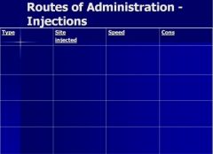
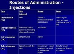
Routes of drug administration that aren't injections...
|
Oral
Sublingual Intrarectal Inhalation Topical Intracerebral Intracerebralventricular |
|
|
How _____ _____ a drug is affects how quickly it can pass through the blood-brain barrier and start working
|
lipid soluble
|
|
|
What is it called when drugs bind with molecules that are not the intended sites of action?
|
Depot binding
|
|
|
A protein in the blood that is responsible for depot binding of some drugs...
|
Albumin
|
|
|
What effect does depot binding have on the action of drugs?
|
Delays and prolongs
|
|
|
What is responsible for most deactivation and excretion of drugs?
|
Enzymes are responsible for deactivation; kidneys are responsible for most excretion
|
|
|
Point of maximum effect...
|
Level of dosage beyond which larger doses do not result in greater effects of the drug, but do result in more bad side effects
|
|
|
Margin of safety...
|
Difference between the dose-response curves for intended effects and side effects of a drug
|
|
|
Therapeutic index...
|
Ratio between the dose that produces desired effect in 50% of animals and the dose that produces a toxic effect in 50% of animals
|
|
|
Two reasons why drugs vary in their effectiveness...
|
1) Different drugs have different sites of action
2) Affinity |
|
|
Definition of affinity...
|
Readiness with which a drug binds to its receptor
|
|
|
What happens when you reach the point of tolerance?
|
Symptoms opposite those of the drug effects can occur when you stop taking the drug
|
|
|
Sensitization...
|
When repeated administration will make the drug more and more effective--not as common as tolerance effects
|
|
|
How does our body compensate for more drugs in the system?
|
1) Making receptors less sensitive to a drug (lowering their affinities)
2) Reducing number of receptors 3) Decreasing effectiveness of receptors so they can't bind to ion channels or make second messengers |
|
|
What may sometimes produce physiological and psychological effects of a placebo?
|
The administration of the placebo
|
|
|
Placebo...
|
An inert substance that has no direct physiological effect
|
|
|
Ways in which agonists and antagonists can act...
|
1) By affecting the production of neurotransmitters
2) By affecting the storage and release of neurotransmitters 3) By affecting the receptors for neurotransmitters 4) By affecting the reuptake or destruction of neurotransmitters |
|
|
If an agonist affects the production of new neurotransmitters, how does it work?
|
Increase the rate of creation and release of neurotransmitters
|
|
|
If an antagonist affects the production of new neurotransmitters, how does it work?
|
Inactivate an enzyme needed to create neurotransmitters
|
|
|
What do transporter molecules do?
|
Bring old neurotransmitters that have returned to the terminal button via reuptake and fill synaptic vesicles by pumping these neurotransmitters through their membranes
|
|
|
How do antagonists affect the storage and release of neurotransmitters?
|
Binding to transporter molecules, deactivating them; synaptic vesicles don't get filled, and when the rupture, nothing is released
|
|
|
How can an antagonist work to affect the storage or release of neurotransmitters that doesn't have anything to do with transporter molecules?
|
Deactivating the proteins that cause docked synaptic vesicles to fuse with presynaptic membrane
|
|
|
How can an agonist work to affect the storage or release of neurotransmitters?
|
Triggering the proteins that cause docked synaptic vesicles to fuse with the presynaptic membrane
|