![]()
![]()
![]()
Use LEFT and RIGHT arrow keys to navigate between flashcards;
Use UP and DOWN arrow keys to flip the card;
H to show hint;
A reads text to speech;
40 Cards in this Set
- Front
- Back
|
The perception of flavor involves: |
Gustatory, Trigeminal, and Olfactory inputs |
|
|
Taste buds are located on these structures: |
Papilae |
|
|
Taste buds are innervated by which of the following cranial nerves? |
X (Vagus) |
|
|
Second order gustatory neurons are located in this nucleus: |
Nucleus of the solitary tract |
|
|
The tastant Salty's primary signaling mechanisms is: |
Sodium channels |
|
|
The tastant Sweet's primary signaling mechanisms is: |
G-protein coupled receptor |
|
|
The tastant Umami's primary signaling mechanisms is: |
G-protein coupled receptor |
|
|
The tastant Sour's primary signaling mechanisms is: |
Non-selective cation channel |
|
|
(TRUE/FALSE): Fibers reaching gustatory cortex arise from the VPM of the thalamus |
True |
|
|
(TRUE/FALSE): Olfaction is mediated by receptors that project directly to the thalamus |
False |
|
|
(might be wrong) Chemosensory transduction in olfactory receptor cells involves: |
G-protein-coupled receptors |
|
|
Name three central projections of the olfactory bulb: |
Amygdala |
|
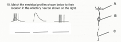
Match the electrical profiles shown beflow to their location in the olfactory neurons shown on the right. |
B, A, C |
|
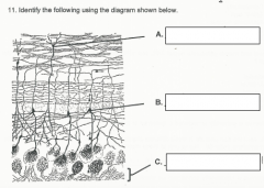
Identify the following using the diagram shown below: |
A. Granule cell |
|
|
(TRUE/FALSE): Gustatory and olfactory information converge in the orbital cortex |
True |
|
|
Sensory cells of the vestibular and auditory systems are found in the ________ labrynth |
Membranous |
|
|
Movement of this fluid (endolymph/perlymph) causes corresponding movements of the eyes via the _________ reflex |
Endolymph |
|
|
Sound waves of high frequency are detected at the (apex/base) of the cochlea by hair cells in the _________. |
Base |
|
|
To compensate for hearing loss, sound (intensity/frequency) must be increased. |
Intensity |
|
|
Place in order starting with periphery |
CN VII |
|
|
What two structures and two nucleuses receive input form the vestibular nuclei? |
Cerebellum |
|
|
The laterla and medial geniculate nuclei, VPL, VPM all belong to which major division of the thalamus? |
Lateral thalamus |
|
|
The thalamus is generally considered a 'gateway' to the _______ |
cortex |
|
|
Thalamic neurons can operate in three different physiological states (modes) depending on the resting membrane potential. Name of these states. |
Tonic firing mode (inactive) |
|
|
Inputs form cerebral cortex, other thalamic nuclei, basal ganglia, and spinal cord are all functionally connected via: |
Thalamus |
|
|
Fibers leaving the thalamus reach the cerebral cortex via the __________ |
Internal capsule |
|
|
Fibers destined for the occipital lobe from the thalamus would be located in which part of the internal capsule? |
Caudal |
|

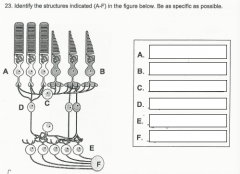
Identify the structures indicated in the figure below. |
A. Rod |
|
What is the direction of light? |
Up |
|
|
List three specialization of the fovea that enable it to convey vision of the highest acuity |
A. All cones/high density of cones |
|
|
Retinal ganglion cells have receptive fields known as _____________ |
Center surround receptive fields |
|
|
Cells of the nasal retina receive info from the (lateral/nasal) portion of the visual field and send (crossed/uncrossed) fibers to the thalamus |
Lateral |
|
|
Lesion of the left optic nerve would result in what visual field deficit |
Ipsilateral blindness |
|
|
Lesion of the optic chiasm would result in what visual field deficit |
Bitemporal hemianopia |
|
|
Lesion of the left ventral Meyer's loop would result in what visual field deficit |
Contralateral superior quadrantinopia |
|
|
Lesion of the left occipital lobe would result in what visual field deficit |
Contralateral hemianopia with macular sparing |
|
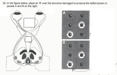
Match structure damage box with deficit produced (A or B) |
Top to bottom |
|
|
Each lower motor neuron innervates a group of muscle fibers, forming a ________ |
motor unit |
|
|
These types of muscle fibers are capable of generating the greatest amount of force: |
Fast twitch, fatigable |
|
|
Upper motor neurons are not found in the: |
Spinal cord |