Reading...
![]()
Play button
![]()
Play button
![]()
Use LEFT and RIGHT arrow keys to navigate between flashcards;
Use UP and DOWN arrow keys to flip the card;
H to show hint;
A reads text to speech;
49 Cards in this Set
- Front
- Back
|
The esophagus measures approximately ___ cm in length in adults.
|
23-25cm
|
|
|
The esophagus begins and ends approximately ____ cm and ___cm from the adult incisor teeth.
|
15-40cm
15 cm start of cervical segment 40 cm is pyloric sphincter |
|
|
The mid thoracic segment of the esophagus begins at the ____ (anatomic landmark).
|
Tracheal bifurcation
|
|
|
The cervical portion of the esophagus is bounded by the ____ nerves and overlapped by which gland?
|
Recurrent laryngeal nerves.
Thyroid gland. |
|
|
The angle of ___ forms a sharp angle between the distal esophagus and the fundus of the stomach.
|
His
|
|
|
What is the Z line?
|
A grossly visible line formed at the junction of esophagus and stomach.
|
|
|
Glycogenic Acanthosis presents as ___ ___ or ____ ____ on the mucosal folds, and is a variant of normal gross anatomy.b
|
White nodules or small plaques.
(May be confused with monilial plaques or leukoplakia). |
|
|
T/F. Heterotopias of the esophagus can include stomach, sebacious glands, thyroid, pancreatic tissue.
|
True
|
|
|
A narrowing of the lower part of the esophagus near the GE junction is called?
|
Schatzki's ring
Type A: above the Z line Type B: below the Z line. |
|
|
The esophagus mucosa consists of a
a) nonkeratinizing, stratified squamous mucosa b) simple columnar mucosa c) transitional mucosa |
A
|
|
|
What are the four layers of the esophagus?
|
Mucosa, submucosa, muscularis propria, serosa.
|
|
|
The mucosa layer consists of (name 3 layers)
|
1) Non keratinizing stratified squamous epithelium
2) Lamina propria 3) Muscularis mucosa |
|
|
Normal basal cell layer comprises ___% of the epithelium.
|
10-15%, may be more in the most distal segment.
|
|
|
The upper extent of the basal layer is defined as the layer in which the nuclei are separated by a distance of ___.
|
equal to the nuclei's diameter.
|
|
|
PAS stain shows that the basal layer of the epithelium is
a) glycogen rich b) glycogen poor |
B
|
|
|
T/F
Occasional lymphocytes within the basal cell layer of the epithelium is a normal finding. |
True
Like the rest of the GI tract, lymphocytes are (CD3+/CD8+) cytotoxic T cells. |
|
|
Progressing superficially, epithelial cells become more flattened and nuclei more ___.
|
pyknotic.
|
|
|
Patients with GERD typically have an (increased/decreased) proliferative activity of the basal cells.
|
increased
|
|
|
Stem cells in the esophagus consist of a single layer of cells and have a (high/low) proliferative activity, being Ki67 (+/-)
|
They have a low proliferative activity and are Ki67 negative.
Ki67 positive cells have high proliferative activity |
|
|
T/F
The lamina propria consist of areolar connective tissue, constains vascular structures, scattered inflammatory cells, and mucus secreting cells. |
True
|
|
|
T/F
Presence of inflammatory cells in the lamina propria is consistent with acid reflux |
False, they are a normal finding
|
|
|
Lymphocytes in the lamina propria consist of
|
Mostly CD4 T cells with some IgA B cells and IgG and igM B cells.
|
|
|
The Muscularis Mucosae typically becomes (thinner, thicker) distally in the esophagus
|
thicker
|
|
|
Esophageal cardiac type glands are diffusely scattered in the ___ layer through all levels of the esophagus, predominating in distal and proximal regions.
|
Lamina propria.
They function to secrete neutral mucin and resemble pyloric glands lined by gastric foveolar like cells. |
|
|
The submucosa consists of loose connective tissue containing vessels, nerve fibers (including the ____ plexus), and submucosal glands.
|
Meissner
|
|
|
T/F
In contrast with glands in the lamina propria, submucosal glands stain with PAS-D and Alcian blue. |
True. Cardiac-type glands in the lamina propria are typically neutral mucin producing glands.
|
|
|
The proximal 5% of muscularis propria is composed entirely of (striated, smooth) muscle.
|
Striated
|
|
|
Auerbach's plexus is found between which layers?
|
Circular muscle and Longitudinal muscle layers.
|
|
|
Scleroderma and Achalasia affect which muscular layer and in what way?
|
Circular muscle layer
(atrophy in scleroderma and hypertrophy in achalasia). |
|
|
T/F
The majority of the esophagus has a serosa layer. |
False, most are lined with fascia.
|
|
|
T/F
Lymphatics in muscular layer are oriented in a longitudinal direction, facilitating intramucosal and submucosal spread of tumor. |
True
Metastasis patterns are often varied and unpredictable |
|
|
The layer of ganglion cells between the circular and longitudinal layers of muscle are called
|
Auerbach's plexus
|
|
|
Barrett's esophagus is defined as replacement of normal epithelium with ____ epithelium.
|
Columnar
|
|
|
What type of mucin producing cells are typical in Barrett's esophagus? What do they stain with?
|
Goblet cells, stains positive with Alcian blue at ph 2.5.
|
|
|
Endoscopic abnormality (macro and microscopic) in Barretts esophagus is useful except in (3 cases)
|
1) patients with highly irregular Z line
2) in children of 1st decade (rarely exhibit metaplasia) 3) patients with long term PPI may not yield goblet cells |
|
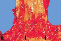
Describe the pathology
|
Barrett's esophagus, with red velvety mucosa which are columnar epithelium extending well into the tubular esophagus.
|
|
|
Goblet cells in Barrett's may be associated with mucin antigens ___ and ___.
|
MUC1, MUC6
|
|
|
Multilayered epithelium consist of apical ___ cells overlying a squamous epithelium.
|
Columnar
(mucin producing) -may represent early/transitional phase of columnar metaplasia in Barrett's esophagus |
|
|
Diagnosis is Barrett's esophagus is established from taking samples from where to where?
|
Stomach just distal to upper end of gastric folds then up every 1-2 cm until squamous epithelium is reached.
|
|
|
Barrett's esophagus is defined as less than ___ cm of columnar lined esophagus with goblet cells.
|
3
|
|
|
Intestinal Metaplasia of the stomach is usually caused by ___ and ___.
|
GERD and Helicobacter
|
|
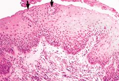
Describe the Pathology
|
Reflux esophagitis, with basal cell hyperplasia and lengthening of the papillae, extending almost to the surface of the mucosa (arrows).
|
|
|
Reflux-associated squamous hyperplasia (RASH) consists of basal cell hyperplasia of greater than __% and extension of paillae into ___ of the epithelium.
|
15%,
upper third Remember that RASH is a nonspecific reaction of esophageal injury |
|
|
T/F
RASH (reflux assoc squamous hyperplasia) can be seen in distal 3cm of esophagus in majority of patients without evidence of reflux. |
True
|
|
|
Other than RASH, what feature could be an early evidence of GERD?
|
dilated intercellular spaces, intraepithelial eosinophils.
|
|
|
Intraepithelial eosinophils are indicative of
|
a) alkaline reflux
b) allergic disorders c) infections d) GERD |
|
|
Intraepithelial neutrophils can be indicative of
|
a) acute esophagitis
b) GERD (less sensitive) |
|
|
The use of DIS (Dilated intercellular spaces), IEE (intraepithelial eosinophils), IEN (intraepithelial neutrophils) as histologic criteria of GERD has what advantage?
|
Localization and orientation of the biopsy is not important.
|
|
|
Barrett's mucosa has a risk of evolving into
|
Adenocarcinoma
|