![]()
![]()
![]()
Use LEFT and RIGHT arrow keys to navigate between flashcards;
Use UP and DOWN arrow keys to flip the card;
H to show hint;
A reads text to speech;
22 Cards in this Set
- Front
- Back
|
What are causes of diabetes mellitus ? |
Destruction of islet cells secondary to pancreatitis. Amyloid deposition in the islets of langerhans in cats. Idiopathic pancreatic atrophy. Hypoplasia. |
|
|
Pathology of diabetes mellitus? |
Reduced insuln--> hyperglycaemia Impaired leucocyte function so a reduced resistance to infection. Hepatic fatty change. Cataracts due to metabolism of glucose by the lens. Renal glomerular sclerosis- deposits of glycoprotein in glomeruli. |
|
|
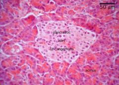
What do normal islets of langherhans appear to look like? |
|
|
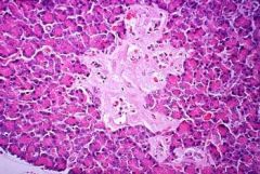
What has occurred in these islets? |
Amyloid has been deposited in them and impairs their function. |
|
|
Why is diabetes insipidus different to diabetes mellitus? |
Diabetes insipidus does not involve the pancreas. Animals with diabetes insipidus have marked polyuria and polydipsia. |
|
|
Causes of diabetes insipidus? |
Central diabetes insipidus- inadequate productions by the neurohypophysis of ADH Peripheral renal epithelial cells can't respond to ADH- congenital abscence of ADH receptors. Blockage of ADH receptors. |
|
|
Types of pancreatic neoplasm we get? |
Insulinoma Gastronoma Glucagonoma |
|
|
What are features of an animal that has an insulinoma? |
Hypoglycaemia |
|
|
What do gastrinomas do ? |
Gastrin--> hypersecretion of gastric acid--> ulceration of the mucosa |
|
|
Glucagonoma clinical signs? |
Hyperglycaemia |
|
|
What are developmental disorders of the thyroid gland? |
Accessory thyroid tissue may develop in the base of the heart or in the mediastinum due to remnants of embryological tissue. Thyroglossal cysts. Both these disorders have the potential to become neoplastic. |
|
|
Causes of hypothyrodism? |
Idiopathic follicular atrophy- replacement with adipose tissue. Lymphocytic thyroiditis-> infiltration of lymphocytes into the gland as a consequence of autoantibody formation. |
|
|
Clinical signs of hypothroidism? |
Reduced basal metabolic rate. Bilaterally symmetrical alopecia. Hyperpigmentation, hyperkeratis. Reduced spermatozoa count. Increase cholesterol levels leading to atheroscelerosis. |
|
|
What species is hyperthyrodism more common in and give symptoms? |
More common in cats. Hyperexcitability. Polyuria. Polydipsia. Weightloss. Heat intolerance. Hypertrophic cardiomyopathy. |
|
What is shown here and what causes it? |
Iron deficient animals may develop goitre. |
|
|
What are the features of follicular adenoma of the thyroid gland? |
Discrete capsule and usually solitary. Common in cat. |
|
|
Features of a follicular carcinoma? |
Locally invasive and often metastatise. |
|
|
Features of C cell neoplasia? |
C cells produce calcitonin in response to hypercalcaemia. Common in bulls. Often occur in association with the phaeochromocytoma and pituatary adenomas (Multiple endocrine neoplasia) |
|
|
Causes and features of primary hyperparathyroidism? |
Chief cell neoplasm usually an adenoma and common in dogs. Lead to bone resorption, hypercalcaemia and may cause pathological fractures of the bone. |
|
|
Causes of secondary hyperparathyroidism? |
Renal failure Nutritonal secondary hyperparathyroidism low calcium to phosphorus ratio |
|
|
What breeds is hypoparathyroidism more common in? Cause? |
Small breeds of dog. Usually caused by lymphocytic thyroiditis. Leads to neuromuscular excitability. |
|
|
What is a chemodectoma? Most commonly seen in? |

Adenoma or carcinoma of the aortic or carotid bodies that act as space occupying lesions. Brachycephalic dogs. |