![]()
![]()
![]()
Use LEFT and RIGHT arrow keys to navigate between flashcards;
Use UP and DOWN arrow keys to flip the card;
H to show hint;
A reads text to speech;
59 Cards in this Set
- Front
- Back
|
Cytotrophoblast |
An inner layer of mononucleated cells, derived from the trophoblast (day 8) |
|
|
Syncytiotrophoblast |
an outer multinucleated zone without distinct cells boundaries, formed from trophoblast around day 8
Initiates invasion of the uterine wall |
|
|
Embryoblast |
Cells of the internal mass of the blastocyst
|
|
|
Hypoblast |
small layer of cells adjacent to the blastocyst cavity, day 8, derives from embryoblast |
|
|
Epiblast layer |
layer of cells adjacent to the amniotic cavity. From embryoblast, day 8, together with hypoblast form a flat disc. Small cavity forms which becomes amniotic cavity |
|
|
7.5 day human blastocyst (picture) |

|
|
|
Primative Yolk Sack |
Forms around day 9, made via a fusion of the hypoblast and the exocoelomic membrane |
|
|
Day 4 |
Blastocoel: ICM surrounded by the trophoblast |
|
|
Day 8-11 |
Primative Yolk Sac Hypoblast proliferates and migrates out to surrounding trophoblast, forming primative yolk sac |
|
|
Day 12 |
Amniotic Cavitiy (cavity inside ICM), mesoderm begins to form |
|
|
Day 19-20 |
Amniotic cavity, yolk sac, chorionic cavity (space within amniotic mesoderm which surround the embryo) |
|
|
Relationship of different cavities |

|
|
|
T/F Maternal and Fetal Blood never exchange directly |
T. Protected by the placental barrier |
|
|
Fetal Villi |
Nutrient Exchange occurs on these, genetic info for screening can also be taken from these |
|
|
3 Types of Abnormal Implantation |
Rejection on the conceptus by the uterus Abnormal sites (ex Fallopian tubes) Hydatidiform mole (placenta keeps growing) |
|
|
Methods of Prenatal Diagnosis |
Ultrasonography Amniocentesis (>14 weeks) Chorionic Villus Sampling |
|
|
When is a functional placenta formed? |
Roughly week 3 |
|
|
Gastrulation |
The reorgannization of the epiblast through cell movements resulting in a three-layered embryonic disc (week 3). Most susceptible to environment and genetic insults that caause birth defects) The body axis are established, and the mother may not be aware of the pregnancy
Epiblast --> --> Ectoderm, Mesoderm, Endoderm |
|
|
Summary of the First Two weeks of Development (4 things) |
1. Cell division called cleavage produces a morula, individual cells are called blastomeres 2. Formation of a blastocyst signifies the 1st lineage specification 3.Differentiation of the innner cell mass into epiblast and hypoblast (2nd lineage differentiation) 4. Development of the extramembryonic tissues to support and protect the embryo |
|
|
Endoderm gives rise to what tissues? |
Internal organs (gut, liver, lung pancreas) |
|
|
Mesoderm gives rise to what tissues? |
Skeleton, muscles, heart, and blood
|
|
|
Ectoderm gives rise to what tissues? |
Neurons, glial cells, skin, and hair |
|
|
Primitive Streak |
characterizes the beginning of gastrulation, a slightly elevated group of cells on dorsal aspect of epiblast. Establishes body polarity |
|
|
primitive groove |
A shallow furrow of cells which separates 2 groups of cells in the primitive streak |
|
|
Primitive node |
where the primitive streak ends cranially |
|
|
Mesoderm vs Mesenchyme |
Mesoderm: embryonic term Mesenchyme: histological term/ describes cell morphology |
|
|
Induction |
the effect of one tissue on the development of adjacent tissue |
|
|
Loss of Primitive streak means |
abnormal fetal development of the lower spine |
|
|
Persistant Primative streak means that the fetus will |
generate a tumor |
|
|
NODAL |
Member of TGF beta family. Inhibited by signals from the hypoblast Binds to a cell receptor to initiate dimerization and enable a signal transduction Required for Left and right determinization (cillia control conc) |
|
|
duplication of primitive streak results in |
conjoined twins |
|
|
Neurulation |
formation of the neural tube |
|
|
Neural Tube |
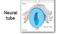
produces the entire cns |
|
|
Neural crest cells |

give rise to peripheral nervous system (red dots in pic) migrate throughout the body and differentiate into diverse cell types |
|
|
Epidermis |
gives rise to skin appendages |
|
|
Anencephaly |
neural tube closue skips a step at the head, folic acid helps in prevention |
|
|
Spina Bifida |
neural tube does not fully close on the dorsal posterior end, can lead to a fluid filled sac on back |
|
|
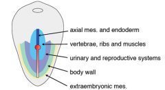
Differenntiation of the mesoderm (from axial outwards) |
axial --> paraxial --> intermediate --> lateral axial is composed of the notochord |
|
|
Paraxial mesoderm |
forms somites |
|
|
somites |
two spheres which form next to the neural tube give rise to muscle, dermis, vertbrae and ribbs |
|
|
Intermediate mesoderm |
forms urinary systems and major portions of the reproductive system |
|
|
Lateral Plate mesoderm |
forms the body cavity, has somatic mesoderm and splanchnic (or visceral mesoderm) |
|
|
Somatic (pariental) mesoderm |
forms the chest cavity, pelvis area (aka the embryonic cavity |
|
|
Splanchnic (visceral) mesoderm |
forms the gut tube (lined with enndoderm) and connected to the enbryonic cavity formed by somatic mesoderm via a mesentary ` |
|
|
The fate of cells is correlated with what in the epiblast? |
Their position |
|
|
Summary of Embryogensis |
1. Fertilization to blastpocyst formation (week 1), development of polarity 2. Formation of bilaminar disc (week 2), development of extramembryonic tissues (nutritive support and signalling) 3. Gastrulation (week 3), formation of germ layers and body axis, cells aquire temporo-spatial information for their fate determination during gastrulation 4. Organogensis (weeks 4-8), differentiation of three germ layers, neurulation, and somite formation |
|
|
Major events during embryogensis |
1. Cell division: generation of multicellular organisms (cleavage and growth) 2. Cell differentiation: generation of different and specialized cell types (germ layers and mesoderm) 3. Morphogenesis: cell movement leading to embryonic organization (gastrulation and cavitiation 4. Induction: a process by which the presence of one tissue influences the development of others |
|
|
Most common birth defects |
Congential heart defects cleeft lip or palate down syndrome (increases with age) spina bifida |
|
|
Types of birth defects (3 classes) |
Malformations Disruptions Deformation |
|
|
Causes of birth defects (2 classes) |
1. genetic factors 2.. environmental factors |
|
|
Teratogen |
subtstance able to cause birth defects |
|
|
teratogenesis |
development of defects in an embryo
|
|
|
After 8 weeks teratogenesis commonly results in ____________ |
mental retardation (heart, upper and lower limbs are done with development that could result in major morphological development by 8 weeks |
|
|
FGF |
Fibroblast growth factor, critical for limb expression |
|
|
Thalidomide |
reduces expression of FGF, causes severe birth defects |
|
|
Syndrome |
a well characterized group of anomlies that occur together in a predicatbale manner, probably due to an underlying etiology |
|
|
Association |
A group of anomlies that occur more frequently together than by chance, but the caus eis unkwon, we do not know the underlying mechanism |
|
|
Sequence |
A group of related anomlies that stem from an initial major anomoly that alters the development of other related tissues |
|
|
Determinants of tertogenic effect |
dose and duration embryonic factors: stage of development; genetic susceptibility Maternal factors: metabolism, excretion |